Correction to: Acta Pharmacologica Sinica 10.1038/s41401-020-00517-z; published online 24 September 2020
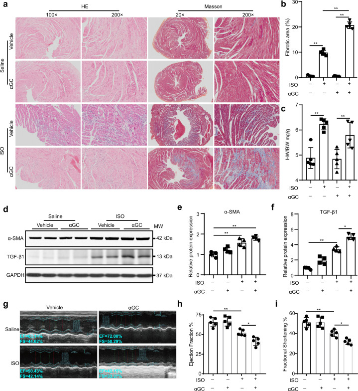
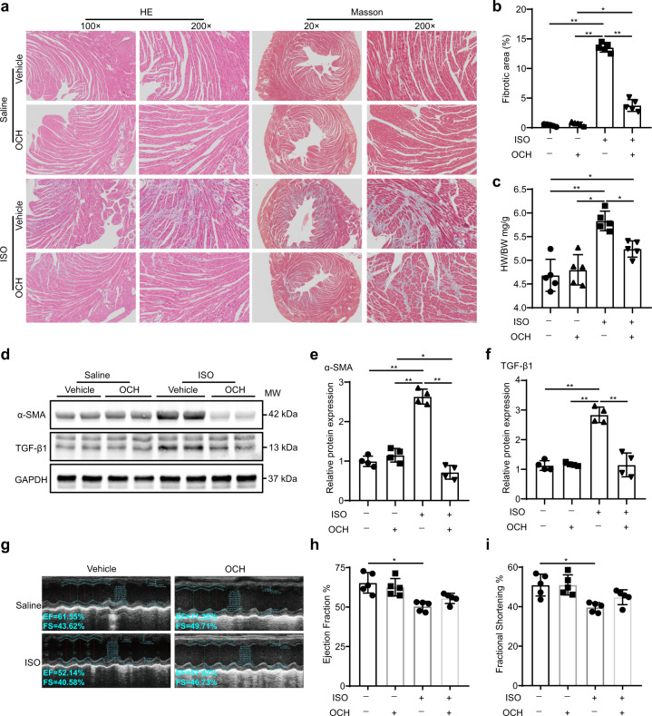
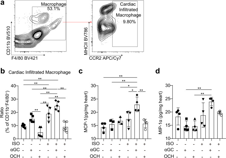
During our recent check of our published article entitled “α-Galactosylceramide and its analog OCH differentially affect the pathogenesis of ISO-induced cardiac injury in mice” published in Acta Pharmacologica Sinica. We realized that there were several mistakes in the final assemble of figures: (1) in Figs. 2a and 4a, representative images of Masson staining were obtained at 20× rather than 2×; (2) in Figs. 2g and 4g, representative images of echocardiography were mis-placed in the process of assembling figures; (3) in Fig. 8d, the chemokine determined is MIP-1α rather than MCP-1α. However, these mistakes do not affect the conclusions of the original article nor the text of the article and the figure legends. We apologize sincerely for any inconvenience caused.
Fig. 2. iNKT cells activated by αGC accelerate progressive ISO stimulation-induced cardiac fibrosis in mice.
WT C57BL/6 mice received ISO (5 mg/kg per day) alone for 7 days or together with αGC (3 μg/mouse) every other day. a HE staining and Masson’s trichrome staining were performed, and the fibrotic area/LV percentage (b) and ratio of heart weight to body weight (c) were calculated. d, e, f Whole-heart lysates were subjected to immunoblotting with the indicated antibody. GAPDH was used as the loading control. The data are expressed as a ratio of the experimental value to the mean value of the saline group. g, h, i Echocardiography was performed to evaluate cardiac function. The data are expressed as the mean ± SD. *P < 0.05 and **P < 0.01.
Fig. 4. iNKT cells activated by OCH attenuate progressive ISO stimulation-induced cardiac fibrosis in mice.
WT C57BL/6 mice received ISO (5 mg/kg per day) alone for 7 days or with OCH (3 μg/mouse) every other day. a HE staining and Masson’s trichrome staining were performed, and the fibrotic area/LV percentage (b) and ratio of heart weight to body weight (c) were calculated. d, e, f Whole-heart lysates were subjected to immunoblotting with the indicated antibody. GAPDH was used as the loading control. The data are expressed as a ratio of the experimental value to the mean value of the saline group. g, h, i Echocardiography was performed to evaluate cardiac function. The data are expressed as the mean ± SD. *P < 0.05 and **P < 0.01.
Fig. 8. Cardiac macrophages in αGC-induced cardiotoxicity.
a–d Hearts were harvested from mice stimulated with ISO (5 mg/kg per day) alone for 7 days or together with αGC or OCH every other day. a FACS was used to analyze the proportions of macrophages and adult monocyte-derived macrophages (infiltrated) in the hearts. b The percentage of CCR2-positive macrophages among CD11b+ F4/80+ cells was calculated. c, d Whole-heart lysates were subjected to ELISA immunoassays to determine the protein concentrations of MCP-1 and MIP-1α. The data are expressed as the mean ± SD. *P < 0.05 and **P < 0.01.
The corrected Figures are as follow:
Contributor Information
Da-yan Cao, Email: dayancao@outlook.com.
Xiao-hui Li, Email: lps008@aliyun.com.


