Abstract
Background
There is no consensus regarding markers of optimal treatment or timing between glucocorticoid intake and assessment of hormone levels in the follow-up of female 21-hydroxylase deficient patients.
Objective
To examine visit-to-visit repeatability in levels of adrenal hormones in adult female patients, to identify predictors of repeatability in hormone levels and to examine concordance between levels of different adrenal hormones.
Method
All patients with confirmed 21-hydroxylase deficiency treated with glucocorticoids, were included. The two most recent blood samples collected on a stable dose of glucocorticoid replacement were compared. Complete concordance was defined as all measured adrenal hormones either within, below or above normal range evaluated in a single-day measurement.
Results
Sixty-two patients, median age of 35 (range 18–74) years were included. All hormone levels showed moderate to excellent repeatability with an intraclass correlation coefficient between 0.80 and 0.99. Repeatability of hormone levels was not affected by the use of long-acting glucocorticoids or time of day for blood sample collection. The median difference in time between the two sample collections was 1.5 (range 0–7.5) h. Complete concordance between 17-hydroxyprogesterone, androstenedione, and testosterone was found in 21% of cases.
Conclusion
During everyday, clinical practice hormone levels in adult female patients with 21-hydroxylase deficiency showed a moderate to excellent repeatability, despite considerable variation in time of day for blood sample collection. We found no major predictors of hormone level variation. Future studies are needed to address the relationship between the timing of glucocorticoid intake vs adrenal hormone levels and clinical outcome in both adults and children.
Keywords: 21-hydroxylase deficiency, 17-hydroxyprogesterone, androstenedione, testosterone, DHEAs, CAH
Introduction
Congenital adrenal hyperplasia (CAH) is a group of autosomal recessive disorders caused by enzyme defects in the steroid biosynthesis of the adrenal cortex. In approximately 95% of cases, the disorder is caused by a pathogenic variant in the CYP21A2 gene resulting in reduced activity of the 21-hydroxylase enzyme (1, 2). Impaired activity of the CYP21A enzyme results in increased circulating levels of adrenal cortex hormones and steroid precursors upstream of CYP21A activity. 21-hydroxylase deficiency is characterised by a wide spectrum of phenotypes determined by the residual 21-hydroxylase activity, ranging from the classic, severe form, to mild non-classic forms. Classic 21-hydroxylase deficiency occurs in 1:10,000 to 1:20,000 live births, is often diagnosed in infants and is conventionally subdivided into salt-wasting and simple virilizing, indicating the degree of aldosterone deficiency (3). The non-classic form is much more common with an estimated prevalence of 1:200 to 1:1000 (4), and is typically diagnosed during workup of early puberty or due to symptoms of mild hyperandrogenism including hirsutism and acne in females (5).
The goal of treatment is first, to replace deficient hormones, and second, to suppress adrenocorticotropic hormone (ACTH) production thereby reducing excessive androgen levels. The treatment, especially in females, represents a challenging balance between iatrogenic hypercortisolism vs endogenous hyperandrogenism. Steroid hormone replacement is tailored according to symptoms and biochemical markers. There is a large number of circulating hormones (adrenal cortex hormones, precursors, ACTH and renin) which potentially can serve as markers of the steroid replacement efficacy. There might also be differences between classic vs non-classic 21-hydroxylase deficiency for example, it has been questioned if DHEAs is a valid disease marker in classic 21-hydroxylase deficiency (6). Adding to the complexity, 24-h hormone profiles in adult patients with 21-hydroxylase deficiency have shown a substantial circadian rhythm highly influenced by glucocorticoid replacement (6). Most recent clinical guideline from the US Endocrine Society suggests levels of androstenedione and 17-hydroxyprogesterone in the upper normal to mildly elevated normal range as a treatment goal (3), but currently, there is no consensus in relation to optimal disease markers or timing between replacement therapy and assessment of hormone levels. In the guideline by the US Endocrine Society, it is recommended that treatment should be monitored by consistently timed hormone measurements in relation to medication schedule and time of day (3). Others suggest that the disease is best monitored by measurements of maximal endogenous hormone levels, corresponding to early morning evaluation before medication (6, 7, 8, 9). Current recommendations, however, seem greatly based on expert opinion and clinical experience rather than empirical evidence.
The purpose of this study was (i) to investigate the repeatability of levels of adrenal hormones in adult female 21-hydroxylase deficient patients on a stable dose of glucocorticoid replacement; (ii) to identify predictors of repeatability of hormone levels focusing on time of day for blood sample collection, phenotype and long-acting vs short-acting glucocorticoid replacement; (iii) to assess concordance between serum levels of 17-hydroxyprogesterone, androstenedione and testosterone in relation to normal range in the same blood sample.
Methods
Patients
Patients were recruited from the Department of Endocrinology and Metabolism, Rigshospitalet, Copenhagen University Hospital. The department takes care of all adult female patients diagnosed with CAH in eastern Denmark, which has 2.7 million inhabitants, corresponding to roughly half of the Danish population. Male CAH patients are followed in the Department of Growth and Reproduction, Rigshospitalet which takes care of andrology. Male CAH patients were not included in the present study. Consecutive female patients treated at Rigshospitalet from 2016 to 2021 with genetically confirmed 21-hydroxylase deficiency were eligible for inclusion in the study. Diagnosis and phenotypic classification were determined by a review of medical records.
Study design
Patients who did not receive glucocorticoid replacement were excluded. Data regarding clinical features (e.g. hirsutism, menstrual abnormalities, bone mineral density and metabolic parameters), diagnosis, phenotypic classification, type of glucocorticoid replacement, and laboratory data were collected by review of medical records. Total glucocorticoid dose was calculated using the following conversion factors: 20 mg hydrocortisone equals 5 mg prednisolone and 0.75 mg dexamethasone, respectively (10). Surface area was calculated using the Du Bois formula (11). The laboratory data included the time of blood sample collection and circulating levels of 17-hydroxyprogesterone, androstenedione, testosterone and DHEAs. For each patient, we systematically identified the two most recent blood samples collected on a stable dose of glucocorticoid replacement, and with no changes in potential oestrogen supplementation or pregnancy. Patients were not recommended blood sampling at specific times and were advised to take the medication in the morning as prescribed.
Biochemical analyses
Blood samples were analysed at the Department of Clinical Biochemistry at Rigshospitalet.
Serum (s) 17-hydroxyprogesterone, androstenedione, testosterone and DHEAs were quantified by liquid chromatography-tandem mass spectrometry, performed by the Waters UPLC-TQS LC-MSMS system. The coefficients of variation (CV) for three levels of quality control were 8% for all hormones. The standard measuring ranges were as follows: 17-hydroxyprogesterone 0.5–75 nmol/L (normal range <10 nmol/L), androstenedione 0.5–60 nmol/L (normal range 2.4–8.9 nmol/L), testosterone 0.2 to 40 nmol/L (normal range 0.55–1.8 nmol/L) and DHEAs 0.1–25 µmol/L (normal range 1.2–9.5 µmol/L). For ten individual samples, testosterone was quantified by competitive electrochemiluminescence immunoassay (ECLIA), performed by the Cobas 8000, e801 module with a standard measuring range from 0.4 to 52 nmol/L. The CV for both low and high levels were 6%.
Statistical analyses
Statistical analyses and data collection were performed in IBM SPSS 27. Baseline characteristics were reported as n (%) for categorical data and as mean with s.d. for normally distributed continuous variables and median with interquartile range (IQR) for non-normally distributed variables. Test for normal distribution was done by visual inspection of histograms. Changes (first vs second measurement) in levels of 17-hydroxyprogesterone, androstenedione, testosterone and DHEAs were evaluated by a paired samples t-test, since the differences in hormone levels were normally distributed. After log transforming all hormone levels to approximate normal distribution, the intraclass correlation coefficient (ICC) was calculated, using a two-way mixed-effect absolute agreement model (12). Mean estimations and 95% CIs were reported for each ICC. ICC reflects both the degree of correlation and agreement between measurements. By convention ICC values less than 0.5 are indicative of poor repeatability, values between 0.5 and 0.75 indicate moderate repeatability, values between 0.75 and 0.9 indicate good repeatability and values greater than 0.9 indicate excellent repeatability (12).
Predictors of differences in serum adrenal hormone levels were assessed by linear regression analyses. The impact of age, type of glucocorticoid replacement (short-acting vs long-acting), surface area adjusted glucocorticoid dose, phenotype and time between adrenal hormone measurements was evaluated in univariate analyses. P values ≤ 0.05 were considered statistically significant. Statistically significant results were subsequently analysed in multivariate analyses.
Ethics
The study was approved by the executive committee at Rigshospitalet October 8, 2020. Due to the retrospective design informed consent was not required.
Results
Patient characteristics
A total of 66 patients with CAH due to 21-hydroxylase deficiency were treated at the department. Four were excluded because they did not receive glucocorticoid replacement, leaving a cohort of 62 patients with a median age of 35 (range 18–74) years. Clinical data are presented in Table 1. Classic 21 hydroxylase deficiency was diagnosed in 34/62 (55%) patients and 28/62 (45%) were diagnosed with the non-classic form. The median age of diagnosis for patients with the classic form was 0 (IQR 0–1) years compared to 10 (IQR 6–17) years for patients with the non-classic form. In both classic and non-classic 21-hydroxylase deficiency the majority of patients did not experience symptoms of hyperandrogenism (classic 71%, non-classic 61%). The remaining group of patients reported symptoms of hirsutism, acne, menstrual irregularities and infertility issues (Table 1). Total mean BMI was 27 ± 8 kg/m2 and was higher in patients with the classic form (31 ± 10 kg/m2 vs 24 ± 4 kg/m2, P = 0.002). Mean systolic blood pressure in the entire cohort was 123 ± 14 and diastolic 80 ± 11 mmHg. Eight patients had blood pressure >140/90 (six classic forms, two non-classic forms) and two (one classic, one non-classic) were treated with antihypertensive drugs. All patients, except two with the classic form, had normal glucose levels based on Hba1c (<48 mmol/mol). These two patients were treated with antidiabetic drugs.
Table 1.
Baseline characteristics of female 21-hydroxylase patients treated at the Department of Endocrinology, Rigshospitalet, Denmark. The classic form includes both the simple virilizing and the salt-wasting form.
| Total | Classic | Non-classic | P-value | |
|---|---|---|---|---|
| Patients included, n (%) | 62 | 34 (55) | 28 (45) | |
| Age at first blood sample collection median (IQR) | 35 (24–49) | 41 (26–50) | 31 (23–47) | 0.37 |
| Age at diagnosis (years) median (IQR) | 3 (0–10) | 0 (0–1) | 10 (6–17) | <0.001 |
| Symptoms of hyperandrogenism | ||||
| Hirsutism, n (%) | 9 (15) | 4 (12) | 5 (18) | 0.55 |
| Acne, n (%) | 2 (3) | 1 (3) | 1 (4) | 0.92 |
| Menstrual irregularities, n (%) | 4 (6) | 2 (6) | 2 (7) | 0.88 |
| Infertility, n (%) | 2 (3) | – | 2 (7) | |
| Height (cm) mean ± s.d. | 161 ± 7 | 160 ± 7 | 162 ± 8 | 0.13 |
| BMI (kg/m2) mean ± s.d. | 27 ± 8 | 31 ± 10 | 24 ± 4 | 0.002 |
| BMD z-score hip mean ± s.d. (n = 29) | −0.29 ± 0.80 | −0.4 ± 0.74 | −0.42 ± 0.96 | 0.29 |
| BMD z-score L2-L4 mean ± s.d. (n = 29) | −0.24 ± 1.51 | 0.37 ± 1.57 | −0.90 ± 0.97 | 0.017 |
| Systolic blood pressure (mmHg) mean ± s.d. | 123 ± 14.0 | 126 ± 14 | 119 ± 13 | 0.024 |
| Diastolic blood pressure (mmHg) mean ± s.d. | 80 ± 11 | 82 ± 11 | 77 ± 10 | 0.045 |
| HbA1c (mmol/mol) median (IQR) | 33 (32–38) | 35 (33–39) | 33 (32–36) | 0.076 |
| Sodium at first sample (mmol/l) median (IQR) | 140 (138–142) | 140 (139–142) | 140 (138–141) | 0.83 |
| Potassium at first sample (mmol/l) median (IQR) | 3.8 (3.7–4.1) | 3.8 (3.7–4.1) | 3.9 (3.7–4.1) | 0.52 |
BMD, bone mineral density; HbA1c, haemoglobin A1c; IQR, interquartile range.
Bone mineral density data were available for 29 patients (20 classic form, 9 non-classic form). In patients with the classic form, none had z-scores below −2SD in hip or lumbar regions. For patients with the non-classic form, 1/9 had z-score below −2SD in the lumbar region. Patients with the non-classic form had significantly lower z-scores in the lumbar region compared to patients with the classic form (P = 0.017, Table 1).
Glucocorticoid therapy
Patients were treated with a variety of glucocorticoid preparations. The majority received hydrocortisone 58/62 (94%). Four patients received long-acting glucocorticoids in monotherapy (n = 2 dexamethasone, n = 2 prednisolone). Combination treatment with both short-acting and long-acting glucocorticoids was significantly more frequent in patients with classic 21-hydroxylase deficiency vs patients with non-classic form (P = 0.004) (Table 2). Patients with the classic form received higher doses of glucocorticoids (P < 0.001), with the difference remaining significant after surface area adjustment (P = 0.002) (Table 2). The median duration of glucocorticoid treatment in the entire cohort was 24 (IQR 16 to 46) years. Treatment duration was longer for patients with the classic form (41 (24–51) vs 16 (8–23) years, P < 0.001).
Table 2.
Glucocorticoid therapy in 62 patients with 21-hydroxylase deficiency treated at the Department of Endocrinology, Rigshospitalet, Denmark. The classic form includes both the simple virilizing and the salt-wasting form.
| Glucocorticoid treatment | Total (n = 62) | Classic (n = 34) | Non-classic (n = 28) | P-value |
|---|---|---|---|---|
| Glucocorticoid treatment years median (IQR) | 24 (16–46) | 41 (24–51) | 16 (8–23) | <0.001 |
| Hydrocortisone treatment | ||||
| n (%) | 58 (94) | 31 (91) | 27 (96) | 0.40 |
| Median dose, mg/day (range) | 20 (5–50) | 20 (5–50) | 15 (5–25) | 0.002 |
| Dexamethasone treatment | ||||
| n (%) | 24 (39) | 18 (53) | 6 (21) | 0.011 |
| Median dose (range) (mg/day) | 0.1 (0.05–0.5) | 0.1 (0.05–0.5) | 0.1 (0.05–0.2) | 0.23 |
| Prednisolone treatment | ||||
| n (%) | 3 (5) | 3 (9) | – | |
| Median dose (range) (mg/day) | 7.5 (2.0–7.5) | 7.5 (2.07.5) | – | |
| Combination of glucocorticoid preparations n (%) | 23 (37) | 18 (53) | 5 (18) | 0.004 |
| Total glucocorticoid dose (mg HC) ± s.d. | 20 ± 9 | 25 ± 9 | 15 ± 7 | <0.001 |
| Total glucocorticoid dose (mg HC/m2/day) (IQR) | 11.7 (8.0–14.7) | 13.4 (10.6–16.6) | 10.7 (6.2–12.4) | 0.002 |
HC, hydrocortisone; IQR, interquartile range.
Repeatability of hormone levels
The median time of day for the first sample collection was 11 h (IQR 09:30 h to 12:45 h) vs 11:30 h (IQR 09:30 h to 13:30 h) for the second sample collection (P = 0.32). The median time distance between the two measurements was 8 (IQR 6–12) months. Levels of serum adrenal hormones are presented in Table 3. The mean levels of hormones assessed on the two different occasions did not significantly differ (all P - values >0.26). Median s-testosterone was 0.77 (IQR 0.35–1.54) at the first assessment and 0.82 (IQR 0.31–1.64) at the second assessment, normal range of 0.55–1.8 nmol/L, whereas s-androstenedione was 3.5 (IQR 1.0–6.6) and 3.2 (IQR 1.2–6.8), normal range of 2.4–8.9 nmol/L. Mean ICC varied from 0.82 to 0.99. Serum DHEAs had the highest ICC (0.99 (95% CI 0.98–0.99)) corresponding to excellent repeatability between the two measurements, whereas s-17-hydroxyprogesterone had the lowest (0.82 (95% CI 0.69–0.89) corresponding to a moderate to good repeatability. ICC for androstenedione and testosterone were 0.89 (95% CI 0.80–0.94) and 0.88 (95% CI 0.79–0.93), respectively, corresponding to good to excellent repeatability. Mean ICC varied from 0.81 to 0.98 for patients on long-acting glucocorticoids and from 0.80 to 0.99 for patients on monotherapy with short-acting glucocorticoids.
Table 3.
Serum hormone levels and intraclass correlation coefficients in 21-hydroxylase deficiency patients treated at the Department of Endocrinology, Rigshospitalet, Denmark. Sample 1 and sample 2 – the two most recent blood samples collected on a stable dose of glucocorticoid treatment and with no changes in potential oestrogen supplementation or pregnancy. The mean levels of hormones assessed on the two different occasions did not significantly differ (all P - values >0.26).
| Sample 1 median (IQR) | Sample 2 median (IQR) | ICC (95% CI) all patients | ICC (95% CI) long-acting GC | ICC (95% CI) short-acting GC | |
|---|---|---|---|---|---|
| 17-OH, n = 57 | 13.4 (5.7–37.2) | 11.2 (3.2–34.3) | 0.82 (0.69–0.89) | 0.82 (0.61–0.92) | 0.80 (0.59–0.90) |
| ASD, n = 51 | 3.5 (1.0–6.6) | 3.2 (1.2–6.8) | 0.89 (0.80–0.94) | 0.81 (0.54–0.92) | 0.92 (0.83–0.96) |
| Testosterone, n = 57 | 0.77 (0.35–1.54) | 0.82 (0.31–1.64) | 0.88 (0.79–0.93) | 0.85 (0.66–0.93) | 0.89 (0.77–0.95) |
| DHEAs, n = 49 | 0.2 (0.1–1.6) | 0.2 (0.1–1.8) | 0.99 (0.98–0.99) | 0.98 (0.96–0.99) | 0.99 (0.97–0.99) |
NR, normal range, 17-OH, 17-hydroxyprogesterone (NR: <10 nmol/L); ASD, androstenedione (NR: 2.4–8.9 nmol/L); testosterone (NR: 0.55–1.8 nmol/L); DHEAs: (NR: 1.2–9.5 µmol/L); IQR: interquartile range; ICC, intraclass correlation coefficient; GC, glucocorticoid.
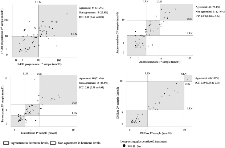
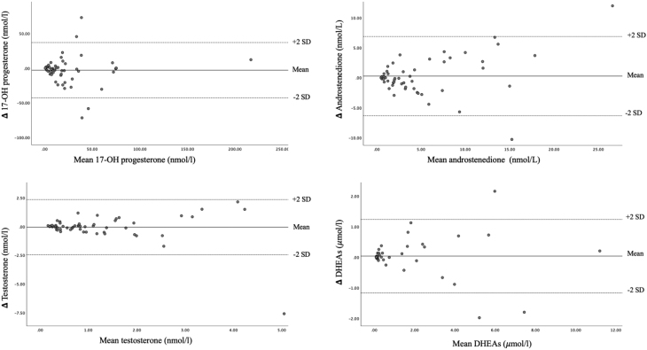
The association between hormone levels from the first and the second visit is provided in Fig. 1. When evaluated according to the normal range the highest agreement was found for s-DHEAs with 100% repeatability (32 pairs below normal range, 15 pairs within normal range and 1 pair above normal range). Serum testosterone levels below normal range in both samples (<0.55 nmol/L) were found in 16/56 (29%) of patients, 18/56 (32%) of patients had s-testosterone levels within normal range in both samples and 6/56 (11%) had increased levels in both samples (> 1.8 nmol/L). The corresponding numbers for androstenedione were 18/51 (32%), 14/51 (27%), and 8/51 (16%) (Fig. 1). Increased levels of s-17-hydroxyprogesterone (>10 nmol/L) in both samples were found in 26/57 (46 %) of patients. The agreement between the two measurements is also shown in Bland–Altman plots (Fig. 2).
Figure 1.
Repeatability of hormone levels in the two samples. Comparison of hormone levels in the two samples. For s-androstenedione, s-testosterone and s-DHEAs the shaded areas represent hormone levels below, within or above normal range in both measurements. For s-17-hydroxyprogesterone the shaded areas represent hormone levels within and above normal range for both measurements. The ICC is reported with the 95% CI. ICC, intraclass correlation coefficient; LLN, lower limit of normal; ULN, upper limit of normal.
Figure 2.
Bland–Altman plots. Bland–Altman plots displaying agreement between serum hormone levels in the first and second blood sample.
Predictors of repeatability of hormone levels
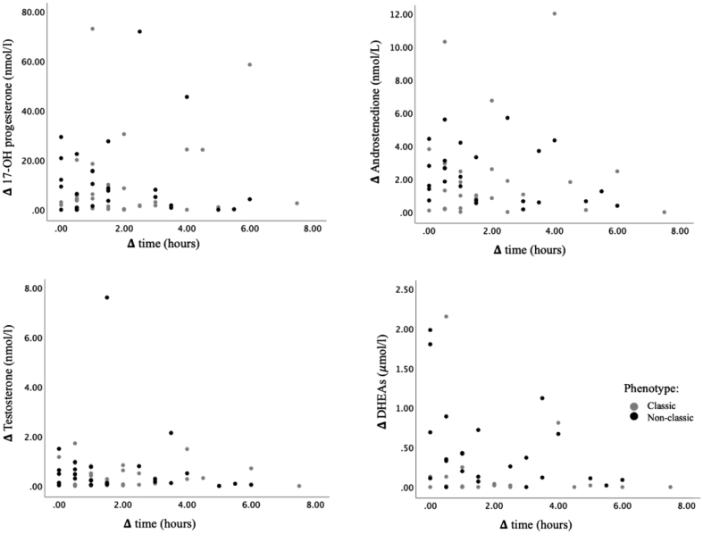
The association between difference in time of day of sample collection vs difference in hormone levels in the two samples is shown in Fig. 3. As seen, difference in time of day between sample collections did not significantly influence differences in hormone levels for any of the four hormones P - values 0.13–0.62. The median difference in time between the two sample collections were 1.5 (IQR 0.5–3), range 0–7.5 h.
Figure 3.
Difference in time of sample collection vs difference in hormone levels. Difference in time (hours) of sample collection vs difference in levels of s-adrenal hormones between the two blood samples.
Using linear regression analyses predictors of repeatability of hormone levels between the two measurements for all four evaluated hormones were examined. Explanatory variables included difference in time of day between sample collections, age, type of glucocorticoid replacement, surface area adjusted glucocorticoid dose and phenotype. The median difference in levels of s-DHEAs between the two measurements was higher in patients with non-classic 21-hydroxylase deficiency compared to the classic form 0.01 (IQR 0.00–0.06) vs 0.33 (IQR 0.11–0.69) µmol/L, P = 0.028). Furthermore, difference in s-DHEAs was inversely associated with age, with a decrease of 0.13 µmol/L per 10 years (P = 0.009). In multivariate analysis both variables (phenotype and age) remained significant predictors of variation in s-DHEAs (both P < 0.04). Difference in s-androstenedione was positively associated with surface area adjusted glucocorticoid dose, with an increase in difference of 1.66 nmol/L per 10 mg hydrocortisone/m2/day (P = 0.003). The remaining explanatory variables were not significantly associated with any of the hormones.
Concordance between 17-hydroxyprogesterone, androstenedione and testosterone
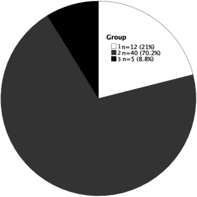
The patients were divided into three groups according to concordance between hormone levels measured in the same blood sample. Group 1: all three hormones either below, within or above normal range. Group 2: two hormones either below, within, or above normal range and one hormone differing. Group 3: two hormones below normal range and one above, or hormones distributed both below, above and within normal range. As seen in Fig. 4, 12/57 (21%) of patients were in group 1 with complete concordance between all three hormones, while 5/57 patients (9%) were in group three with the highest degree of discordance. The remaining 70% of patients were in group 2.
Figure 4.
Concordance between levels of s-17-hydroxyprogesterone, s-androstenedione and stestosterone at the first sampling day. Concordance between levels of 17-hydroxyprogesterone, androstenedione and testosterone at the first sampling day. Group 1: the highest degree of concordance with all three hormones either below, within, or above the normal range. Group 2: two hormones either below, within, or above the normal range and one hormone differing. Group 3: two hormones below normal range and one above, or hormones distributed both below, above and within the normal range.
Discussion
The main result from the present study was that potential biochemical markers of treatment efficacy showed a moderate to excellent visit-to-visit repeatability in female patients with 21-hydroxylase deficiency considered to be well controlled on a stable glucocorticoid dose. We did not identify any major predictors of repeatability of hormone levels. Finally, we found a substantial discordance in levels of the different adrenal hormones when evaluated in accordance with normal ranges on a single day. Only 21% of patients showed complete concordance with levels of 17-hydroxyprogesterone, androstenedione and testosterone either below, within or above the normal range.
The clinical presentation including, blood pressure, hirsutism, acne, menstrual irregularity, BMI, and cushingoid features plays an important role in the assessment of optimal treatment in female patients with 21-hydroxylase deficiency. However, biochemical markers are essential for disease control as well because some clinical features, for example, osteoporosis, only occurs after years of glucocorticoid overtreatment, and there may be unknown long-term consequences of hyperandrogenism in women (13, 14). It is therefore important to have access to reliable and stable biochemical markers that reflect treatment efficacy, to help guide glucocorticoid dose titration.
As inclusion criterion patients should be well controlled on a stable steroid dose. A minority of patients had some minor remnant symptoms of hyperandrogenism but did not justify the increase in glucocorticoid dose. Patients with the classic form needed significantly higher doses of steroids to control symptoms and it may be a contributing factor to higher BMI and higher blood pressure in the classic form. However, in general and despite a median treatment duration of 24 years the patients did not show evidence of a cushingoid phenotype based on glucose metabolism, blood pressure and levels of sodium and potassium. In the classic form mean BMI was 30.5 kg/m2 and according to Statistics Denmark mean BMI among Danish women in 2010 was 24,7 kg/m2 (15). Thus, it is a possibility that steroid treatment may contribute to adiposity in patients with classic 21-hydroxylase deficiency. In recent guidelines from 2018 (3), it is recommended that patients with the non-classic form are treated with oral contraceptives and/or anti-androgens instead of steroids. However, the majority of our patients have a rather severe form of non-classic 21-hydroxylase deficiency, diagnosed long ago during childhood and initially treated with steroids at the pediatric department. Our ambition is to reduce and if possible, withdraw steroid treatment but due to iatrogenic adrenal insufficiency and reluctance among the patients, it is challenging. Despite long-term glucocorticoid treatment none of the patients had overt signs of steroid side effects based on metabolic parameters and bone mineral density.
To the best of our knowledge, this is the first study to report data on visit-to-visit repeatability in hormone levels in female 21-hydroxylase deficient patients on a stable glucocorticoid replacement dose. Based solely on stability, our data suggest that s-DHEAs is the preferable marker of biochemical efficacy with an excellent repeatability and 100% agreement between the two samples when evaluated according to normal levels. The long half-life of 19 to 22 h (16) may contribute to stable serum levels. The remaining adrenal hormones have half-lives of less than 20 min (17). However, it is obviously essential that the marker accurately represents optimal glucocorticoid treatment, and that a therapeutic target range can be identified. Low levels of s-DHEAs have previously been described in patients with classic 21-hydroxylase deficiency even when the disease was uncontrolled (6, 18, 19), questioning its usefulness as a treatment efficacy marker in classic 21-hydroxylase deficiency, but it cannot be excluded, that it might be useful in non-classic 21-hydroxylase deficiency (6). ICC for s-DHEAs in patients with the non-classic form was 0.98 (95% CI 0.96 to 0.99) corresponding to excellent repeatability. Serum androstenedione and s-testosterone showed a good and comparable repeatability, superior to s-17-hydroxyprogesterone, and based on our results these two hormones might be the best candidates for monitoring treatment in 21-hydroxylase deficiency.
Patients were encouraged to take their medication in the morning before blood sampling. Another suggested approach to achieve stable hormone measurements is to measure maximal hormone levels (6, 7, 8, 9). Based on studies investigating circadian rhythm in relation to glucocorticoid intake in 21-hydroxylase deficient patients this corresponds to early morning (before glucocorticoid administration) evaluation for patients receiving hydrocortisone or night-time prednisone, or late afternoon evaluation for patients receiving morning prednisone or night-time dexamethasone (6, 8, 9). Since we did not advise our patients to abstain from glucocorticoid replacement before measurement of hormone levels, we are not able to elucidate this hypothesis. It should be emphasised that the evaluation of the stability of hormone levels when aiming for maximum hormone concentration has never been investigated in clinical studies. In general, the hormone levels showed a good repeatability, despite considerable variation in the time of day the blood samples were collected. We did not document the timespan from the last glucocorticoid intake to hormone assessment, which is a major study limitation. Therefore, we are not able to elucidate the exact impact of exogenous steroids vs variation in adrenal hormone levels.
We also investigated the impact of other potential predictors of hormone variation (phenotype, age, type of glucocorticoid replacement, surface area adjusted glucocorticoid dose). We did not find any impact of long-acting steroids on hormone level variation for any of the hormones, with comparable ICC in patients treated with vs without long-acting glucocorticoids. Serum DHEAs was more stable in patients with classic 21-hydroxylase deficiency compared to patients with the non-classic form. Most likely, the low variation in s-DHEAs in classic 21-hydroxylase deficiency reflects that s-DHEAs is not primarily influenced by glucocorticoid treatment in the classic form. Serum DHEAs showed larger variation in younger patients and variation in s-androstenedione was positively associated with glucocorticoid dose. We do not have any plausible explanation for these observations, and it might be a chance finding. Taken together, despite exploring many different factors that might influence hormone variation we did not find any major predictors.
Ideally, treatment efficacy should be monitored by one stable biomarker reflecting treatment efficacy and long-term prognosis. Such biomarker has not been identified in 21-hydroxylase deficiency. Our data highlight that having multiple biomarkers can be a challenge in everyday clinical practice since it potentially leads to discordant results. In around 20% of cases, we found good agreement between serum levels of three adrenal hormones when evaluated according to normal ranges. However, in approximately 9% of patients, hormone levels were distributed both below, within and above the normal range measured in the same blood sample. In this analysis we excluded DHEAs. The different hormones showed different patterns. Androstenedione and testosterone were mainly below or within normal range whereas s-17-hydroxyprogesterone were mainly within or above the normal range. It should be emphasised that achieving hormone levels within normal range may not be the optimal goal of treatment (3).
This retrospective study has several limitations. It is a major limitation that we do not have the exact time for the last intake of steroids. For some of the hormones (especially 17OH-progesterone), the ICC 95% CI are quite broad covering relevant differences (from moderate to good repeatability), and therefore a larger cohort would have been preferable to reduce the CIs. It cannot be excluded that a larger variation in hormone levels could have been identified if the time interval for blood sample collection was expanded (all samples were collected between 07:30 h and 15:30 h). Moreover, the study only included adult women exposed to different treatment regimens, implying that caution should be taken when extrapolating the results to other cohorts including males and children Finally, the measured hormones are not exclusively secreted from the adrenals. We do not routinely do an ovarian ultrasound and hormonal contribution from a concomitant diagnosis of polycystic ovarian syndrome cannot be excluded.
In conclusion, hormone levels in adult females with 21-hydroxylase deficiency showed a moderate to excellent repeatability, and we did not identify any major predictors of repeatability of in hormone levels. This is a novel observation that needs to be confirmed in future studies focusing on the relationship between the timing of glucocorticoid intake vs adrenal hormone levels and clinical outcome Based solely on stability, s-androstenedione and s-testosterone are preferable as disease markers compared to s-17-hydroxyprogesterone. This, of course, presupposes that a level representing treatment efficacy and long-term prognosis can be determined. Serum DHEAs might be useful in monitoring treatment efficacy for patients with the non-classic form, but this topic merits further investigations. Future research should investigate the association between clinical symptoms of over or under treatment and hormone values.
Declaration of interest
The authors declare that there is no conflict of interest that could be perceived as prejudicing the impartiality of the research reported.
Funding
This work did not receive any specific grant from any funding agency in the public, commercial or not-for-profit sector.
References
- 1.White PC, New MI, Dupont B. HLA-linked congenital adrenal hyperplasia results from a defective gene encoding a cytochrome P-450 specific for steroid 21-hydroxylation. PNAS 1984817505–7509. ( 10.1073/pnas.81.23.7505) [DOI] [PMC free article] [PubMed] [Google Scholar]
- 2.Krone N, Dhir V, Ivison HE, Arlt W. Congenital adrenal hyperplasia and P450 oxidoreductase deficiency. Clinical Endocrinology 200766162–172. ( 10.1111/j.1365-2265.2006.02740.x) [DOI] [PubMed] [Google Scholar]
- 3.Speiser PW, Arlt W, Auchus RJ, Baskin LS, Conway GS, Merke DP, Meyer-Bahlburg HFL, Miller WL, Murad MH, Oberfield SE, et al. Congenital adrenal hyperplasia due to steroid 21-hydroxylase deficiency: an Endocrine Society clinical practice guideline. Journal of Clinical Endocrinology and Metabolism 20181034043–4088. ( 10.1210/jc.2018-01865) [DOI] [PMC free article] [PubMed] [Google Scholar]
- 4.Hannah-Shmouni F, Morissette R, Sinaii N, Elman M, Prezant TR, Chen W, Pulver A, Merke DP. Revisiting the prevalence of nonclassic congenital adrenal hyperplasia in US Ashkenazi Jews and Caucasians. Genetics in Medicine 2017191276–1279. ( 10.1038/gim.2017.46) [DOI] [PMC free article] [PubMed] [Google Scholar]
- 5.Nordenström A, Falhammar H. MANAGEMENT OF ENDOCRINE DISEASE: Diagnosis and management of the patient with non-classic CAH due to 21-hydroxylase deficiency. European Journal of Endocrinology 2019180R127–R145. ( 10.1530/EJE-18-0712) [DOI] [PubMed] [Google Scholar]
- 6.Debono M, Mallappa A, Gounden V, Nella AA, Harrison RF, Crutchfield CA, Backlund PS, Soldin SJ, Ross RJ, Merke DP. Hormonal circadian rhythms in patients with congenital adrenal hyperplasia: identifying optimal monitoring times and novel disease biomarkers. European Journal of Endocrinology 2015173727–737. ( 10.1530/EJE-15-0064) [DOI] [PMC free article] [PubMed] [Google Scholar]
- 7.Merke DP, Bornstein SR. Congenital adrenal hyperplasia. Lancet 20053652125–2136. ( 10.1016/S0140-6736(0566736-0) [DOI] [PubMed] [Google Scholar]
- 8.Charmandari E, Matthews DR, Johnston A, Brook CG, Hindmarsh PC. Serum cortisol and 17-hydroxyprogesterone interrelation in classic 21-hydroxylase deficiency: is current replacement therapy satisfactory? Journal of Clinical Endocrinology and Metabolism 2001864679–4685. ( 10.1210/jcem.86.10.7972) [DOI] [PubMed] [Google Scholar]
- 9.Dauber A, Feldman HA, Majzoub JA. Nocturnal dexamethasone versus hydrocortisone for the treatment of children with congenital adrenal hyperplasia. International Journal of Pediatric Endocrinology 20102010 347636. ( 10.1155/2010/347636) [DOI] [PMC free article] [PubMed] [Google Scholar]
- 10.Meikle AW, Tyler FH. Potency and duration of action of glucocorticoids. Effects of hydrocortisone, prednisone and dexamethasone on human pituitary-adrenal function. American Journal of Medicine 197763200–207. ( 10.1016/0002-9343(7790233-9) [DOI] [PubMed] [Google Scholar]
- 11.Du Bois D, Du Bois EF. A formula to estimate the approximate surface area if height and weight be known. Nutrition 19895303–311; discussion 12–13. [PubMed] [Google Scholar]
- 12.Koo TK, Li MY. A guideline of selecting and reporting intraclass correlation coefficients for reliability research. Journal of Chiropractic Medicine 201615155–163. ( 10.1016/j.jcm.2016.02.012) [DOI] [PMC free article] [PubMed] [Google Scholar]
- 13.Falhammar H, Filipsson Nyström H, Wedell A, Brismar K, Thorén M. Bone mineral density, bone markers, and fractures in adult males with congenital adrenal hyperplasia. European Journal of Endocrinology 2013168331–341. ( 10.1530/EJE-12-0865) [DOI] [PubMed] [Google Scholar]
- 14.Ceccato F, Barbot M, Albiger N, Zilio M, De Toni P, Luisetto G, Zaninotto M, Greggio NA, Boscaro M, Scaroni C, et al. Long-term glucocorticoid effect on bone mineral density in patients with congenital adrenal hyperplasia due to 21-hydroxylase deficiency. European Journal of Endocrinology 2016175101–106. ( 10.1530/EJE-16-0104) [DOI] [PubMed] [Google Scholar]
- 15.Toft U, Vinding AL, Larsen FB, Hvidberg MF, Robinson KM, Glümer C. The development in body mass index, overweight and obesity in three regions in Denmark. European Journal of Public Health 201525273–278. ( 10.1093/eurpub/cku175) [DOI] [PubMed] [Google Scholar]
- 16.Legrain S, Massien C, Lahlou N, Roger M, Debuire B, Diquet B, Chatellier G, Azizi M, Faucounau V, Porchet H, et al. Dehydroepiandrosterone replacement administration: pharmacokinetic and pharmacodynamic studies in healthy elderly subjects. Journal of Clinical Endocrinology and Metabolism 2000853208–3217. ( 10.1210/jcem.85.9.6805) [DOI] [PubMed] [Google Scholar]
- 17.Holst JP, Soldin OP, Guo T, Soldin SJ. Steroid hormones: relevance and measurement in the clinical laboratory. Clinics in Laboratory Medicine 200424105–118. ( 10.1016/j.cll.2004.01.004) [DOI] [PMC free article] [PubMed] [Google Scholar]
- 18.Sellers EP, MacGillivray MH. Blunted adrenarche in patients with classical congenital adrenal hyperplasia due to 21-hydroxylase deficiency. Endocrine Research 199521537–544. ( 10.1080/07435809509030471) [DOI] [PubMed] [Google Scholar]
- 19.Helleday J, Siwers B, Ritzén EM, Carlström K. Subnormal androgen and elevated progesterone levels in women treated for congenital virilizing 21-hydroxylase deficiency. Journal of Clinical Endocrinology and Metabolism 199376933–936. ( 10.1210/jcem.76.4.8473408) [DOI] [PubMed] [Google Scholar]


 This work is licensed under a


