Abstract
A rapid, quantitative PCR assay (TaqMan assay) which quantifies Clostridium botulinum type E by amplifying a 280-bp sequence from the botulinum neurotoxin type E (BoNT/E) gene is described. With this method, which uses the hydrolysis of an internal fluoregenic probe and monitors in real time the increase in the intensity of fluorescence during PCR by using the ABI Prism 7700 sequence detection system, it was possible to perform accurate and reproducible quantification of the C. botulinum type E toxin gene. The sensitivity and specificity of the assay were verified by using 6 strains of C. botulinum type E and 18 genera of 42 non-C. botulinum type E strains, including strains of C. botulinum types A, B, C, D, F, and G. In both pure cultures and modified-atmosphere-packaged fish samples (jack mackerel), the increase in amounts of C. botulinum DNA could be monitored (the quantifiable range was 102 to 108 CFU/ml or g) much earlier than toxin could be detected by mouse assay. The method was applied to a variety of seafood samples with a DNA extraction protocol using guanidine isothiocyanate. Overall, an efficient recovery of C. botulinum cells was obtained from all of the samples tested. These results suggested that quantification of BoNT/E DNA by the rapid, quantitative PCR method was a good method for the sensitive assessment of botulinal risk in the seafood samples tested.
Botulinum neurotoxin (BoNT) produced by Clostridium botulinum is responsible for food-borne botulism, and the nonproteolytic C. botulinum (group II) has been reported to grow and produce toxin at temperatures as low as 3.3 to 4°C (12, 39), although a recent report (29) indicated that toxin was produced at an even lower temperature (2°C). Accordingly, there is a great concern over the growth and toxin production of these organisms in low-temperature-storage food such as vacuum- or modified-atmosphere (MA)-packaged seafood (35) or refrigerated processed foods of extended durability (REPFED) (33). Currently, mouse assay is the only universally acknowledged method for the detection of C. botulinum. It is highly sensitive and specific, but costly, time-consuming, laborious, and requires handling of laboratory animals; thus, only a limited number of samples can be analyzed at one time. If a more rapid and convenient risk assessment could be established, the development of new packaged food products such as REPFED, which is at present potentially discouraged because of concerns over C. botulinum, would be stimulated. To date, several alternative in vitro assays have been developed (11, 47), and among them, enzyme-linked immunosorbent assay (ELISA) has been most widely used for food analysis (36). However, ELISA has several deficiencies, including sensitivity, complexity in handling, and accuracy. Recently, a novel in vitro bioassay (47) has been developed for the detection of BoNT type B (BoNT/B) which seems to have solved these deficiencies. Multiple alternative methods should be developed and evaluated for their performance, cost, and adaptability to automation before they are fully integrated into the food industry.
Because of their high specificity and sensitivity, PCR-based assays have advantages for rapid, accurate identification of pathogenic bacteria. However, PCR assays with electrophoresis are primarily qualitative techniques and not appropriate for accurate quantification of a target sequence. Quantification of target gene numbers by PCR has been attempted with most probable number PCR (MPN PCR) (18), competitive PCR (21, 24), and PCR-ELISA (16, 41). These methods require the multiple handling of culture tubes (MPN PCR), an internal standard with identical PCR efficiency which must be analyzed during the log phase of the reaction (competitive PCR), and post-PCR treatment (PCR-ELISA). The approach to botulinal risk assessment would be greatly simplified and advantageous, if quantitative PCR could be accomplished in an automated system.
Recently 5′ nuclease assays (TaqMan assay) have been described as a unique detection system for PCR products (20, 26). The 5′ nuclease assay exploits the 5′→3′ nuclease activity of Taq DNA polymerase, which digests an internal probe labeled with a quencher dye and a reporter dye. The probe is designed to hybridize to an internal region of the amplified sequence. For the intact probe, the fluorescence from the reporter dye is suppressed efficiently by the quencher dye due to its spatial proximity to the reporter. As the PCR amplification proceeds, the 5′ nuclease activity of Taq DNA polymerase cleaves the probe, separating the two dyes and thus resulting in an increase in reporter fluorescence signal that can be detected on a fluorescence spectrometer. Therefore, the increase in the reporter dye fluorescence is a direct consequence of target amplification. The method has been applied to detect Listeria monocytogenesis (2), Shiga-like toxin-producing Escherichia coli (48), Salmonella (6, 22), and E. coli O157:H7 (31). These studies have mainly addressed “yes-or-no” detection rather than the quantification of pathogens, which relies on end point data collection with a microplate reader format (TaqMan LS-50B PCR detection system).
A truly automated, quantitative PCR of target genes in a large number of samples has been successfully realized with a rapid, quantitative PCR assay with the ABI PRISM 7700 sequence detection system (SDS) employing a 5′ nuclease assay. This method has been shown to be rapid and sensitive for the quantification of PCR or reverse transcription-PCR products (7, 15). This system, a combination of a thermal cycler, laser, and a detection-software system, allows for the quantification of PCR products in real time as revealed by the fluorescence increase produced by 5′ nuclease activity during the amplification process. When the threshold cycle, Ct, for each standard is plotted on the y axis and the starting quantity for each standard is plotted on the x axis, a standard curve can be obtained. This standard curve can be used to quantify the template in unknown samples. This system has been applied for quantification of Mycobacterium tuberculosis (9), Yersinia pestis (19), and Salmonella (30).
In this paper, we describe a rapid, quantitative PCR assay employing the ABI PRISM 7700 SDS for the estimation of C. botulinum type E populations. The complete nucleotide sequences of the neurotoxin genes for type E were published (34), allowing the design of PCR primers for detection (5, 14, 40, 42, 43). However, the reported primers cannot be applied to the rapid, quantitative PCR method, because the method requires designing an internal probe region that must have a higher annealing temperature than the primers. In this study, we have designed a new primer set and a probe suitable for the rapid, quantitative PCR method and applied the assay to the quantification of C. botulinum type E in MA-packaged fish.
MATERIALS AND METHODS
Bacterial strains and culture conditions.
The Clostridium strains and non-Clostridium strains tested in this study are listed in Table 1. The Clostridium cultures were grown at 30°C overnight in Gifu anaerobic medium (GAM) broth (Nissui Pharmaceutical Co., Tokyo, Japan) anaerobically (BBL anaerobic jar; BBL Microbiology Systems, Cockeysville, Md.). Non-Clostridium strains were grown at 37 to 25°C overnight in Trypticase soy broth (TSB) (BBL) under aerobic conditions, with NaCl (2.5%) added when necessary.
TABLE 1.
Strains tested by the rapid, quantitative PCR method
| Species | Type | Strain | Sourcea | TaqMan assay result |
|---|---|---|---|---|
| Clostridium botulinum | A | 62A | SCNIH | − |
| 97 | SCNIH | − | ||
| Renkon-1 | SCHU | − | ||
| Hall | SCHU | − | ||
| Kyoto | SCHU | − | ||
| B | 9B | SCNIH | − | |
| Okra | SCNIH | − | ||
| QC | SCHU | − | ||
| Karashi | SCHU | − | ||
| C | 003-9 | SCHU | − | |
| D | Karugamo | SCHU | − | |
| E | Iwanai | SCNIH | + | |
| Tenno 2 | SCNIH | + | ||
| 5545 | SCNIH | + | ||
| 164-1 | SCNIH | + | ||
| 35396 | SCHU | + | ||
| Biwako | SCHU | + | ||
| F | Langeland | SCHU | − | |
| Yaeyama | SCHU | − | ||
| 9H-01F | SCHU | − | ||
| Cardella | SCHU | − | ||
| 4257 | SCHU | − | ||
| G | G2734 | SCHU | − | |
| G2741 | SCHU | − | ||
| Clostridium perfringens | BBC 2401 | BBC | − | |
| Clostridium sporogenes | IFO 13950 | IFO | − | |
| Alteromonas macleodii | NCIMB 1963 | NCIMB | − | |
| Bacillus cereus | IFO 13494 | IFO | − | |
| Escherichia coli | IFO 3806 | IFO | − | |
| Morganella morganii | Fish | − | ||
| Photobacterium phosphoreum | ATCC 11040 | ATCC | − | |
| Pseudomonas fluorescene | IAM 12022 | IAM | − | |
| Pseudomonas putida | Fish | − | ||
| Staphylococcus aureus | IFO 3761 | IFO | − | |
| Salmonella enterica serovar Typhimurium | IFO13245 | IFO | − | |
| Vibrio parahaemolyticus | IFO 12711 | IFO | − | |
| Vibrio alginolyticus | Kyushu University (Japan) | − | ||
| Vibrio hollisae | JCM 1283 | JCM | − | |
| Klebsiella pneumoniae | Roast chicken | − | ||
| Hafnia alvei | Meat products | − | ||
| Serratia marcescens | IAM 12142 | IAM | − | |
| Proteus vulgaris | IAM 12542 | IAM | − | |
| Enterobacter cloacae | Meat products | − | ||
| Listonella anguillarum | NCIMB 2286 | NCIMB | − | |
| Photobacterium angustum | NCIMB 1895 | NCIMB | − | |
| Photobacterium damsela | ATCC 33539 | ATCC | − | |
| Photobacterium leiognathi | NCIMB 2193 | NCIMB | − | |
| Salinivibrio costicola | NCIMB 701 | NCIMB | − |
SCNIH, G. Sakaguchi's Collection at National Institute of Health, Japan (given by S. Igimi); SCHU, G. Sakaguchi's Collection at Hiroshima University, Hiroshima, Japan, ATCC, American Type Culture Collection, T Manassas, Va.; NCIMB, National Collection of Industrial Bacteria; IFO; Institute for Fermentation, Osaka, Japan; BBC, Japanese Association of Veterinary Biologics; JCM, Japan Collection of Microorganisms, Saitama, Japan.
C. botulinum type E spore preparations.
Spore suspensions of C. botulinum were prepared as described previously (25). The spores were suspended in a small volume of cold sterile distilled water and stored at or below 4°C. The number of spores per milliliter was determined on a poured medium of Clostridia count agar (Nissui Pharmaceutical Co, Ltd., Tokyo, Japan) consisting of peptone (15.0 g), soy peptone (8.0 g), yeast extract (8.0 g), meat extract (7.5 g), l-cysteine hydrochloride (0.75 g), ferric ammonium citrate (1.0 g), sodium metabisulfite (1.0 g), and agar (30 g) dissolved in 1 liter of distilled water (final medium pH of 7.6). Ten milliliters of the 10-fold dilutions of samples and 15 ml of the medium (heated at 50°C) were mixed into a P. T. Pouch (Sakami Medical Instruments, Tokyo, Japan) and sealed, preventing contamination with air bubbles. After incubation at 30°C for 24 h, black colonies formed by sulfite-reducing clostridia were counted. Based on spore counts in each stock suspension, appropriate volumes of suspensions of the same type were mixed and diluted with water to make the desired spore concentration with equal numbers of spores of each strain. Spore suspensions were heat shocked (60°C, 10 min) before they were inoculated on the fish fillets. A spore mixture of four strains was used for both growth and toxin production experiments in fish fillets. A single strain (Iwanai) was used in pure culture experiments in preference to a mixture of strains to allow the growth curve to be determined more accurately, because in a mixed suspension, growth can be dominated by the strain best able to grow under each condition, making it difficult to obtain reproducible data on growth.
For DNA extraction from clean spores, clean spore suspensions without vegetative cells and sporangia were prepared by the enzymatic method as described previously (1). After confirmation that vegetative cells and sporangia were completely digested, the spores were washed 10 times by centrifugation with sterile distilled water and used for DNA extraction experiments.
Seafood samples.
All of the seafood samples used in this study were purchased from a local retail shop. They were transported to the laboratory on ice, processed immediately in the laboratory, and frozen at −20°C until used. Before the experiments, three fillets were randomly selected for the initial presence of C. botulinum type E by both mouse assay and the PCR method after enrichment in Trypticase-peptone-glucose-yeast extract (TPGY) broth (Trypticase from BBL Microbiology Systems Cockeysville, Md.; all others from Difco Products, Detroit, Mich.). The absence of C. botulinum type E was confirmed.
PCR primers and fluorogenic probe.
PCR primers and a fluorogenic probe based on the nucleotide sequence data of the BoNT type E (BoNT/E) gene were designed by using Primer Express, version 1.0 (Applied Biosystems) from the GenBank database (34) (accession no. X62089). Selection of primers and a probe allowed for adjustment of the melting temperatures to those optimal for the ABI 7700 SDS. The TaqMan probe consists of an oligonucleotide with a 5′-reporter dye and a downstream, 3′-quencher dye. The fluorescent reporter dye 6-carboxy-fluorescein (FAM) is covalently linked to the 5′ end of the oligonucleotide. This reporter dye is quenched by 6-carboxy-tetramethyl rhodamine (TAMRA) located at the 3′ end. A 280-bp region was selected for specific real-time PCR amplification, after the nucleotide sequences of BoNT/A, -B, -C, -D, and -E (3, 17, 34, 44, 46) were aligned by using the Genetyx-Mac computer program (Software Development Co., Tokyo, Japan). Information regarding the primers and probe sequences is given in Table 2. The specificity and efficiency of the PCR primers and probe was determined by PCR with purified DNA templates from C. botulinum type E and other bacteria strains. Chromosomal DNA of cultures grown overnight was purified by standard methods (37). Approximately 5 ng of DNA from each strain of bacteria was amplified by using these primers.
TABLE 2.
Primers and fluorogenic probe specific for the amplification of the BoNT/E gene
| Primer or probe | Sequence (5′→3′) | Denaturation temp (°C)a | Location within the BoNT/E geneb |
|---|---|---|---|
| Primers | |||
| BE1430F | 5′-GTGAATCAGCACCTGGACTTTCAG-3′ | 66.5 | 1430–1453 |
| BE1709R | 5′-GCTGCTTGCACAGGTTTATTGA-3′ | 64.7 | 1709–1688 |
| Probe | |||
| BE1571FP | 5′-R-ATGCACAGAAAGTGCCCGAAGGTGA-Q-p-3′c | 73.6 | 1571–1595 |
Calculated by nearest-neighbor algorithm.
BoNT/E gene as described in GenBank accession no. X62089. Positions correspond to the nucleotide numbers downstream from the ATG start codon of the BoNT/E gene.
R, 6-FAM; Q, TAMRA; p, phosphate cap.
Rapid, quantitative PCR assays.
All amplification reactions were performed in a total volume of 50 μl. Thermal cycling was performed with a two-step PCR protocol: 50°C for 2 min, 95°C for 10 min, and 60 cycles of 95°C for 15 s and 65°C for 1 min. Reaction volumes (50 μl) for the PCR consisted of 5 μl of template DNA; 5.0 mM MgCl2; 5 μl of 10×TaqMan buffer A; 200 nM each primer; 200 μM each dATP, dCTP, and dGTP; 400 μM dUTP; 1.25 U of AmpliTaq Gold DNA polymerase (Applied Biosystems); 0.5 U of uracil-N-glycosidase (AmpErase UNG; Applied Biosystems); and 100 nM TaqMan probe. Amplification and detection were performed with the ABI PRISM 7700 sequence detector (Applied Biosystems). The use of the sequence detector allows measurement of the fluorescent spectra of all 96 wells of the thermal cycler. During amplification, the fluorescence intensities of the fluorescent reporter dye (FAM; emission wavelength at 518 nm), the fluorescent quencher dye (TAMRA; emission wavelength at 582 nm), and the reference dye (6-carboxy-X-rhodamine; ROX; emission wavelength at 602 nm; included in TaqMan buffer A) were determined in real time for each well. The fluorescence signal was normalized by dividing the emission intensity of the reporter dye (FAM) by the emission intensity of a reference dye (ROX) to obtain a ratio defined as Rn (normalized reporter) for a given reaction tube. The ABI 7700 employs a computer algorithm to calculate a value termed ΔRn as follows: ΔRn = Rn+ − Rn−, where Rn+ is Rn at any given time during the reaction, and Rn− is the baseline Rn during cycles 3 to 15. Data for individual reactions are graphically displayed as ΔRn on the y axis, and the cycle number is on the x axis. The threshold ΔRn is set at 10 times the standard deviation of the mean baseline emission calculated for PCR cycles 3 to 15. A reaction is considered positive if its ΔRn curve exceeds the threshold at the completion of 60 cycles. For quantification of PCR products, a fluorescent threshold was manually set across all samples in the experiment such that it bisected the exponential phase of the fluorescent signal increase. The cycle threshold (Ct) was defined as the cycle number at which a sample's ΔRn fluorescence crossed the threshold. Data collection and multicomponent analysis were performed with the Sequence Detection Software version 1.6.3 (Applied Biosystems) programs on a personal computer. Target DNA content in 96 samples, including the linear standard samples, was measured simultaneously in one assay run according to the manufacturer's protocol. The PCR product was verified with ethidium bromide-stained agarose gels by procedures described elsewhere.
Standard curve and amplification efficiency.
Standard curves of Ct versus log10 of the copy number were used to estimate the copy number of DNA targets in unknown samples. For a comparison of PCR amplification efficiencies among different strains of C. botulinum type E, slopes of the standard curve lines constructed for each strain were calculated by performing a linear regression analysis with the computer software Microsoft Excel 98 (Microsoft, Redmond, Wash.). From this slope, the amplification efficiency (e) was estimated by the formula e = 10−1/s − 1, where s is the slope. The e value can also be defined as Xn = Xo × (1 + e)n, where Xn is the number of target molecules at cycle n, Xo is the initial number of target molecules, and n is the number of cycles. Thus, when the amplification efficiency is 100%, the e value becomes 1.0.
Sequence of the amplicon region.
DNA segments of about 570 bp (positions 1350 to 1920, corresponding to the nucleotide numbers downstream from the ATG start codon of the BoNT/E gene) including the upstream and downstream regions of the amplicon were sequenced for various strains of C. botulinum type E. These regions were amplified with primers BE1350F (5′-TCCTAAAGAAATTGACGATACAGTAAC-3′) and BE1920R (5′-AAGCTCGGGTTCAAATTCTAATA-3′). The amplified products were cloned in pCR II vector plasmids by using the TA cloning kit (Invitrogen). These plasmids were used to transform competent E. coli JM109 cells. The plasmid DNAs were purified by the standard method (37). The purified plasmid DNAs were sequenced with the Thermo Sequence II dye terminator cycle sequencing kit (Amersham Pharmacia Biotech, Buckinghamshire, United Kingdom) according to the manufacturer's instructions.
DNA extraction procedures.
For C. botulinum DNA extraction from pure culture and food samples, two extraction protocols, the Chelex method (45) and a method based on the lysing and nuclease-inactivating properties of the chaotropic agent guanidine isothiocyanate (GuSCN) (8), were used as previously described (22) with a slight modification. Briefly, with protocol 1 (Chelex extraction method), 1 ml of bacterial culture fluid or food slurry was placed into a microcentrifuge tube and centrifuged for 5 min at 15,000 × g. The resulting pellet was resuspended with 200 μl of 5% Chelex 100 (Bio-Rad Laboratories, Hercules, Calif.) solution. After vigorous mixing, the bacterial suspension was boiled for 10 min and then placed on ice and centrifuged for 5 min at 15,000 × g to pellet the particulate matter. Five microliters of the supernatant was used as PCR template. With protocol 2 (GuSCN method), 1 ml of culture fluid or food slurry was centrifuged for 5 min at 15,000 × g. After the supernatant was discarded, the pellet was resuspended in 500 μl of 4 M guanidine isothiocyanate solution (Gibco BRL, Gaithersburg, Md.) added with Tween 20 (2% [wt/vol]) (Wako Pure Chemical Industries, Ltd., Osaka, Japan). After vigorous mixing, the bacterial suspension was boiled for 10 min and then placed on ice and centrifuged for 10 min at 15,000 × g to pellet the particulate matter. A portion (400 μl) of the supernatant was transferred to a new tube containing 400 μl of 100% isopropanol. After vigorous mixing, the mixture was centrifuged for 10 min at 15,000 × g, the supernatant was discarded, and the pellet was rinsed with 75% isopropanol. The pellet was then dissolved in 160 μl of distilled water by heating for 3 min at 70°C. Prior to being used, the template DNA solution was centrifuged for 5 min at 15,000 × g to further remove water-insoluble impurities according to Chen et al. (6). Five microliters of the solution was used as a PCR template.
Recovery of BoNT/E DNA from various samples spiked with C. botulinum type E.
The ability to recover BoNT/E DNA in fish fillets was evaluated by spiking fish samples with different numbers of C. botulinum type E cells, recovering the DNA from each spiked sample, and subjecting it to the rapid, quantitative PCR assay. Subsamples (25 g) of jack mackerel fillet, either fresh or spoiled (after 9 days of storage at 10°C) were combined with 75 ml of phosphate-buffered saline (PBS) in sterile stomacher bags and pummeled for 2 min in a stomacher 400 Lab Blender (Seward, London, United Kingdom). After stomaching, the slurries were poured into sterile microcentrifuge tubes and immediately inoculated with 100 μl of 10-fold serial dilutions of C. botulinum type E (Iwanai) culture grown anaerobically in GAM broth (30°C, 18 h). Subsamples inoculated with 100 μl of sterile buffer served as a negative control. Inoculated food slurries were mixed vigorously for 1 min on a gyratory shaker to assure an even distribution of cells. For determination of inoculum populations, 1-ml 10-fold serial dilutions of the undiluted, original cultures were taken for C. botulinum type E enumeration (CFU per milliliter) by the anaerobic pouch method as described above and incubated at 30°C for 48 h.
Experiments with recovery of C. botulinum type E cells from a variety of seafood samples were performed as described above, except that the spoiled samples used were those stored at 10°C for 7 days. In these experiments, the recovery of two concentrations (8.5 × 10⁴ and 8.5 × 101 CFU per PCR tube) of C. botulinum type E cells deposited into the food slurries were calculated with a universal standard curve produced from a pure culture. Thus, when a delay in the Ct was observed in the food samples, the interpolation from the Ct value resulted in a lower cell number calculated.
Growth curve of C. botulinum type E measured by the rapid, quantitative PCR method in pure culture.
Erlenmeyer flasks containing 800 ml of anaerobic GAM broth were inoculated with 2.9 × 101 spores of C. botulinum type E (Iwanai) and incubated at 30°C for 24 h. The number of C. botulinum cells during growth was determined both by culture and the rapid, quantitative PCR method. Enumeration by culture was carried out on poured medium of Clostridia count agar as described above.
Calibration of the copy numbers of BoNT/E DNA in fish samples.
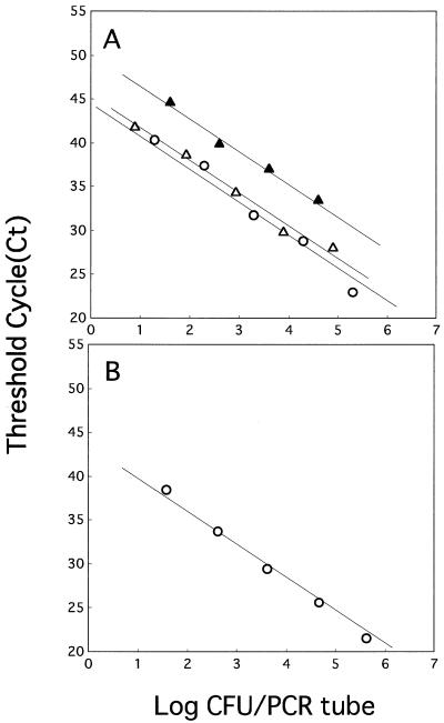
For quantification of the copy number of BoNT/E DNA in fish samples, a calibration was made by depositing a known amount of C. botulinum type E cells (106 CFU) into 1 ml of control fish slurries. This calibration was made for each sample. The fish fillets used for this purpose were those which had not been inoculated with C. botulinum at the beginning of storage and had been stored under the same conditions for the same periods as the samples. For preparation of the cells for calibration, cell culture in the exponential growth phase (107 CFU/ml) in TPGY broth at 30°C was diluted 10-fold with PBS, and portions (1 ml) of the suspension were transferred to 1.5-ml Eppendorf tubes. The cells in the tubes were harvested by centrifugation and stored at −30°C until they were used. For each analysis, these tubes containing the cell pellets of 106 CFU were thawed and poured with 1 ml of control fish slurry. After vigorous mixing, the slurries were extracted in the same way as the samples. A standard curve was produced for every 96-well analysis. The cells used for this purpose were prepared in the same way as the cells used for calibration, except that the thawed cell pellets of the tube containing 106 CFU were extracted directly with extraction reagents. Four serial dilutions of of BoNT/E DNA solution (i.e., corresponding to 106, 105, 104, or 103 CFU/ml) were made after the extraction. These four serial dilutions of BoNT/E DNA were analyzed in triplicate for every 96-well analysis to make a standard curve. After PCR amplification, the calibrated number of BoNT/E DNA per milliliter of fish slurry, X, was determined by the formula X = X′ × I/I′ where X′ is the amount of BoNT/E DNA in samples given by Ct values on a standard curve (Fig. 2A), I is the known amount of C. botulinum type E cells (106 CFU/g) deposited into the control fish slurries in each reaction, and I′ is the recovered amount of cells in the control fish slurries determined by interpolation from the Ct values on the standard curve.
FIG. 2.
Relationship between Ct values and numbers of C. botulinum type E (Iwanai) vegetative cells (A) or spores (B) in the rapid, quantitative PCR assay. DNA was extracted by the GuSCN method from vegetative cells or spores from pure culture (○), from a post spiked fresh fish fillet slurry (▵), and from a postspiked spoiled (after 9 days of storage at 10°C) fish fillet slurry (▴).
Growth curve of C. botulinum type E measured by the rapid, quantitative PCR method in MA-packaged fish.
Fish fillets, each 25 g, placed on sterile petri dishes were either uninoculated or inoculated with the spore suspensions (100 μl) of C. botulinum type E (a mixture of the four strains Iwanai, Tenno 2, 5545, and 164-1). The spore suspensions were inoculated and spread on the fish fillets with sterile glass spreaders (2.0 × 101 spores per g of sample). Immediately after inoculation, petri dishes containing one fillet were packed in gas-impenetrable laminate film bags (300 by 200 mm; Mitsubishi Chemical Industry Co. Ltd., Japan), flushed with N2 at 100% after evacuation. The O2 transmission rate of the film was 8.7 cm3/m2/24 h/atm. The details of the packaging methods and gas analyses have been described previously (23). Packaged samples were incubated at 10°C. Total aerobic counts, toxin levels, and BoNT/E DNA were analyzed daily. Fillets sampled once used were not used again in the investigation. After opening the bags, inoculated and uninoculated samples were asceptically put into stomacher bags with 75 ml of sterile salt (0.85% NaCl) water. The suspensions were then pummeled for 2 min in a stomacher 400 Lab Blender (Seward, London, United Kingdom). After the necessary serial dilutions were made with the sterile salt water, total aerobic counts were determined with a standard agar medium (Nissui Pharmaceutical Co., Ltd., Tokyo, Japan) plate (50% artificial seawater–tap water). The plates were incubated aerobically at 25°C for 48 h. Extraction and quantitative analysis of BoNT/E DNA were performed as described above. To verify whether the amplification product was a target gene, the amplified PCR product of one sample was subjected to direct sequencing as described previously (38).
Toxin detection.
The procedure to assay C. botulinum toxin in fish samples followed the FDA protocol (13), with a slight modification. Briefly, fish samples (25 g) were defrosted and then extracted with an equivalent volume of gelatin phosphate buffer, pH 6.2. The mixture was centrifuged at 15,000 × g for 10 min. The pH of the supernatant liquid was adjusted to 6.0 to 6.2 with 1 N HCl. A portion (0.2 ml) of a 10% trypsin solution was added to 1.8 ml of the supernatant liquid, and this mixture was incubated at 37°C for 1 h Trypsinization was done for all samples. A pair of mice were infected with 0.5 ml of the supernatant liquid. All mice were observed for 96 h for symptoms of botulism.
Nucleotide sequence accession number.
The sequences determined in this study have been deposited in the DDBJ nucleotide sequence database under the following accession numbers: bont/E (Iwanai), AB040123; bont/E (Tenno2), AB040124; bont/E (Biwako), AB040125, bont/E (5545), AB040126; bont/E (35396), AB040127; and bont/E (164-1), AB040128.
RESULTS
Designing C. botulinum type E-specific primers and fluorogenic probe.
The BoNT/E DNA is highly AT rich (GC ratio, 25%), and it is generally difficult to design PCR primers for it. Several specific PCR primers have been reported (5, 14, 40, 42, 43). However, the annealing temperature for these primers is low, resulting in low PCR efficiency, and they could not be adapted to the rapid, quantitative PCR assay. Also, we could not find a proper region for TaqMan probe design in the internal region between the primers previously reported. Therefore, we have decided to design our own specific primers and a fluorogenic TaqMan probe. The software primer express (version 1.0) (ABI) was used following the recommended guidelines for designing fluorogenic probes for rapid, quantitative PCR assays. Briefly, PCR quantification needs high PCR efficiency and specificity; thus, primers should have a high melting temperature (Tm) (above 65°C) in the BoNT/E gene. The PCR amplicon should not exceed 350 bp in length to increase PCR efficiency. The fluorogenic probe should have a Tm higher than that of the primers. Consequently, only one region (1430 to 1709, nucleotide numbering downstream from the ATG start codon of BoNT/E gene) was proposed as a possible region for the primer and probe design. From eight combinations of primers and probes recommended by the software, which differed slightly in sequence, but were located in the same region, we have designed two forward and reverse primers each and one probe in the BoNT/E gene considering the sequence specificity for the BoNT/E DNA. We tested the PCR efficiency with various combinations of the primers and probes and chose the combination most efficient in PCR amplification (Table 2).
Specificity of PCR primers and a TaqMan probe in the detection of C. botulinum type E.
The specificity of the assay was tested against a panel of bacterial templates from 6 strains of C. botulinum type E and 18 genera of 42 non-C. botulinum type E strains, including strains of C. botulinum types A, B, C, D, F, and G (Table 1). Only C. botulinum type E (strains Iwanai, 5545, Tennno 2, 35396, Biwako, and 164-1) reacted with the probe. These samples gave Ct values of <25, which we designated as the upper limit of positivity. All of the other strains tested, including C. botulinum types A, B, C, D, F, and G were essentially nonreactive to the probe (Table 1), with Ct values of >60 cycles, confirming the species-specific nature of the assay.
PCR amplification pattern.
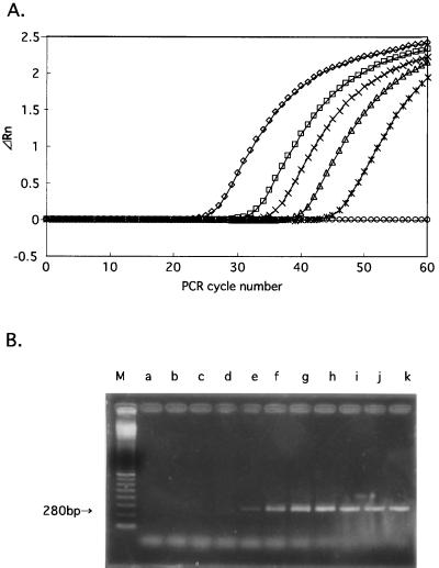
Quantification of the PCR-amplified product from serial dilutions of C. botulinum type E (Iwanai) DNA revealed sigmoid-shaped curves typical of PCR samples (Fig. 1A). A comparison of the fluorescence intensities of the samples at any given cycle indicated that the intensity was greater for samples containing higher DNA templates than for those containing lower DNA templates. The PCR product concentration from samples containing 0.5 pg of genomic DNA could be determined after 45 cycles. The number of cycles at which the fluorescence intensity rose above the threshold levels ranged from 22 (5 ng/PCR tube) to 45 (0.5 pg/PCR tube). With 0.05 pg of genomic DNA per PCR tube, no multiplication of fluorescence occurred. Agarose gel electrophoresis of PCR products amplified from 5 ng of template DNA per PCR tube is also shown in Fig. 1B. The gel showed that only the toxin gene (280 bp) was being amplified regardless of PCR cycles.
FIG. 1.
(A) Real-time PCR amplification plots for reactions with a starting DNA template concentration of C. botulinum type E (Iwanai) ranging from 50 fg to 5 ng per PCR tube. Cycle number is plotted versus change in normalized reporter signal (ΔRn) defined as described in Materials and Methods. Two replicate plots were made for each standard sample, but the data for only one are shown here. ◊, 5 ng; □, 500 pg; ×, 50 pg; ▵, 5 pg; ∗, 0.5 pg; ○, 0.05 pg of DNA per PCR tube, respectively. (B) Agarose gel electrophoresis of PCR products amplified from 5 ng of template DNA per PCR tube with different cycles. Lanes: M, 100-bp DNA ladder; a, 10 cycles; b, 15 cycles; c, 20 cycles; d, 25 cycles; e, 30 cycles; f, 35 cycles; g, 40 cycles; h, 45 cycles; i, 50 cycles; j, 55 cycles; k, 60 cycles.
PCR amplification efficiency of various strains of C. botulinum type E.
Amplification efficiency was measured by comparing the standard curves constructed with six strains of C. botulinum type E (Iwanai, 5545, Tenno 2, 35396, Biwako, and 164-1) by rapid, quantitative PCR. The Ct of each concentration of BoNT/E DNA was determined. Then, the Ct versus log DNA was plotted to produce a standard curve. The slope of the standard curves allowed us to calculate the average amplification efficiency (Table 3). Our comparison did not reveal any significantly different amplification kinetics among these six strains (Table 3). These results were further confirmed by sequencing the amplicon region of these strains. The sequences in the primer and probe regions were identical in all of the strains tested (data not shown; DDBJ nucleotide sequence database accession no. AB040123 to AB040128). Therefore, a single standard curve generated with DNA of C. botulinum type E (Iwanai) was used for quantification for mixed population analysis.
TABLE 3.
Amplification parameters of six different strains of C. botulinum type E
| Strain | Slopea | Amplification efficiencyb |
|---|---|---|
| Iwanai | −3.633 | 0.885 |
| Tenno 2 | −3.669 | 0.873 |
| 5545 | −3.881 | 0.810 |
| 164-1 | −3.869 | 0.813 |
| 35396 | −3.744 | 0.850 |
| Biwako | −3.778 | 0.840 |
The slope of standard curve line (Ct values versus log10 copy number) constructed for each strain.
Efficiencies of PCR amplification calculated from the slope by the formula described in Materials and Methods.
Recovery of BoNT/E DNA from various samples.
The sensitivity of the assay was evaluated with either a pure culture (Fig. 2A), fish (jack mackerel) slurry (Fig. 2A), or a clear spore suspension (without vegetative cells) (Fig. 2B) of C. botulinum type E (Iwanai) by two DNA extraction methods (Chelex and GuSCN). Recovery of BoNT/E DNA from these samples was evaluated from 10-fold serial dilutions of C. botulinum cell or spore suspensions of pure culture or fish slurry spiked before DNA extraction. For pure culture, a good reverse correlation between cell number and threshold cycle was obtained by both extraction methods (for the GuSCN method, see Fig. 2A) (data not shown for the Chelex method). However, with inoculated fish slurries, only the GuSCN method worked well (Fig. 2A). Extraction was equally successful from clean spores by the GuSCN method (Fig. 2B). By the Chelex method, the presence of food components attenuated PCR signals severely, and a very low correlation was obtained between cell number and fluorescence intensity (data not shown). Therefore, we decided to use the GuSCN method in the subsequent experiments. By the GuSCN method, the fluorescence intensity (PCR product concentrations) in fish samples, both fresh and spoiled, and pure cultures containing comparable bacterial loads was linear over 5 orders of magnitude, with the R2 values of the lines being greater than 0.97 in each case (Fig. 2A). However, the y axis was higher in spoiled fish fillets than fresh fillets and pure cultures (Fig. 2A).
Similar experiments performed with a variety of seafood samples revealed linear relationships between the fluorescence intensity and C. botulinum cell numbers, although in some cases, the Ct values were 1 to 3 cycles higher than those obtained with the pure culture (data not shown). Table 4 shows the results of the recovery of two different concentrations of C. botulinum type E (Iwanai) cells measured by the rapid, quantitative PCR method from a variety of seafood samples. Overall, recovery was good (ranging from 19.1% to 173.5%), and there was no serious difference in the order of the bacterial number between the seafood samples (Table 4). However, in some samples (spoiled samples of salmon and common mackerel), BoNT/E DNA was not recovered unless the supernatant after GuSCN precipitation was extracted with phenol-chloroform.
TABLE 4.
Quantification of C. botulinum type E cell number (per PCR tube) recovered from a variety of fresh and spoiled (stored at 10°C for 7 days) seafood samples via rapid, quantitative PCR assay
| Seafood sample | No. of bacteria recovered (% recovery)a
|
|||
|---|---|---|---|---|
| High inoculation (8.5 × 10⁴)b
|
Low inoculation (8.5 × 101)b
|
|||
| Fresh | Spoiled | Fresh | Spoiled | |
| Flathead | 3.5 × 10⁴ (41.2) | 3.3 × 10⁴ (38.2) | 4.3 × 101 (50.0) | 6.5 × 101 (76.5) |
| Sea bream | 1.5 × 10⁵ (173.5) | 2.4 × 10⁴ (28.5) | 3.5 × 101 (41.2) | 4.3 × 101 (50.0) |
| Salmon | 3.8 × 10⁴ (44.1) | 1.6 × 10⁴ (19.1)c | 8.0 × 101 (94.1) | 8.5 × 101 (100.0)c |
| Common mackerel | 5.3 × 10⁴ (61.8) | 1.7 × 10⁴ (19.7)c | 9.0 × 101 (105.9) | 7.0 × 101 (82.4)c |
| Shrimp | 6.0 × 10⁴ (70.6) | 1.9 × 10⁴ (22.1) | 3.0 × 101 (35.3) | 4.0 × 101 (47.1) |
| Crab | 6.0 × 10⁴ (70.6) | 5.3 × 10⁴ (61.8) | 1.0 × 10² (120.6) | 9.3 × 101 (108.8) |
Determined by a universal standard curve made with a pure culture of C. botulinum type E (Iwanai) as described in the Materials and Methods.
The number of C. botulinum cells (determined by the culture method) actually deposited into the food slurries.
A phenol-chloroform extraction step was required for recovery.
Comparison of growth curves with the PCR assay and culture method.
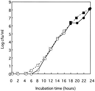
To determine if the rapid, quantitative PCR method could be used for quantifying C. botulinum type E instead of a conventional culture method, the growth curves constructed with the data obtained by the rapid, quantitative PCR method (translated to cell number) and the data obtained by the culture method were compared (Fig. 3). Similar growth curves were obtained by both methods, indicating that the rapid, quantitative PCR method could be used instead of the culture method for monitoring C. botulinum growth. Although the 10-CFU/PCR sample was the most diluted spiked sample used for drawing the standard curve (Fig. 2B), the rapid, quantitative PCR method was able to detect C. botulinum type E (Iwanai) in this experiment at as little as 0.1 CFU/PCR (Ct = 48.3, translated to 4 CFU/ml of culture) in an 8-h incubation, when the culture count obtained at the same time was 10 CFU/ml (Fig. 3). The results of similar experiments with pure culture showed that C. botulinum type E at ≦1 CFU/tube could be detected occasionally, while that at 1 to ∼5 CFU/PCR (10 to ∼200 CFU/ml of culture) could be detected constantly (data not shown). The presence of C. botulinum type E toxin was also tested in 1-ml cultures at each sampling point. The mouse assay required an incubation time of 18 h for toxin detection (corresponding to a cultural count of ca. 106). It was apparent that the rapid, quantitative PCR method was much more sensitive than the mouse assay, making it possible to monitor the early log growth.
FIG. 3.
Growth curves constructed by culture methods (□) and the rapid, quantitative PCR assay (○) with a pure culture of C. botulinum type E (Iwanai). The culture (in TSB) was incubated at 30°C. Solid symbols denote the samples detected with BoNT by mouse assay.
Time course monitoring of C. botulinum levels in MA-packaged fish.
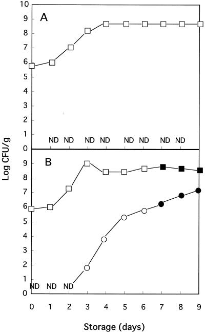
Using the rapid, quantitative PCR method, the time course of C. botulinum type E in fish fillet (jack mackerel) packaged with a modified gas atmosphere (N2 at 100%) was examined (Fig. 4). Spore mixtures of four strains of C. botulinum type E (Iwanai, Tenno 2, 5545, and 164-1) were inoculated to jack mackerel fillets, which were then gas packaged and stored at 10°C. The presence of C. botulinum type E was quantified by the rapid, quantitative PCR method. We have calibrated the amount of BoNT/E DNA per gram of fish fillets by depositing C. botulinum type E (Iwanai) cells into control samples (uninoculated with C. botulinum type E) prior to DNA extraction. By doing so, it was possible to determine the amount of BoNT/E DNA in fish samples, eliminating the effects of both DNA extraction loss and the possible PCR inhibitors in food samples.
FIG. 4.
Time course change in the cell number of C. botulinum type E (a mixture of four strains: Iwanai, Tenno 2, 5545, and 164-1) estimated by the rapid, quantitative PCR assay (○) and viable counts of aerobic spoilage bacteria (□) in MA-packaged fish fillets uninoculated (A) or inoculated (B) with C. botulinum spore mixtures (four strains). The packaged fillets were incubated at 10°C for 9 days. Solid symbols denote the samples detected with BoNT by mouse assay. ND, no BoNT/E DNA detected.
Given the initial spore number of 2.0 × 101 CFU/g, C. botulinum in fish fillets revealed no fluorescence signal by 2 days of storage (Fig. 4B). The signal was first detected at 3 days of storage (calculated as 7.9 × 101 CFU/g), and enhanced signals were obtained thereafter (Fig. 4B). At day 7, the C. botulinum count calculated by the rapid, quantitative PCR method reached 1.6 × 10⁶ CFU/g. No signal was obtained from uninoculated fish fillets, either fresh or spoiled, throughout the experiment (Fig. 4A), confirming that our primers and probe for C. botulinum type E do not cross-react with the natural background bacterial flora of fish. Also, representative PCR products from the inoculated samples were sequenced and confirmed to have the BoNT/E DNA sequence. The mouse toxin assay was done at the same time. Toxin was first detected by mouse assay on day 7 (corresponding to the 1.6 × 10⁶ CFU of C. botulinum per g as measured by the rapid, quantitative PCR method), which was 5 days after the first signals were detected by the rapid, quantitative PCR method.
DISCUSSION
The ability to quantify the BoNT gene from diverse kinds of samples by using an automated procedure could be beneficial to food testing laboratories. The detection of the BoNT gene by PCR has proven to be a sensitive and rapid alternative to the mouse assay (5, 14, 40, 42, 43). However, these methods using PCR are qualitative, not quantitative. In this report, we have developed and evaluated a rapid, quantitative PCR (TaqMan; ABI 7700 system) for automatically quantifying the BoNT/E gene. The method, based on real-time analysis of PCR amplification (7, 15), has several advantages. (i) Unlike PCR-ELISA and MPN PCR, it is a simple and rapid method because it is a real-time assay in a closed tube without a need for any post-PCR manipulations. (ii) Unlike competitive PCR, real-time PCR makes DNA quantification much more precise and reproducible, since it is based on Ct values rather than an end-point measurement of the amount of accumulated PCR product. Consequently, the system allows for a large dynamic range, and we have achieved linearity over 6 log units of input DNA. (iii) Unlike competitive PCR, real-time PCR does not require an internal standard, the design and validation of which are laborious.
The present results suggest that our primers and probe could be used for quantification of various strains of C. botulinum type E. The adaptation of the 5′nuclease quantitative PCR to previously reported PCR primers (5, 14, 40, 42, 43) is not possible due to the requirement of regions with Tm at least 5°C higher than those of the primer regions. Accordingly, we have designed an original primer and probe region (Table 2). Beside the primer and probe design, primer concentrations, and temperature cycling profile were found to be important parameters as reported for the TaqMan PCR assay (2). Also, since the Tm of the TaqMan probe does not increase during PCR amplification because of its inability to be extended by the Taq DNA polymerase, we have chosen a two-step PCR to ensure the hybridization of the TaqMan probe during extension, in which the annealing and extension steps were combined and carried out at 65°C. The results of the study revealed that the primers and probe were highly specific for the C. botulinum type E strains tested (Table 1). Moreover, PCR efficiency was almost the same between the different strains of C. botulinum type E tested in this study (Table 3). Sequencing the amplicon region of these strains revealed that they were identical in the primer and probe regions (data not shown). These results suggest that our primers and probe could be used for quantification of various strains of C. botulinum type E in food and environmental samples, although these aspects should be studied more carefully with additional strains of C. botulinum type E.
The most important advantage of the rapid, quantitative PCR method is the ability to monitor the growth of C. botulinum type E in food where natural microbial competitors are present. At present, laborious and time-consuming studies with the mouse assay are necessary in challenge tests of C. botulinum for risk assessment of food products. The quantitative monitoring of the growth of C. botulinum by conventional culture methods is, if not impossible, time-consuming and erroneous, because isolation agar media such as egg yolk agar would not effectively inhibit certain other members of the genus Clostridium with similar morphologic characteristics of colonies (13). A selective agar medium has been reported (10) and used for monitoring C. botulinum type E in a recent study (27) and our laboratory (B. Kimura et al., unpublished data). However, the selectivity of the medium is still limited, and a confirmation test for representative colonies is necessary. This is laborious and time-consuming as well as erroneous because of the difficulty in discriminating typical colonies. This is true particularly when a large number of background bacteria are present. An MPN technique coupled with PCR has been applied for monitoring C. botulinum type E in fish and the environment (18). However, a large number of dilutions and replicates is required. This makes the rapid analysis of multiple samples difficult by this method. Also, it should be noted that the MPN counts are a compromise, because the equation presumes an even distribution of cells among samples. With the rapid, quantitative PCR method, a more accurate, rapid, and automated monitoring of the copy number of BoNT/E DNA in as many as 96 samples at one time is possible. Thus, a large number of packaged food samples (different components, lots, and storage conditions) could be examined. Also, the data indicated that the rapid, quantitative PCR assay is capable of quantifying C. botulinum type E in fish if ≧102 to 103 CFU/g is present. This means that growth could be monitored much earlier than toxin could be detected by the mouse assay (Fig. 3 and 4).
One limitation of this method is that it is for quantifying the BoNT/E DNA copy number, not the toxin itself. At present, there is only a little information (32) available on the conditions for toxin production by C. botulinum at the cellular level. C. botulinum toxin is generally detected in the late exponential phase in pure culture (4). During the preparation of this manuscript, McGrath et al. (28) reported on the quantification of mRNA of the BoNT/E gene of C. botulinum type E by competitive reverse transcription-PCR. They have demonstrated that gene expression was 100 times stronger in the stationary phase than in the mid-log phase. Also, they found different levels of gene expression in two different types of broth, indicating that culture medium affects toxin production. These results suggest that quantification of mRNA, rather than DNA, would be more appropriate for risk assessment of C. botulinum. However, from a practical point of view, in food testing laboratories, quantification of DNA is simpler to perform. Also, there is no reason to reject the idea that an increase in BoNT/E DNA reflects a potential risk of toxin production. Thus, the sensitive and rapid detection of the increase of BoNT/E DNA in food could serve as an early warning system. Recently, a novel in vitro bioassay for direct detection of BoNT/B, amplifying the enzymatic activity of the neurotoxin light chain, has been described (47). This method seems more sensitive and less expensive than the mouse assay or ELISA method (11). An advantage of the rapid, quantitative PCR method over the in vitro bioassay would be a much higher sensitivity and quantitative accuracy, while an obvious disadvantage is that it is not a method for direct detection of BoNT. It would be interesting to evaluate these methods all at one time in the future. Unfortunately, however, the in vitro bioassay method has not been established for BoNT/E yet. In practical terms, it would be most effective to use the rapid, quantitative PCR methods in combination with other methods for directly detecting BoNT (11, 47) or the mouse assay (when necessary). In such cases, samples demonstrating growth potential would be tested by either of the latter assays for confirmation. By doing so, a much more rapid and convenient risk assessment of C. botulinum would be realized.
When DNA was extracted either by the Chelex method or the GuSCN method from fish samples, a linear relationship between the initial number of template copies and Ct values was obtained only with the GuSCN method. The Chelex method worked well with DNA isolated from pure cultures (data not shown), but only a very low correlation could be obtained between the initial number of template copies and Ct values when DNA was isolated from fish slurries (data not shown). This is likely due to the inhibition of PCR amplification by the large amounts of food components, which could not be eliminated from the PCR tube. In our previous study (22), the Chelex method worked well for “yes-or-no” detection of Salmonella invA from raw meat and shrimp. In that study, subsamples (25 g) were diluted with 225 ml (10× dilution) of Trypticase soy broth (TSB) or Universal preenrichment broth (UB; Difco Laboratories, Detroit, Mich.). In the present study, subsamples (25 g) were diluted with 75 ml of phosphate buffer in order to increase the detection sensitivity. Thus, it appears that the Chelex method does not work well when samples are not diluted enough to eliminate the inhibitory effect of food components. Since the Chelex extract protocol is a one-tube reaction, this seems inevitable. With the GuSCN method, isopropanol precipitation is performed, and the influence of the PCR inhibitor may be less than that for the Chelex extract protocol. A similar phenomenon was described by Chen et al. (6). From these results, it was suggested that the GuSCN method was more appropriate for extraction of DNA from fish samples for quantification.
The application of the rapid, quantitative PCR method to other foods could be limited unless the performance of PCR is carefully evaluated food by food. The present results showed that the GuSCN DNA extraction method was efficient for the quantification of BoNT/E DNA in a variety of fish samples, although in some samples, a phenol-chloroform extraction step was required (Table 4). However, higher Ct values (1 to 3 cycles) than those obtained with the pure culture were obtained in some cases (data not shown), resulting in the variation in recovery (Table 4). The variation may be due to either the extraction efficiency of DNA, the inhibition of PCR amplification by PCR inhibitors, or a large amount of DNA from background spoilage bacteria. Thus, a universal standard curve cannot be used for accurate quantification of BoNT/E DNA in a variety of fish samples (based on fish species and duration of storage). Accordingly, in this study, an accurate quantification of BoNT/E DNA in jack mackerel samples was achieved by calibrating the standard curve by depositing a known amount of C. botulinum type E cells into the fish slurry (uninoculated with C. botulinum) for each analysis before extraction. By doing so, it was possible to determine the copy numbers of BoNT/E DNA, eliminating the effect of both the loss of DNA during extraction and the potential PCR inhibitors (Fig. 4). However, in practical use, it would be more convenient to apply the universal standard curve, since the variation in recovery was not significant enough to make a difference in the order of the bacterial number (Table 4). The merit of using a universal standard curve is that it is simple and saves money, although there are some limitations to the interpretation of the data. The possible variations should be determined before routine analysis of various food samples, and the most practical and time/money-saving calibration method should be selected.
In conclusion, although DNA extraction protocols may need to be optimized for food by food evaluation, the use of the rapid, quantitative PCR assay could provide a rapid, quantitative assay for C. botulinum type E in seafood samples. At present, one of the most serious drawbacks of conventional techniques in food microbiology is the difficulty of determining the growth of a target microorganism in a complex mixture of bacteria by plate count techniques. This method, enabling us to elucidate how bacteria grow in the real environment, where they are typically associated with surfaces and are in dynamic competition with a heterogeneous microflora, could be applicable to other food pathogens or certain target microbes in the environment.
ACKNOWLEDGMENTS
This work was supported by a Grant-in-Aid for Scientific Research (A 08556034) from the Ministry of Education, Science, Sports and Culture of Japan and a Grant-in-Aid from the Japan Food Industry Center.
We thank S. Igimi (the National Institute of Health, Japan) and G. Sakaguchi for kindly supplying C. botulinum strains. We also thank K. Venkateswaran (Jet Propulsion Laboratory, NASA) for useful suggestions.
REFERENCES
- 1.Ando Y, Iida H. Factors affecting the germination and spores of Clostridium botulinum type E. Jpn J Microbiol. 1970;14:361–370. doi: 10.1111/j.1348-0421.1970.tb00536.x. [DOI] [PubMed] [Google Scholar]
- 2.Bassler H A, Flood S J A, Livak K J, Marmaro J, Knorr R, Batt C A. Use of a fluorogenic probe in a PCR-based assay for the detection of Listeria monocytogenes. Appl Environ Microbiol. 1995;61:3724–3728. doi: 10.1128/aem.61.10.3724-3728.1995. [DOI] [PMC free article] [PubMed] [Google Scholar]
- 3.Binz T, Kurazono H, Popoff M R, Eklund M W, Sakaguchi G, Kozaki S, Krieglstein K, Henschen A, Gill D M, Niemann H. Nucleotide sequence of the gene encoding Clostridium botulinum neurotoxin type D. Nucleic Acids Res. 1990;18:5556. doi: 10.1093/nar/18.18.5556. [DOI] [PMC free article] [PubMed] [Google Scholar]
- 4.Call J E, Cooke P H, Miller A J. In situ characterization of Clostridium botulinum neurotoxin synthesis and export. J Appl Bacteriol. 1995;79:257–263. doi: 10.1111/j.1365-2672.1995.tb03135.x. [DOI] [PubMed] [Google Scholar]
- 5.Campbell K D, Collins M D, East A K. Gene probes for identification of the botulinal neurotoxin gene and specific identification of neurotoxin types B, E, and F. J Clin Microbiol. 1993;31:2255–2262. doi: 10.1128/jcm.31.9.2255-2262.1993. [DOI] [PMC free article] [PubMed] [Google Scholar]
- 6.Chen S, Yee A, Griffiths M, Larkin C, Yamashiro C T, Behari R, Paszko-Kolva C, Rahn K, Grandis S A D. The evaluation of a fluorogenic polymerase chain reaction assay for the detection of Salmonella species in food commodities. Int J Food Microbiol. 1997;35:239–250. doi: 10.1016/s0168-1605(97)01241-5. [DOI] [PubMed] [Google Scholar]
- 7.Christian A H, Stevens J, Livak K J, Williams P M. Real time quantitative PCR. Genome Res. 1996;6:986–994. doi: 10.1101/gr.6.10.986. [DOI] [PubMed] [Google Scholar]
- 8.Ciulla T A, Sklar R M, Hauser S L. A simple method for DNA purification from peripheral blood. Anal Biochem. 1988;174:485–488. doi: 10.1016/0003-2697(88)90047-4. [DOI] [PubMed] [Google Scholar]
- 9.Desjardin L E, Chen Y, Perkins M D, Teixeira L, Cave M D, Eisenach K D. Comparison of the ABI 7700 system (TaqMan) and competitive PCR for quantification of IS6110 DNA in sputum during treatment of tuberculosis. J Clin Microbiol. 1998;36:1964–1968. doi: 10.1128/jcm.36.7.1964-1968.1998. [DOI] [PMC free article] [PubMed] [Google Scholar]
- 10.Dezfulian M, McCroskey L M, Hatheway C L, Dowell V R., Jr Selective medium for isolation of Clostridium botulinum from human feces. J Clin Microbiol. 1981;13:526–531. doi: 10.1128/jcm.13.3.526-531.1981. [DOI] [PMC free article] [PubMed] [Google Scholar]
- 11.Doellgast G J, Triscott M X, Beard G A, Bottoms J D, Cheng T, Roh B H, Roman M G, Hall P A, Brown J E. Sensitive enzyme-linked immunosorbent assay for detection of Clostridium botulinum neurotoxins A, B, and E using signal amplification via enzyme-linked coagulation assay. J Clin Microbiol. 1993;31:2402–2409. doi: 10.1128/jcm.31.9.2402-2409.1993. [DOI] [PMC free article] [PubMed] [Google Scholar]
- 12.Eklund M W, Wieler D I, Poysky F T. Outgrowth and toxin production of nonproteolytic type B Clostridium botulinum at 3.3 to 5.6°C. J Bacteriol. 1967;93:1461–1462. doi: 10.1128/jb.93.4.1461-1462.1967. [DOI] [PMC free article] [PubMed] [Google Scholar]
- 13.Food and Drug Administration. Bacteriological analytical manual. 8th ed. Gaithersburg, Md: AOAC International; 1992. [Google Scholar]
- 14.Franciosa G, Ferreira J L, Hatheway C L. Detection of type A, B, and E botulism neurotoxin genes in Clostridium botulinum and other Clostridium species by PCR: evidence of unexpressed type B toxin genes in type A toxigenic organisms. J Clin Microbiol. 1994;32:1911–1917. doi: 10.1128/jcm.32.8.1911-1917.1994. [DOI] [PMC free article] [PubMed] [Google Scholar]
- 15.Gibson UEM, Heid C A, Williams P M. A novel method for real time quantitative RT-PCR. Genome Res. 1996;6:995–1001. doi: 10.1101/gr.6.10.995. [DOI] [PubMed] [Google Scholar]
- 16.Gonzalez I, Garcia T, Fernandez A, Sanz B, Hernandez P E, Martin R. Rapid enumeration of Escherichia coli in oysters by a quantitative PCR-ELISA. J Appl Microbiol. 1999;86:231–236. doi: 10.1046/j.1365-2672.1999.00659.x. [DOI] [PubMed] [Google Scholar]
- 17.Hauser D, Eklund M W, Kurazono H, Binz T, Niemann H, Gill D M, Boquet P, Popoff M R. Nucleotide sequence of Clostridium botulinum C1 neurotoxin. Nucleic Acids Res. 1990;18:4924. doi: 10.1093/nar/18.16.4924. [DOI] [PMC free article] [PubMed] [Google Scholar]
- 18.Hielm S, Hyytiä E, Ridell J, Korkeala H. Detection of Clostridium botulinum in fish and environmental samples using polymerase chain reaction. Int J Food Microbiol. 1996;31:357–365. doi: 10.1016/0168-1605(96)00984-1. [DOI] [PubMed] [Google Scholar]
- 19.Higgins J A, Ezzell J, Hinnebusch B J, Shipley M, Henchal E A, Ibrahim M S. 5′nuclease PCR assay to detect Yersinia pestis. J Clin Microbiol. 1998;36:2284–2288. doi: 10.1128/jcm.36.8.2284-2288.1998. [DOI] [PMC free article] [PubMed] [Google Scholar]
- 20.Holland P M, Abramson R D, Watson R, Gelfand D H. Detection of specific polymerase chain reaction product by utilizing the 5′→3′ exonuclease activity of Thermus aquaticus. Proc Natl Acad Sci USA. 1991;88:7276–7280. doi: 10.1073/pnas.88.16.7276. [DOI] [PMC free article] [PubMed] [Google Scholar]
- 21.Janse BJH, Gaskell J, Akhtar M, Cullen D. Expression of Phanerochaete chrysosporium genes encoding lignin peroxidases, manganese peroxidases, and glyoxal oxidase in wood. Appl Environ Microbiol. 1998;64:3536–3538. doi: 10.1128/aem.64.9.3536-3538.1998. [DOI] [PMC free article] [PubMed] [Google Scholar]
- 22.Kimura B, Kawasaki S, Fujii T, Kusunoki J, Itoh T, Flood S J A. Evaluation of TaqManTM PCR assay for detecting Salmonella in raw meat and shrimp. J Food Prot. 1999;62:329–335. doi: 10.4315/0362-028x-62.4.329. [DOI] [PubMed] [Google Scholar]
- 23.Kimura B, Kuroda S, Murakami M, Fujii T. Growth of Clostridium perfringens in fish fillets packaged with carbon dioxide controlled atmosphere at abusive temperatures. J Food Prot. 1996;59:704–710. doi: 10.4315/0362-028X-59.7.704. [DOI] [PubMed] [Google Scholar]
- 24.Lee S-Y, Bollinger J, Bezdicek D, Ogram A. Estimation of the abundance of an uncultured soil bacterial strain by a competitive PCR method. Appl Environ Microbiol. 1996;62:3787–3793. doi: 10.1128/aem.62.10.3787-3793.1996. [DOI] [PMC free article] [PubMed] [Google Scholar]
- 25.Lindbroth S E, Genigeorgis G A. Probability of growth and toxin production by nonproteolytic Clostridium botulinum in rockfish stored under modified atmospheres. Int J Food Microbiol. 1986;3:167–181. [Google Scholar]
- 26.Livak K J, Flood S J A, Marmaro J, Giusti W, Deetz K. Oligonucleotides with fluorescent dyes at opposite ends provide a quenched probe system useful for detecting PCR product and nucleic acid hybridization. PCR Methods Appl. 1995;4:357–362. doi: 10.1101/gr.4.6.357. [DOI] [PubMed] [Google Scholar]
- 27.Lyver A, Smith J P, Nattress F M, Austin J W, Blanchfield B. Challenge studies with Clostridium botulinum type E in a value-added surimi product stored under a modified atmosphere. J Food Saf. 1998;18:1–23. [Google Scholar]
- 28.McGrath S, Dooley J S G, Haylock R W. Quantification of Clostridium botulinum toxin gene expression by competitive reverse transcription-PCR. Appl Environ Microbiol. 2000;66:1423–1428. doi: 10.1128/aem.66.4.1423-1428.2000. [DOI] [PMC free article] [PubMed] [Google Scholar]
- 29.Moorhead S M, Bell R G. Psychrotrophic clostridia mediated gas and botulinal toxin production in vacuum-packed chilled meat. Lett Appl Microbiol. 1999;28:108–112. doi: 10.1046/j.1365-2672.1999.00490.x. [DOI] [PubMed] [Google Scholar]
- 30.Nogva H K, Lillehaug D. Detection and quantification of Salmonella in pure cultures using 5′-nuclease polymerase chain reaction. Int J Food Microbiol. 1999;51:191–196. doi: 10.1016/s0168-1605(99)00122-1. [DOI] [PubMed] [Google Scholar]
- 31.Oberst R D, Hays M P, Bohra L K, Phebus R K, Yamashiro C T, Paszko-Kolva C, Flood S J A, Sargeant J M, Gillespie J R. PCR-based DNA amplification and presumptive detection of Escherichia coli O157:H7 with an internal fluorogenic probe and the 5′ nuclease (TaqMan) assay. Appl Environ Microbiol. 1998;64:3389–3396. doi: 10.1128/aem.64.9.3389-3396.1998. [DOI] [PMC free article] [PubMed] [Google Scholar]
- 32.Patterson-Curtis S I, Johnson E A. Regulation of neurotoxin and protease formation in Clostridium botulinum Okra B and Hall A by arginine. Appl Environ Microbiol. 1989;55:1544–1548. doi: 10.1128/aem.55.6.1544-1548.1989. [DOI] [PMC free article] [PubMed] [Google Scholar]
- 33.Peck M W. Clostridium botulinum and the safety of refrigerated processed foods of extended durability. Trends Food Sci Technol. 1997;8:186–192. [Google Scholar]
- 34.Poulet S, Hauser D, Quanz M, Niemann H, Popoff M R. Sequences of the botulinal neurotoxin derived from Clostridium botulinum type E (strain Beluga) and Clostridium butyricum (strains ATCC 43181 and ATCC 43755) Biochem Biophys Res Commun. 1992;183:107–113. doi: 10.1016/0006-291x(92)91615-w. [DOI] [PubMed] [Google Scholar]
- 35.Reddy N R. Shelf life and toxin development by Clostridium botulinum during storage of modified-atmosphere-packaged fresh aquacultured salmon fillets. J Food Prot. 1997;60:1055–1063. doi: 10.4315/0362-028X-60.9.1055. [DOI] [PubMed] [Google Scholar]
- 36.Rowman M G, Humber J Y, Hall P A, Reddy N R, Solomon H M, Triscott M X, Beard G A, Bottoms J D, Cheng T, Doellgast G J. Amplified immunoassay ELISA-ELICA for measuring Clostridium botulinum type E neurotoxin in fish fillets. J Food Prot. 1994;57:985–990. doi: 10.4315/0362-028X-57.11.985. [DOI] [PubMed] [Google Scholar]
- 37.Sambrook J, Fritsch E F, Maniatis T. Molecular cloning: a laboratory manual. 2nd ed. Cold Spring Harbor, N.Y: Cold Spring Harbor Laboratory; 1989. [Google Scholar]
- 38.Satomi M, Kimura B, Mizoi M, Sato T, Fujii T. Tetragenococcus muriaticus sp. nov., a new moderately halophilic lactic acid bacterium isolated from fermented squid liver sauce. Int J Syst Bacteriol. 1997;47:832–836. doi: 10.1099/00207713-47-3-832. [DOI] [PubMed] [Google Scholar]
- 39.Schmidt C F, Lechowich R V, Folinazzo J F. Growth and toxin production by type E Clostridium botulinum below 40°F. J Food Sci. 1961;26:626–630. [Google Scholar]
- 40.Sciacchitano C J. Molecular detection of Clostridium botulinum type E neurotoxin gene in smoked fish by polymerase chain reaction and capillary electrophoresis. J AOAC Int. 1996;79:861–865. [PubMed] [Google Scholar]
- 41.Stevens J, Yu F S, Hassoun P M, Lanzillo J J. Quantification of polymerase chain reaction products: enzyme immunoassay based systems for digoxigenin and biotin-labelled products that quantify either total or specific amplicons. Mol Cell Probes. 1996;10:31–41. doi: 10.1006/mcpr.1996.0005. [DOI] [PubMed] [Google Scholar]
- 42.Szabo E A, Pemberton J M, Desmarchelier P M. Detection of the genes encoding botulinum neurotoxin types A to E by the polymerase chain reaction. Appl Environ Microbiol. 1993;59:3011–3020. doi: 10.1128/aem.59.9.3011-3020.1993. [DOI] [PMC free article] [PubMed] [Google Scholar]
- 43.Takeshi K, Fujinaga Y, Inoue K, Nakajima H, Oguma K, Ueno T, Sunagawa H, Ohyama T. Simple method for detection of Clostridium botulinum type A to F neurotoxin genes by polymerase chain reaction. Microbiol Immunol. 1996;40:5–11. doi: 10.1111/j.1348-0421.1996.tb03310.x. [DOI] [PubMed] [Google Scholar]
- 44.Thompson D E, Brehm J K, Oultram J D, Swinfield T J, Shone C C, Atkinson T, Melling J, Minton N P. The complete amino acid sequence of Clostridium botulinum type A neurotoxin, deduced by nucleotide sequence analysis of the encoding gene. Eur J Biochem. 1990;189:73–81. doi: 10.1111/j.1432-1033.1990.tb15461.x. [DOI] [PubMed] [Google Scholar]
- 45.Walsh P S, Metzger D A, Higuchi R. Chelex 100 as a medium for simple extraction of DNA for PCR-based typing from forensic material. Bio Techniques. 1991;10:506–513. [PubMed] [Google Scholar]
- 46.Whelan S M, Elmore M J, Bodsworth N J, Brehm J K, Atkinson T, Minton N P. Molecular cloning of the Clostridium botulinum structural gene encoding the type B neurotoxin and determination of its entire nucleotide sequence. Appl Environ Microbiol. 1992;58:2345–2354. doi: 10.1128/aem.58.8.2345-2354.1992. [DOI] [PMC free article] [PubMed] [Google Scholar]
- 47.Wictome M, Newton K, Jameson K, Hallis B, Dunnigan P, Mackay E, Clarke S, Taylor R, Gaze J, Foster K, Shone C. Development of an in vitro bioassay for Clostridium botulinum type B neurotoxin in foods that is more sensitive than the mouse bioassay. Appl Environ Microbiol. 1999;65:3787–3792. doi: 10.1128/aem.65.9.3787-3792.1999. [DOI] [PMC free article] [PubMed] [Google Scholar]
- 48.Witham P K, Yamashiro C T, Livak K J, Batt C A. A PCR-based assay for the detection of Escherichia coli Shiga-like toxin genes in ground beef. Appl Environ Microbiol. 1996;62:1347–1353. doi: 10.1128/aem.62.4.1347-1353.1996. [DOI] [PMC free article] [PubMed] [Google Scholar]


