Abstract
Xanthomonadins are yellow, membrane-bound pigments produced by members of the genus Xanthomonas. We identified an ethyl methanesulfonate-induced Xanthomonas oryzae pv. oryzae mutant (BXO65) that is deficient for xanthomonadin production and virulence on rice, as well as auxotrophic for aromatic amino acids (Pig− Vir− Aro−). Reversion analysis indicated that these multiple phenotypes are due to a single mutation. A genomic library of the wild-type strain was used to isolate a 7.0-kb clone that complements BXO65. By transposon mutagenesis, marker exchange, sequence analysis, and subcloning, the complementing activity was localized to a 849-bp open reading frame (ORF). This ORF is homologous to the aroE gene, which encodes shikimate dehydrogenase in various bacterial species. Shikimate dehydrogenase activity was present in the wild-type strain and the mutant with the complementing clone, whereas no activity was found in BXO65. This clone also complemented an Escherichia coli aroE mutant for prototrophy, indicating that aroE is functionally conserved in X. oryzae pv. oryzae and E. coli. The nucleotide sequence of the 2.9-kb region containing aroE revealed that a putative DNA helicase gene is located adjacent to aroE. Our results indicate that aroE is required for normal levels of virulence and xanthomonadin production in X. oryzae pv. oryzae.
Xanthomonas oryzae pv. oryzae causes bacterial leaf blight, a serious disease of rice. Worldwide at least 350 different plant diseases are known to be caused by various xanthomonads (17). A characteristic feature of the genus Xanthomonas is the production of yellow, membrane-bound pigments called xanthomonadins (28). The xanthomonadins were initially thought to be carotenoids, but later they were characterized as a unique group of halogenated aryl polyene pigments (2, 3). The functional role of xanthomonadins is poorly understood. The vast majority of pigment-deficient mutants that have been isolated from several xanthomonads are prototrophs (20, 29) and virulence proficient upon wound inoculation (9, 20, 29). Pigment-deficient mutants of Xanthomonas juglandis and X. oryzae pv. oryzae have been reported to be more sensitive to photobiological damage than the wild-type strains are (13, 22), suggesting that the pigment may provide protection against photodamage.
An 18.6-kb region containing seven transcriptional units required for xanthomonadin biosynthesis has been isolated from Xanthomonas campestris pv. campestris (20, 21). One of the transcriptional units, pigB, encodes a diffusible factor that is involved in both pigment and extracellular polysaccharide production (21). The pigB mutants have also been shown to be impaired for epiphytic survival and host infection (22).
We isolated an ethyl methanesulfonate (EMS)-induced pigment-deficient mutant of X. oryzae pv. oryzae that is also virulence deficient and auxotrophic for aromatic amino acids. A genomic clone that restores pigmentation, prototrophy, and virulence to this mutant was isolated by functional complementation. Characterization of this clone revealed that shikimate dehydrogenase, an enzyme in the aromatic amino acid biosynthetic pathway, is required for normal levels of pigment production and virulence in X. oryzae pv. oryzae.
MATERIALS AND METHODS
Bacterial strains and media.
All bacterial strains and plasmids used are listed in Table 1. X. oryzae pv. oryzae strains were grown at 28°C in either peptone sucrose (PS) medium (29) or modified Miller's minimal medium M4 (15). Escherichia coli strains were grown in Luria-Bertani medium (18) at 37°C. The following concentrations of antibiotics were used: rifampin, 50 μg ml−1; spectinomycin, 50 μg ml−1; ampicillin, 100 μg ml−1; kanamycin, 50 μg ml−1; chloramphenicol, 20 μg ml−1; streptomycin, 50 μg ml−1; and cycloheximide, 75 μg ml−1.
TABLE 1.
Strains and plasmids used in this study
| Strain or plasmid | Relevant characteristicsa | Reference or source |
|---|---|---|
| E. coli strains | ||
| DH5α | F′ endA1 hsdR17 (rk− mk+) supE44 thi-1 recA1 gyrA relA1 f80dlacZDM15 (lacZYA-argF)U169 | Lab collection |
| S17-1 | RP4-2Tc::Mu-Kn::Tn7 pro hsdR recA | 26 |
| AB2835 | aroE353 | 30 |
| Plasmids | ||
| pBluescript (KS) | Apr | Stratagene, La Jolla, Calif. |
| pHM1 | Spr Smrcos parA IncW derivative of pRI40 | 11 |
| pUFR034 | IncW Nmr Tra− Mob+mob(P) LacZα+ Par+cos | 8 |
| pAG4 | pUFR034 + 7.0-kb EcoRI fragment encoding aroE | This study |
| pAG5 | pBluescript (KS) + 7.0-kb EcoRI fragment encoding aroE | This study |
| pAG7 | pUFR034 + 1.2-kb EcoRI-PstI fragment encoding aroE | This study |
| pAG8 | pAG4-aroE1::Tn3-HoHo1 | This study |
| pAG9 | pAG5-aroE2::mTn7 | This study |
| pAG10 | pAG5-aroE3::mTn7 | This study |
| pAG11 | pHM1 + aroE2::mTn7 (in the 7.0-kb EcoRI fragment) | This study |
| pAG12 | pHM1 + aroE3::mTn7 (in the 7.0-kb EcoRI fragment) | This study |
| X. oryzae pv. oryzae strains | ||
| BXO1 | Laboratory wild type, Indian isolate | Lab colection |
| BXO43 | rif-2, derivative of BXO1 | Lab colection |
| BXO62 | pig-10, obtained by EMS mutagenesis of BXO1 | This study |
| BXO65 | rif-5, derivative of BXO62 | This study |
| BXO1704 | BXO65/pAG4 | This study |
| BXO1706 | aroE1::Tn3-HoHo1 rif-2 | This study |
| BXO1707 | aroE2::mTn7 rif-2 | This study |
rif-2 and rif-5 indicate mutations that confer resistance to rifampin; pig-10 indicates pigment deficiency.
EMS mutagenesis and isolation of pigment-deficient mutants.
EMS (Sigma Chemical Co., St. Louis, Mo.) mutagenesis of X. oryzae pv. oryzae was done as described for E. coli by Miller (18). Twenty microliters of a cell suspension from a mutagenized and washed X. oryzae pv. oryzae cell preparation was inoculated into 2 ml of PS medium and incubated at 28°C for 24 h before the cells were dilution plated to obtain single colonies on PSagar (PSA) plates. Pigment-deficient mutants were obtained at a frequency of 1% of the surviving cells. One pigment-deficient mutant failed to grow on minimal medium, and the auxotrophic phenotype was diagnosed as a deficiency of all three aromatic amino acids by using the pool plate method (7). Spontaneous prototrophic revertants and rifampin-resistant (Rifr) derivatives of X. oryzae pv. oryzae strains were obtained by plating saturated cultures (approximately 108 CFU/plate) on plates containing minimal medium and plates containing PSA plus rifampin, respectively.
Virulence assays with rice plants.
Forty-day-old greenhouse-grown rice plants of susceptible rice cultivar Taichung Native-1 were inoculated by clipping leaf tips with sterile scissors dipped in saturated cultures (109 cells/ml) of X. oryzae pv. oryzae (14). When this method was used, approximately 106 cells were deposited at the site of inoculation. The plants were incubated in a greenhouse with minimum and maximum temperatures of approximately 25 and 30°C, respectively, and a relative humidity of approximately 60%. Lesion lengths were measured at regular intervals. No lesions were observed in control experiments in which the leaves were inoculated with scissors dipped in water.
Extraction and quantification of pigment.
The procedure described previously (2) for extraction of xanthomonadin from X. juglandis was used, with some modifications. X. oryzae pv. oryzae cultures were grown to the stationary phase, and xanthomonadin was extracted in chloroform-methanol (2:1) by shaking for 3 h at room temperature. The amount of pigment produced per 100 mg (dry weight) of bacterial cells was expressed as the absorbance (optical density at 445 nm) of the crude pigment extracts (21). We also quantified the pigment by assuming that the structure and molar extinction coefficient of xanthomonadin from X. oryzae pv. oryzae are identical to those of a xanthomonadin from X. juglandis (3). Similar conclusions could be drawn by using either method for pigment estimation.
Functional complementation.
A partial EcoRI-digested genomic library of our laboratory wild-type X. oryzae pv. oryzae strain, having an average insert size of 30 kb, was prepared (L. Rajgopal, Y. Jameer, S. Dharmapuri, M. Karunakaran, R. Ramanan, A. P. K. Reddy, and R. V. Sonti, Abstr. Rice Genet. III Symp., p. 939–944, 1995) in the broad-host-range Kmr cosmid cloning vector pUFR034 (8). A total of 960 clones from this library were transferred from E. coli DH5α to S17-1 by using pRK600 as a helper (24). Genomic clones from the library in S17-1 were mobilized in pools of 12 clones each into the pigment-deficient mutant by performing biparental mating. Transconjugants that appeared on PSA-rifampin-kanamycin selection plates were replica plated onto minimal medium plates containing kanamycin to identify prototrophic and pigment-proficient colonies.
Transposon mutagenesis and marker exchange.
Transposon 3 insertions were obtained with the pAG4 clone by using Tn3-HoHo1 as described by Stachel et al. (27), with some modifications. Plasmids were isolated, and the insertions were localized on either the insert or the vector DNA by restriction analysis.
The 7.0-kb EcoRI fragment in pAG4 was also cloned into the EcoRI site of pBluescript(KS) to obtain pAG5 and was mutagenized by mini-transposon 7 (mTn7) derivatives by using an in vitro transposition kit (Genome Priming System; New England Biolabs [NEB], Beverly, Mass.). The mTn7 element encodes kanamycin resistance. Two different mTn7 insertions on the cloned DNA in plasmids pAG9 and pAG10 (Table 1) were found to be in the aroE gene (see below). These two insertions were cloned into the shuttle vector pHM1 (Spr) (Table 1) by using EcoRI restriction sites (EcoRI does not cut within the mTn7 element) to obtain plasmids pAG11 and pAG12. Plasmids pAG11 and pAG12 and the derivatives of pAG4 containing Tn3-HoHo1 insertions were mobilized individually into BXO43 for marker exchange. Marker exchange was done by growing the cells in either PS-ampicillin medium (for Tn3-HoHo1) or PS-kanamycin medium (for mTn7) for more than 30 generations by serial passage. Colonies that were either Apr Kms (for Tn3-HoHo1) or Kmr Sps (for mTn7) were analyzed by Southern hybridization to confirm that marker exchange had occurred as expected.
Protein extraction, quantification, and enzyme assay.
Cell lysates were prepared as described by Chaudhuri and Coggins (6), and protein contents were estimated by the Bradford method (5). Shikimate dehydrogenase activities in cell lysates of X. oryzae pv. oryzae strains were assayed as described previously for E. coli (32). Shikimic acid and NADP were used as substrates, and reduction of NADP to NADPH was assayed by monitoring increase in absorbance at 340 nm. One unit of enzyme activity was defined as 1 μmol of NADPH produced per min.
Plasmid preparation and DNA sequencing.
Plasmid DNA was isolated by the alkaline lysis method (25). Restriction digestions were performed by using enzymes obtained from NEB as recommended by the supplier. Primers that are outwardly directed from Tn3-HoHo1 were used to obtain the sequence of DNA flanking the Tn3-HoHo1 insertions. mTn7-specific primers provided in the Genome Priming System kit from NEB were used to sequence the mTn7 insertion sites. M13 forward and reverse primers were used to sequence the ends of a 7.0-kb insert in clone pAG5. The sequencing reactions, electrophoresis, and sequence data analysis were performed with an ABI Prism 377 automated DNA sequencer (Perkin-Elmer, Foster City, Calif.). A homology search in the database was performed through the National Center for Biotechnology Information by using the BLAST algorithm (1). Promoter prediction was performed by using software at the Baylor College of Medicine search launcher (www.hgsc.bcm.tmc.edu).
Nucleotide sequence accession number.
The sequence of a 2.953-kb region containing aroE was determined. This sequence has been submitted to GenBank with accession no. AF258797.
RESULTS
Isolation of a pigment- and virulence-deficient aromatic amino acid auxotroph of X. oryzae pv. oryzae.
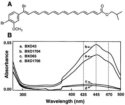
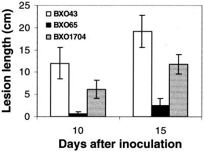
A pigment-deficient mutant (BXO62) that is auxotrophic for all three aromatic amino acids was isolated after EMS mutagenesis of the BXO1 strain (see Materials and Methods). To aid in the subsequent analysis, spontaneous Rifr derivatives of BXO1 and BXO62 designated BXO43 and BXO65, respectively, were obtained as described in Materials and Methods. The pigment production, prototrophic-auxotrophic, and virulence properties of the Rifr derivatives were similar to those of their respective parent strains (data not shown). Pigment was extracted from strains BXO43 and BXO65 and quantified as described in Materials and Methods. BXO65 produces approximately 23% of the pigment produced by BXO43 (Table 2). An absorption spectrum of the pigment extracted from the BXO43 strain showed a characteristic structure with peak at 445 nm and shoulders at 425 and 470 nm (Fig. 1). These features were missing from the absorption spectrum of the BXO65 strain (Fig. 1). The virulence characteristics of these strains were assayed with rice leaves as described in Materials and Methods. Lengths of lesions caused by BXO43 and BXO65 were measured 10 and 15 days after inoculation (Fig. 2). It is apparent that BXO65 is severely virulence deficient (Vir−). Prototrophic revertants of BXO65 were isolated and were found to have regained pigment and virulence proficiency (data not shown). Simultaneous reversion suggests that a single mutation is responsible for the pleiotropic phenotype of BXO65.
TABLE 2.
Pigment production and shikimate dehydrogenase activity in various X. oryzae pv. oryzae strains
| Straina | Pigment production (optical density at 445 nm)b | Shikimate dehydrogenase activity (mU/mg of protein)c |
|---|---|---|
| BXO43 | 0.632 ± 0.006d | 5.35 ± 0.57 |
| BXO65 | 0.15 ± 0.018 | NDAe |
| BXO1704 | 0.816 ± 0.03 | 65.3 ± 6.07 |
BXO43 is the wild-type strain, BXO65 is an EMS-induced Pig− Aro− Vir− mutant, and BXO1704 is BXO65 with pAG4 (a complementing plasmid).
Pigment production was measured by determining absorbance at 445 nm as described in Materials and Methods.
Protein was extracted and shikimate dehydrogenase activity was assayed as described in Materials and Methods.
Mean ± standard error based on three independent experiments.
NDA, no detectable activity.
FIG. 1.
Structure of xanthomonadin I and absorption spectra of crude pigment extracts from X. oryzae pv. oryzae cultures. (A) Structure of isobutyl derivative of xanthomonadin isolated from X. juglandis, a walnut pathogen (3). (B) Absorption spectra of crude pigment extracts from BXO431 (wild-type strain), BXO65 (an EMS-induced Pig− Aro− Vir− mutant), BXO1706 an aroE1::Tn3-HoHo1 marker exchange mutant), and BXO1704 (BXO65 with a complementing plasmid pAG4). The dashed vertical lines indicate the wavelengths corresponding to the characteristic peak and shoulders in the absorption spectrum of xanthomonadin.
FIG. 2.
Virulence of X. oryzae pv. oryzae strains for rice. Inoculations were performed with greenhouse-grown plants of susceptible rice cultivar Taichung Native-1 as described in Materials and Methods. Lesion lengths were measured 10 and 15 days after inoculation. The values are means and standard deviations based on three independent experiments. BXO43, wild-type strain; BXO65, EMS-induced Pig− Aro− Vir− mutant; BXO1704, BXO65 with complementing plasmid pAG4. Similar results were obtained following inoculation of growth chamber-grown rice plants (data not shown).
Isolation of a genomic clone that restores prototrophy, pigmentation, and virulence to BXO65.
Clones from a cosmid genomic library of BXO1 were mobilized into BXO65 in 55 pools containing 12 clones each (see Materials and Methods). We identified three donor pools which yielded Aro+ Pig+ exconjugants. Individual clones from the three pools were mobilized into BXO65, which led to identification of three independent complementing clones, pAG1, pAG2, and pAG3. The EcoRI restriction patterns of the three plasmids revealed that there were three common EcoRI fragments (7.0, 4.0, and 1.5 kb). When the 7.0-kb fragment was subcloned (pAG4) and mobilized into BXO65, it was found to contain the complementing activity. The absorption spectrum of the pigment extracted from BXO1704 (BXO65/pAG4) was identical to that of BXO43 (Fig. 1), and the amount of pigment produced was estimated to be 30% more than the amount produced in the wild type (Table 2). This could have been due to the presence of the aroE gene on a multicopy plasmid in BXO1704. The pAG4 plasmid also complemented BXO65 partially (∼60%) for virulence on rice (Fig. 2). This partial complementation could have been a result of instability of the clone because of the absence of antibiotic selection inside the plant.
Transposon mutagenesis of pAG4 and marker exchange.
The 7.0-kb EcoRI fragment in the pAG4 clone was subjected to mutagenesis by using Tn3-HoHo1 and mTn7 (see Materials and Methods). Seven independent Tn3-HoHo1 insertions were obtained on pAG4, and one of them, aroE1::Tn3-HoHo1, affected the ability to complement BXO65 (data not shown). This insertion was marker exchanged in the BXO43 background. The marker exchange mutant BXO1706 was found to be Pig− (Fig. 1), Vir− (data not shown), and auxotrophic for aromatic amino acids. Introduction of pAG4 into BXO1706 restored the Pig+ Vir+ Aro+ phenotype (data not shown). This indicated that aroE1::Tn3-HoHo1 disrupted a transcriptional unit that is required for pigmentation, virulence, and prototrophy. Marker exchange mutants obtained with six other Tn3-HoHo1 insertions were Pig+ Vir+ Aro+. Twenty-nine mTn7 insertions were obtained for the 7.0-kb DNA cloned in pAG5 (see Materials and Methods). Two of these insertions, aroE2::mTn7 and aroE3::mTn7 in plasmids pAG9 and pAG10, respectively, were found to be in the aroE open reading frame (ORF) (see below). These two insertions along with flanking 7.0-kb sequences were cloned in the EcoRI site of the plasmid shuttle vector pHM1 to give plasmids pAG11 and pAG12 (see Materials and Methods). Both pAG11 and pAG12 failed to complement BXO65, indicating that aroE2::mTn7 and aroE3::mTn7 abolished the complementing ability of the 7.0-kb genomic fragment. When the aroE2::mTn7 insertion was marker exchanged in the BXO43 background, it produced the Pig− Vir− Aro− mutant strain BXO1707 (data not shown). The aroE3::mTn7 insertion could not be marker exchanged, most likely because of limited homology on one side of the insertion (Fig. 3).
FIG. 3.
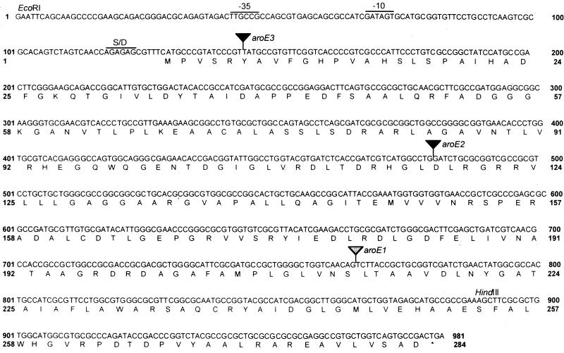
Nucleotide sequence of X. oryzae pv. oryzae aroE gene and deduced amino acid sequence. The solid and shaded triangles indicate mTn7 and Tn3-HOHO1 insertions, respectively. The aroE3::mTn7 insertion could not be marker exchanged. The predicted ribosome binding site (S/D) and -35 and -10 promoter regions are indicated. Restriction sites for EcoRI and HindIII are also indicated.
Sequence analysis.
A 2.953-kb region cloned in pAG5 was sequenced by using transposon-specific primers, as well as primer walking (see Materials and Methods). The insertions aroE1::Tn3-HoHo1, aroE2::mTn7, and aroE3::mTn7 were found to be present in a 849-bp ORF with the potential to encode a 283-amino-acid protein (Fig. 3). A homology search using the BLAST algorithm revealed that the ORF is homologous to aroE, which encodes shikimate dehydrogenase, an enzyme in the aromatic amino acid biosynthetic pathway of various bacterial species. Maximum homologies were found with aroE from Pseudomonas aeruginosa (EMBL/GenBank/DDBJ database accession no. X85015), Neisseria meningitidis (33), E. coli (4), and Haemophilus influenzae (10), as shown in Table 3. Promotor prediction, as described in Materials and Methods, indicated the presence of −35 and −10 promoter regions 100 and 75 nucleotides upstream of the putative ATG start codon, respectively. Within the ORF the aroE1::Tn3-HoHo1, aroE2::mTn7, and aroE3::mTn7 insertions are located at the 634th, 350th, and 15th nucleotides, respectively (Fig. 3). A PstI restriction site was identified 200 bp downstream of the aroE stop codon. This site was used to clone a 1.2-kb EcoRI-PstI fragment that included the aroE ORF to obtain plasmid pAG7. When pAG7 was introduced into BXO65, this minimal fragment was sufficient to confer the Pig+ Vir+ Aro+ phenotype (data not shown).
TABLE 3.
Homology of the X. oryzae pv. oryzae aroE gene with the aroE genes of other bacterial species
| Organism | Length of gene product (amino acids) | % Similarity (% identity) | E valuea | Accession no. |
|---|---|---|---|---|
| P. aeruginosa | 274 | 58 (45) | e-54 | X85015 |
| N. meningitidis | 269 | 57 (41) | e-50 | U82840 |
| E. coli | 272 | 58 (44) | e-50 | U18997 |
| H. influenzae | 272 | 53 (38) | e-35 | U32748 |
E values were obtained by using the BLAST algorithm.
In the 2.953-kb sequence, another ORF downstream of aroE was identified by using the BLAST algorithm. This incomplete ORF started at 1,546th nucleotide and showed very strong homology in the 1,407 bp sequenced (55% similarity and 36% identity at the amino acid level) to dinG (DNA damage-inducible gene G), which encodes an ATP-dependent helicase in E. coli (16).
Complementation of E. coli aroE mutant by pAG4.
E. coli aroE mutant strain AB2835 (30) was transformed individually with pAG4 and pAG5 and with plasmids pAG8, pAG9, and pAG10 having transposon insertions in the aroE ORF (Table 1). Plasmids pAG4 and pAG5 complemented AB2835 for growth on minimal medium, whereas the other plasmids having an insertion in the aroE ORF were not able to do so. All strains grew on minimal medium supplemented with the three aromatic amino acids and the aromatic vitamins p-aminobenzoic acid and p-hydroxybenzoic acid. Plasmid curing from strain AB2835/pAG4 resulted in a loss of prototrophy (data not shown).
Assay of shikimate dehydrogenase activity.
Protein extracts were prepared from the wild-type and aroE mutant strains of X. oryzae pv. oryzae, and shikimate dehydrogenase activity was measured (see Materials and Methods). The Michaelis constant of this enzyme for shikimic acid was calculated to be 5 × 10⁻⁵ M, a value very similar to that in E. coli (32). The enzyme activity was found to be 5.4 mU/mg of protein in wild-type strain BXO43 (Table 2). No activity was detected in aroE mutant-strain BXO65 or in BXO1706 (aroE1::Tn3-HoHo1) or BXO1707 (aroE2::mTn7). The BXO1704 strain (BXO65/pAG4) had an activity of 65 mU/mg of protein, approximately 12 times that of BXO43 (Table 2).
X. oryzae pv. oryzae aroE mutants are unable to synthesize a diffusible factor involved in xanthomonadin biosynthesis.
Cross-feeding of BXO65 with 15 independently isolated EMS-induced prototrophic pigment-deficient mutants was tested by streaking the mutant strains adjacent to BXO65 in pairwise combinations on PSA plates. Development of a yellow color in BXO65 at the adjacent colony boundary was considered to indicate cross-feeding for pigmentation. BXO65 could not be cross-fed for pigmentation by wild-type strain BXO43, whereas 8 of 15 EMS-induced prototrophic pigment-deficient mutants could be. None of the 15 prototrophic pigment-deficient mutants could be cross-fed by each other, BXO43, or BXO65 for pigmentation.
DISCUSSION
In this paper we present evidence that mutations in the aroE gene of X. oryzae pv. oryzae result in reduced levels of xanthomonadin production and virulence. To the best of our knowledge, this is the first report of a mutation affecting a specific step in a general metabolic pathway that also affects xanthomonadin biosynthesis. Insertional inactivation of any of the seven transcriptional units previously shown to be required for xanthomonadin biosynthesis in X. campestris pv. campestris has not been reported to cause any nutrient deficiencies (20, 21). Also, clones containing the aroE gene of X. oryzae pv. oryzae do not show any homology with the pig gene cluster of either X. campestris pv. campestris or X. oryzae pv. oryzae (Goel, Rajagopal, and Sonti, unpublished results). This indicates that the aroE gene is from a different genomic locus than the gene cluster that was previously shown to be required for xanthomonadin biosynthesis.
Shikimate dehydrogenase catalyzes the conversion of dehydroshikimate to shikimic acid. How might shikimate production be related to xanthomonadin biosynthesis? One possibility, as previously suggested (12), is that the aromatic ring in xanthomonadin may be derived from the shikimate pathway. This would be interesting because the aromatic ring in aromatic carotenold pigments is derived from cyclization of the polyene chain and not from the shikimate pathway. However, we have observed that shikimic acid facilitates neither growth on minimal medium nor pigmentation in aroE mutants (Goel and Sonti, unpublished results). This might be because aroE mutants of X. oryzae pv. oryzae are deficient in shikimic acid uptake, as reported previously for E. coli aroE mutants (19).
The residual amount of pigment made in the aroE mutants may be derived either from a very small amount of shikimic acid produced by spontaneous conversion of dehydroshikimate to shikimic acid or through a second minor pathway. It is also possible that the pigment produced by the aroE mutants is devoid of the aromatic ring. Detailed structural analysis and biochemical experiments, such as feeding wild-type cells with labelled shikimic acid and monitoring the incorporation of the label, are required to address these issues.
Several EMS-induced pigment-deficient mutants are able to cross-feed aroE mutant strains for pigment production. This suggests that a diffusible compound that can enter the pigment biosynthetic pathway accumulates in these strains. As xanthomonadins are membrane-bound pigments, the compound or intermediate must act prior to commitment to the membrane-bound state. The inability of the wild-type strain and some of the EMS-induced mutants to cross-feed aroE strains suggests that the compound does not accumulate in these strains. The inability of the prototrophic EMS-induced pigment-deficient mutants to cross-feed each other suggests that they are defective in reactions that involve commitment to either the membrane-bound state or subsequent steps in xanthomonadin production and transport.
The virulence deficiency of the aroE mutants is most likely due to a growth defect. The in planta doubling times for the wild type, aroE mutant BXO65, and a mutant with a complementing clone are 8.6, 25.7, and 10.5 h, respectively (data not shown). This is the first report which suggests that one or more aromatic amino acids may be limiting for growth of X. oryzae pv. oryzae in rice plants. A virulence deficiency has previously been reported to be associated with either arginine or leucine auxotrophy in X. oryzae pv. oryzae (31). The results also suggest that shikimate dehydrogenase can be used as a target to develop novel bactericides against X. oryzae pv. oryzae. These bactericides can be compounds that affect the shikimate dehydrogenase of the pathogen without affecting the host. The shikimate pathway may be a good target for this purpose as it is absent in mammals. It is pertinent to note that effective bactericides are not available for use against X. oryzae pv. oryzae and that judicious application of such compounds could help control yield losses due to this devastating rice pathogen.
ACKNOWLEDGMENTS
We thank Mehar Sultana and N. Nagesh for primer synthesis and DNA sequencing and Jan E. Leach, K. Veluthambi, Jim Pittard, J. Gowrishankar, and A. R. Poplawsky for providing bacterial strains.
A.K.G. and L.R. were supported by fellowships from the Council of Scientific and Industrial Research (CSIR), Government of India.
REFERENCES
- 1.Altschul S F, Madden T L, Schaffer A A, Zhang J, Zhang Z, Miller W, Lipman D J. Gapped BLAST and PSI-BLAST: a new generation of protein database search programs. Nucleic Acids Res. 1997;25:3389–3402. doi: 10.1093/nar/25.17.3389. [DOI] [PMC free article] [PubMed] [Google Scholar]
- 2.Andrewes A G, Hertzberg S, Jensen S-L, Starr M P. Xanthomonas pigments. 2. The Xanthomonas “Carotenoids”—non-carotenoid brominated aryl-polyene esters. Acta Chem Scand. 1973;27:2383–2395. doi: 10.3891/acta.chem.scand.27-2383. [DOI] [PubMed] [Google Scholar]
- 3.Andrewes A G, Jenkins C L, Starr M P, Shepherd J, Hope H. Structure of xanthomonadin I, a novel dibrominated aryl-polyene pigment produced by the bacterium Xanthomonas juglandis. Tetrahedran Lett. 1976;45:4023–4024. [Google Scholar]
- 4.Anton I A, Coggins J R. Sequencing and overexpression of the Escherichia coli aroE gene encoding shikimate dehydrogenase. Biochem J. 1988;249:319–326. doi: 10.1042/bj2490319. [DOI] [PMC free article] [PubMed] [Google Scholar]
- 5.Bradford M M. A rapid and sensitive method for the quantitation of microgram quantities of protein utilizing the principle of protein-dye binding. Anal Biochem. 1976;72:248–254. doi: 10.1016/0003-2697(76)90527-3. [DOI] [PubMed] [Google Scholar]
- 6.Chaudhuri S, Coggins J R. The purification of shikimate dehydrogenase from Escherichia coli. Biochem J. 1985;226:217–223. doi: 10.1042/bj2260217. [DOI] [PMC free article] [PubMed] [Google Scholar]
- 7.Davis R W, Botstein D, Roth J R. A manual for genetic engineering: advanced bacterial genetics. Cold Spring Harbor, N.Y: Cold Spring Harbor Laboratory Press; 1980. [Google Scholar]
- 8.DeFeyter R, Kado C I, Gabriel D W. Small stable shuttle vectors for use in Xanthomonas. Gene. 1990;88:65–72. doi: 10.1016/0378-1119(90)90060-5. [DOI] [PubMed] [Google Scholar]
- 9.Durgapal J C. Albino mutation in rice bacterial blight pathogen. Curr Sci. 1996;70:15. [Google Scholar]
- 10.Fleischmann R D, Adams M D, White O, Clayton R A, Kirkness E F, Kerlavage A R, Bult C J, Tomb J-F, Dougherty B A, Merrick J M, McKenney K, Sutton G, Fitzhugh W, Fields C A, Gocayne J D, Scott J D, Shirley R, Liu L-I, Glodek A, Kelley J M, Weidman J F, Phillips C A, Spriggs T, Hedblom E, Cotton M D, Utterback T R, Hanna M C, Nguyen D T, Saudek D M, Brandon R C, Fine L D, Fritchman J L, Fuhrmann J L, Geoghagen N S M, Gnehm C L, McDonald L A, Small K V, Fraser C M, Smith H O, Venter J C. Whole-genome random sequencing and assembly of Haemophilus influenzae Rd. Science. 1995;269:496–512. doi: 10.1126/science.7542800. [DOI] [PubMed] [Google Scholar]
- 11.Innes R, Hirose M, Kuempel P. Induction of nitrogen fixing nodules on clover requires only 32 kilobase pairs of DNA from the Rhizobium trifolii symbiosis plasmid. J Bacteriol. 1988;170:3793–3802. doi: 10.1128/jb.170.9.3793-3802.1988. [DOI] [PMC free article] [PubMed] [Google Scholar]
- 12.Jenkins C L, Starr M P. The pigment of Xanthomonas populi is a nonbrominated aryl-heptaene belonging to xanthomonadin pigment group II. Curr Microbiol. 1982;7:195–198. [Google Scholar]
- 13.Jenkins C L, Starr M P. The brominated aryl polyene (xanthomonadin) pigments of X. juglandis protect against photobiological damage. Curr Microbiol. 1982;7:323–326. [Google Scholar]
- 14.Kauffman H E, Reddy A P K, Hsieh S P Y, Merca S D. An improved technique for evaluation of resistance of rice varieties to Xanthomonas oryzae. Plant Dis Rep. 1973;57:537–541. [Google Scholar]
- 15.Kelemu S, Leach J E. Cloning and characterization of an avirulence gene from Xanthomonas campestris pv. oryzae. Mol Plant-Microbe Interact. 1990;3:59–65. [Google Scholar]
- 16.Lewis L K, Mount D W. Interaction of LexA repressor with the asymmetric dinG operator and complete nucleotide sequence of the gene. J Bacteriol. 1992;174:5110–5116. doi: 10.1128/jb.174.15.5110-5116.1992. [DOI] [PMC free article] [PubMed] [Google Scholar]
- 17.Leyns F, DeCleene M, Swings J G, Deley J. The host range of the genus Xanthomonas. Bot Rev. 1984;50:308–356. [Google Scholar]
- 18.Miller J H. A short course in bacterial genetics: a laboratory manual for Escherichia coli and related bacteria. Cold Spring Harbor, N.Y: Cold Spring Harbor Laboratory Press; 1992. [Google Scholar]
- 19.Pittard J, Wallace B J. Distribution and function of genes concerned with aromatic biosynthesis in Escherichia coli. J Bacteriol. 1966;91:1494–1508. doi: 10.1128/jb.91.4.1494-1508.1966. [DOI] [PMC free article] [PubMed] [Google Scholar]
- 20.Poplawsky A R, Kawalek M D, Schadd N W. A xanthomonadin-encoding gene cluster for the identification of pathovars of Xanthomonas campestris. Mol Plant-Microbe Interact. 1993;6:545–552. [Google Scholar]
- 21.Poplawsky A R, Chun W. pigB determines a diffusible factor needed for extracellular polysaccharide slime and xanthomonadin production in Xanthomonas campestris py. campestris. J Bacteriol. 1997;179:439–444. doi: 10.1128/jb.179.2.439-444.1997. [DOI] [PMC free article] [PubMed] [Google Scholar]
- 22.Poplawsky A R, Chun W. Xanthomonas campestris pv. campestris requires a functional pigB for epiphytic survival and host infection. Mol Plant- Microbe Interact. 1998;11:466–475. doi: 10.1094/MPMI.1998.11.6.466. [DOI] [PubMed] [Google Scholar]
- 23.Rajagopal L, Sivakamasundari C, Balasubramanian D, Sonti R V. The bacterial pigment xanthomonadin provides protection against photodamage. FEBS Lett. 1997;415:125–128. doi: 10.1016/s0014-5793(97)01109-5. [DOI] [PubMed] [Google Scholar]
- 24.Rajagopal L, Dharmapuri S, Sayeepriyadarshini A T, Sonti R V. A genomic library of Xanthomonas oryzae pv. oryzae in the broad host range mobilizing Escherichia coli strain S17–1. Int Rice Res Notes. 1999;24:20–21. [Google Scholar]
- 25.Sambrook J, Fritsch E F, Maniatis T. Molecular cloning: a laboratory manual. 2nd ed. Cold Spring Harbor, N.Y: Cold Spring Harbor Laboratory Press; 1989. [Google Scholar]
- 26.Simon R, Preifer U, Puhler A. A broad host range mobilisation system for in vivo genetic engineering: transposon mutagenesis in gram negetive bacteria. Bio/Technology. 1983;1:784–791. [Google Scholar]
- 27.Stachel S E, An G, Flores C, Nester E W. A Tn3 lacZ transposon for random β-galactosidase gene fusions: application to the analysis of gene expression in Agrobacterium. EMBO J. 1985;4:891–898. doi: 10.1002/j.1460-2075.1985.tb03715.x. [DOI] [PMC free article] [PubMed] [Google Scholar]
- 28.Starr M P. The genus Xanthomonas. In: Starr M P, Stolp H, Truper H G, Balows A, Schlegel H G, editors. The prokaryotes. Vol. 1. Berlin, Germany: Springer-Verlag; 1981. pp. 742–763. [Google Scholar]
- 29.Tsuchiya K, Mew T W, Wakimoto S. Bacteriological and pathological characteristics of wild type and induced mutants of Xanthomonas campestris pv. oryzae. Phytopathology. 1982;72:43–46. [Google Scholar]
- 30.Whipp M J, Camakaris H, Pittard A J. Cloning and analysis of shiA gene, which encodes the shikimate transport system of Escherichia coli K-12. Gene. 1998;209:185–192. doi: 10.1016/s0378-1119(98)00043-2. [DOI] [PubMed] [Google Scholar]
- 31.Yamasaki Y, Murata N, Suwa T. The effect of mutations for nutritional requirements on the pathogenicity of two pathogens of rice. Proc Jpn Acad. 1964;40:226–231. [Google Scholar]
- 32.Yaniv H, Gilvarg C. Aromatic biosynthesis. XIV. 5-Dehydroshikimic reductases. J Biol Chem. 1955;213:787–795. [PubMed] [Google Scholar]
- 33.Zhou J, Bowler L D, Spratt B G. Interspecies recombination and phylogenetic distortions within the glutamine synthetase and shikimate dehydrogenase genes of Neisseria meningitidis and commensal Neisseria species. Mol Microbiol. 1997;23:799–812. doi: 10.1046/j.1365-2958.1997.2681633.x. [DOI] [PubMed] [Google Scholar]


