Abstract
Plasmid transfer between strains of Bacillus thuringiensis subsp. israelensis was studied under a range of environmentally relevant laboratory conditions in vitro, in river water, and in mosquito larvae. Mobilization of pBC16 was detected in vitro at a range of temperatures, pH values, and available water conditions, and the maximum transfer ratio was 10−3 transconjugant per recipient under optimal conditions. Transfer of conjugative plasmid pXO16∷Tn5401 was also detected under this range of conditions. However, a maximum transfer ratio of 1.0 transconjugant per recipient was attained, and every recipient became a transconjugant. In river water, transfer of pBC16 was not detected, probably as a result of the low transfer frequency for this plasmid and the formation of spores by the introduced donor and recipient strains. In contrast, transfer of plasmid pXO16∷Tn5401 was detected in water, but at a lower transfer ratio (ca. 10−2 transconjugant per donor). The number of transconjugants increased over the first 7 days, probably as a result of new transfer events between cells, since growth of both donor and recipient cells in water was not detected. Mobilization of pBC16 was not detected in killed mosquito larvae, but transfer of plasmid pXO16::Tn5401 was evident, with a maximum rate of 10−3 transconjugant per donor. The reduced transfer rate in insects compared to broth cultures may be accounted for by competition from the background bacterial population present in the mosquito gut and diet or by the maintenance of a large population of B. thuringiensis spores in the insects.
Bacillus thuringiensis is a gram-positive bacterium that produces insecticidal crystal protein toxins during sporulation. B. thuringiensis was first discovered in diseased silkworms in 1901 (19, 31) but has since been isolated from a range of environments, including insects, soil, dust from stored grain, and leaves of coniferous and deciduous trees (7, 24, 25, 28). In 1977, B. thuringiensis subsp. israelensis was isolated from a mosquito-breeding pond in the Negev Desert of Israel and was found to be highly active towards dipteran larvae (16). A number of insecticidal protein toxins that are encoded on a single 72-MDa plasmid have been identified in B. thuringiensis subsp. israelensis. Movement of plasmids within B. thuringiensis strains has been proposed to be the main mechanism for generating diversity in toxin genes. In addition to plasmids carrying insecticidal toxin genes, many other plasmids, such as pXO11, pXO13, pXO14, pXO15, pXO16, and pAW63, have been detected in B. thuringiensis, and these plasmids have no known function apart from their conjugative ability (6, 26, 32). Strains of B. thuringiensis can have as few as one plasmid to more than six plasmids.
Movement of plasmids between B. thuringiensis strains has been of interest in the construction of strains with novel toxin combinations, the study of the mechanisms of plasmid transfer in bacteria, and the study of the evolution of toxin combinations. Furthermore, since B. thuringiensis is closely related to the human pathogens Bacillus anthracis and Bacillus cereus, interest has been focused on the genetic exchange systems of these bacteria in relation to biosafety (22). Plasmid movement between B. thuringiensis strains can be monitored directly by plasmid profiling analysis of cells. This is possible when transfer frequencies are high and all recipients in a population become transconjugants. When the transfer frequency is lower, a shuttle plasmid with an antibiotic resistance gene can be used to monitor the movement of conjugative plasmids (3). The shuttle plasmids are either mobilizable or oriT bearing, such as pBC16. Differences in the abilities of conjugative plasmids to move the reporter plasmids have been demonstrated. Green et al. (17) transposon tagged conjugative plasmid pXO12 and demonstrated that it transferred itself and pBC16 at similar rates (10−2 transconjugant per recipient). In contrast, transposon tagging of plasmid pXO16 showed that it was transferred to every recipient cell but mobilized pBC16 only at a frequency of approximately 10−3 to 10−4 transconjugant per recipient (1).
Very few studies have examined plasmid transfer between B. thuringiensis strains and, in particular, between B. thuringiensis subsp. israelensis strains in the environment. Haack et al. (18) reported transfer of the broad-host-range conjugative transposon Tn916 between Bacillus subtilis and B. thuringiensis subsp. israelensis in nonsterile sandy soil. Significantly, although B. thuringiensis is applied to waterways to control pests such as mosquitoes and blackflies, no studies have examined plasmid transfer between B. thuringiensis strains in water yet. In nature B. thuringiensis strains may find themselves in the midgut environment of killed susceptible larvae. This environment has proven to be conducive to gene exchange. Studies have found that plasmids can move between donor and recipient strains of B. thuringiensis in larvae of the lepidopteran insects Galleria mellonella, Spodoptera littoralis, and Lacanobia oleracea at levels similar to that found in broth culture (20, 30) but not in the midgut environment of the coleopteran insect Phaedon cochleriae. So far, there have been no studies of transfer between B. thuringiensis strains in dipteran insects.
The primary objective of this study was to obtain information regarding mobilization and transfer of plasmids between B. thuringiensis subsp. israelensis strains under environmentally realistic conditions. Insecticidal strains of B. thuringiensis subsp. israelensis were constructed with either a mobilizable plasmid (pBC16) or a transposon-tagged conjugative plasmid (pXO16::Tn5401) so that gene transfer in laboratory media, water, and insects could be studied.
MATERIALS AND METHODS
Bacterial strains and plasmids.
The strains used in this study are listed in Table 1. All strains were kept at −80°C in storage medium (Protect; Technical Consultants Limited). To maintain the plasmid compositions of B. thuringiensis strains used frequently in this work, cultures were stored as 5-ml aliquots in sterile bijou bottles at −20°C in 20% (vol/vol) glycerol. Every month, a bijou bottle was removed from the freezer and the contents were thawed, streaked onto nutrient agar (Oxoid, Basingstoke, Hampshire, United Kingdom), and grown at 30°C for 2 days. The plate was kept as a source of inoculum at 5°C for up to 1 month, after which a new inoculum plate was prepared from glycerol stocks.
TABLE 1.
Bacterial strains and plasmids
| Strain | Plasmid | Relevant genotype or characteristics | Source or reference |
|---|---|---|---|
| B. thuringiensis subsp. kurstaki HD1 cry Smr | Cryr Smr | 20 | |
| B. thuringiensis subsp. israelensis IPS82 | cry4A cry4B | 16 | |
| B. thuringiensis subsp. israelensis IPS82(pBC16) | pBC16 | cry11A cyt1A Tcr | This study |
| B. thuringiensis subsp. israelensis IPS78(pXO16) | pXO16::Tn5401 | Tcr | This study |
| B. thuringiensis subsp. israelensis 658 cry Smr | Cryr Smr | P. Jarrett | |
| B. thuringiensis subsp. israelensis AND931(pXO16::Tn5401) | pXO16::Tn5401 | Tcr Smr | 2 |
| B. thuringiensis subsp. israelensis GBJ002/IPS70 | Nalr | 21 |
Construction of B. thuringiensis subsp. israelensis IPS82(pBC16) and IPS78 (pXO16::Tn5401).
Plasmid pBC16 was introduced into B. thuringiensis subsp. israelensis IPS82 by electroporation by using a field strength of 8.75 kV/cm (400 Ω, 25 μF, 1.75 kV) and a Bio-Rad gene pulser (30). B. thuringiensis subsp. israelensis IPS78(pXO16::Tn5401) was constructed by plasmid mating. B. thuringiensis subsp. israelensis AND931(pXO16::Tn5401) contains a selectable tetracycline resistance gene marker on transposon Tn5401. This strain is also resistant to streptomycin (chromosomal mutation), but it does not contain the insecticidal proteins present in the original strain. To create a marked insecticidal strain, B. thuringiensis subsp. israelensis AND931(pXO16::Tn5401) was mated with B. thuringiensis subsp. israelensis IPS78. The cell mixture was plated on nutrient agar containing 5 μg of tetracycline ml−1, which selected for strains harboring pXO16::Tn5401, and included both the donor and transconjugant strains. Since pXO16::Tn5401 transfers at a high frequency, all recipients should have become transconjugants, and these transconjugants could have accounted for up to 50% of the population obtained. One hundred colonies were examined by light microscopy for the presence of ovoid insecticidal protein toxin crystals, which were present only in the recipient strain. All colonies with ovoid inclusions were then retested for the ability to grow on nutrient agar containing 50 μg of streptomycin ml−1. Any colonies unable to grow on media containing streptomycin were confirmed to be B. thuringiensis subsp. israelensis IPS78(pXO16::Tn5401) colonies.
Standard broth mating procedure.
The standard broth mating procedure used was that described by Thomas et al. (30). Briefly, a single colony was used to inoculate separately 50 ml of brain heart infusion (BHI) (Oxoid) broth containing the appropriate antibiotic and incubated at 30°C for 18 h with shaking (40 rpm). The overnight culture was diluted to an optical density at 600 nm of 1.1 (approximately 108 CFU ml−1) in 0.25× Ringer's solution (BDH). Aliquots (0.5 ml) of both donor and recipient cell suspensions were added to three 250-ml conical flasks containing 50 ml of pre-warmed BHI broth and incubated at 30°C for 6 h with shaking (40 rpm). Three broth preparations with donor cells and three preparations with recipient cells were treated in the same fashion and used as controls. After the incubation period, samples were serially diluted in 0.25× Ringer's solution and spread plated onto nutrient agar containing the appropriate antibiotics to select for donor, recipient, and transconjugant CFU. In experiments in which mobilization of pBC16 was studied, the bacteria were enumerated on nutrient agar containing 25 μg of tetracycline ml−1(donor), 50 μg of streptomycin ml−1 (recipient), or both 25 μg of tetracycline ml−1 and 50 μg of streptomycin ml−1 (transconjugant). When the transfer of pXO16::Tn5401 was measured, the bacteria were enumerated on nutrient agar containing 5 μg of tetracycline ml−1(donor), 25 μg of nalidixic acid ml−1 (recipient), or both 5 μg of tetracycline ml−1 and 25 μg of nalidixic acid ml−1 (transconjugant). The effects of time, temperature, pH, and salt concentration on plasmid transfer were measured by using the standard broth mating procedure with appropriate modifications (30).
Survival and gene transfer in water.
Donor and recipient B. thuringiensis strains were used to inoculate individual nutrient agar plates containing the appropriate antibiotics. After incubation at 30°C for 24 h, a single colony was used to inoculate 50 ml of BHI broth, and the inoculated flask was incubated at 30°C for 18 h with shaking (40 rpm). The overnight culture was pelleted by centrifugation at 2,000 × g for 5 min and resuspended and diluted to an optical density at 600 nm of 1.1 (approximately 108 CFU ml−1) in filtered (pore size, 0.22 μm; Whatman) river water. The water was collected from the River Avon at Tiddington, Stratford-upon-Avon, United Kingdom, in August 1997 and October 1999 for experiments to measure the movement of pBC16 and pXO16::Tn5401, respectively. To three 90-ml replicates of fresh nonsterile River Avon water in 250-ml conical flasks, 10 ml of a bacterial suspension containing donor and recipient cells (ratio, 1:1) suspended in filtered river water was added, and the preparations were incubated at 10 and 25°C. Flasks inoculated with only donor and recipient cells and flasks containing only filtered river water were included and treated in the same fashion. Samples were taken, diluted in 0.25× Ringer's solution, and spread plated onto nutrient agar alone and nutrient agar containing the relevant antibiotics to determine the levels of background, donor, recipient, and transconjugant populations. In addition, samples were heated at 70°C for 20 min and dilution plate counted on the same media described above to determine the numbers of donor and recipient spores.
Insect rearing.
Mosquito eggs (Aedes aegypti, rock strain) on filter paper were obtained from the School of Biological Sciences, Manchester University, Manchester, United Kingdom. Eggs were hatched by placing the filter paper on which they were laid in a Buchner flask containing 100 ml of distilled water under a vacuum (−0.65 × 10⁵ Pa) for 1 h to break open the egg casings. The larvae were transferred to a 5-liter polyethylene bowl containing hamster feed pellets and incubated at 25°C for up to 1 week.
Studies with A. aegypti larvae.
Spore and crystal mixtures of donor and recipient strains were prepared from nutrient agar plates that had been incubated at 30°C for 5 days. Spores and crystals were resuspended in sterile 0.25× Ringer's solution to an optical density at 600 nm of 1.1 (approximately 1 × 10⁸ CFU ml−1). Gene transfer was studied in second- or third-instar larvae of the mosquito (A. aegypti). Aliquots (250 ml) of donor and recipient cell suspension were added to 25 ml of distilled water containing 12 mosquito larvae in a plastic autoclavable beaker. Water microcosms were incubated at 25°C with a light cycle consisting of 18 h of light (24 W m−2) and 6 h of darkness and 65% relative humidity. Microcosms containing only mosquito larvae and donor inocula, only mosquito larvae and recipient inocula, and only mosquito larvae and water were included as controls and treated in the same fashion. Samples of three pooled mosquito larvae from each of the three replicate beakers were taken after 4, 24, and 48 h. The three mosquito larvae were washed in 1 liter of fresh sterile distilled water and placed in 1 ml of sterile distilled water in a 1.5-ml Eppendorf tube. A sterile pellet resuspender was used to crush the larvae, and dilutions were made in sterile 0.25× Ringer's solution and plated onto nutrient agar alone and nutrient agar containing the relevant antibiotics to select for transconjugant, donor, recipient, and background populations. In addition, samples were heat treated at 70°C for 20 min and dilution plate counted to determine the numbers of donor and recipient spores in samples.
Statistical analysis.
For time course studies and quantitative-level treatments (e.g., temperature, pH, and available water) a one-way model of analysis of variance was used (29). Differences between times and levels were expressed by using a least significant difference bar. In some instances a two-way analysis of variance was used when two factors were compared.
RESULTS
Strain construction.
B. thuringiensis subsp. israelensis IPS82(pBC16) was shown to contain plasmid pBC16 and was insecticidal to susceptible A. aegypti larvae. In the standard mating broth B. thuringiensis subsp. israelensis IPS82(pBC16) was unable to mobilize pBC16 to a B. thuringiensis subsp. kurstaki HD1 cry Smr recipient. However, plasmid pBC16 was mobilized efficiently from this strain to the recipient B. thuringiensis subsp. israelensis HD658 cry Smr. The transconjugant strain B. thuringiensis subsp. israelensis IPS78(pX016::Tn5401) was also shown to be insecticidal and capable of transferring plasmid pX016::Tn5401 into B. thuringiensis subsp. israelensis GBJ002 (IPS70).
Transfer of pBC16 and pXO16::Tn5401 in liquid culture.
Mobilization of pBC16 from B. thuringiensis subsp. israelensis IPS82 to B. thuringiensis subsp. israelensis 658 cry Smr was studied over a 24-h period (data not shown). The donor and recipient strains had antagonistic effects on each other. There was a slight, nonsignificant increase in the number of donors from 7.1 × 10⁵ CFU ml−1 at the time of inoculation to 1.7 × 10⁶ CFU ml−1 after 2 h. This was followed by a decrease in the number of donors to 4.6 × 10⁵ CFU ml−1 after 6 h of incubation. Subsequently, the number of donors increased to 7.8 × 10⁷ CFU ml−1 after 10 h and remained at this level for the rest of the experiment, whereas there was an initial rise in the number of recipients from 3.9 × 10⁵ CFU ml−1 at the time of inoculation to 1.4 × 10⁷ CFU ml−1 after 6 h of incubation. The highest density of recipients was detected after 6 h, and thereafter the density decreased to 4.1 × 10⁴ CFU ml−1 after 24 h. The number of transconjugants detected initially after 2 h was 5.3 × 101 CFU ml−1, and the number increased to 1.1 × 10⁴ after 10 h of incubation, after which there was a decline in the size of the population to 2.5 × 10² CFU ml−1 after 24 h. The increase in the number of donor cells resulted in an accompanying decrease in the number of recipient cells, which was reflected in the number of transconjugants. The curve for the number of transconjugants produced reflected the curve for the number of recipients. When transfer of pXO16::Tn5401 between B. thuringiensis subsp. israelensis IPS78 (pXO16::Tn5401) and B. thuringiensis subsp. israelensis GBJ002 after as little as 4 h was studied, the transfer ratio was found to be 1.0; in effect, all of the recipients gained a copy of the plasmid (data not shown).
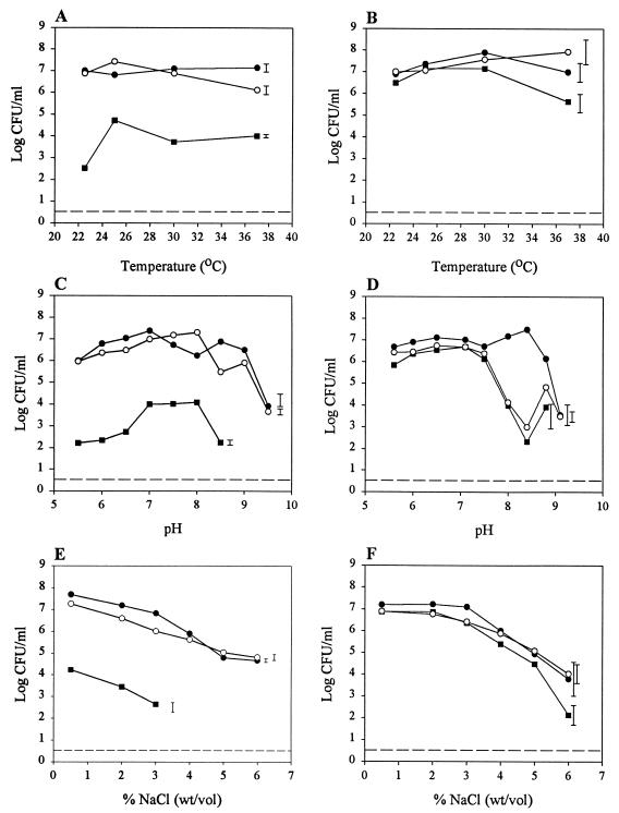
The effect of temperature on transfer of pBC16 between B. thuringiensis subsp. israelensis IPS82(pBC16) and B. thuringiensis subsp. israelensis 658 cry Smr is shown in Fig. 1A. There was not a significant difference between the final population sizes of the donor after incubation at different temperatures, and this was also true for the recipient. The largest number of transconjugants was detected at 25°C (1.2 × 10⁴ CFU ml−1). The lowest transfer ratio was recorded at 22.5°C. The transfer ratio increased to 8.9 × 10⁻³ transconjugant per donor at 25°C. No significant decrease in the transfer ratio was observed between 25 and 37°C although the ratio varied by almost 1 order of magnitude. Over this temperature range the average transfer ratio was 8 × 10⁻² transconjugant per donor. The highest transfer ratio per recipient was observed at 37°C (3.4 × 10⁻⁴ transconjugant per recipient) due to the decrease in the number of recipient cells. When the effect of temperature on the transfer of pXO16::Tn5401 from B. thuringiensis subsp. israelensis IPS78 (pXO16::Tn5401) to B. thuringiensis subsp. israelensis GBJ002 was studied, the results were comparable to the results obtained for transfer of pBC16 (Fig. 1B). The transfer ratio was approximately 1 transconjugant per donor or recipient between 22.5 and 30°C and then decreased to 4.5 × 10⁻² transconjugant per donor and 5 × 10⁻² transconjugant per recipient at 37°C.
FIG. 1.
Effects of various culture conditions on growth of strains and transfer of plasmids pBC16 and pXO16::Tn5401 in B. thuringiensis subsp. israelensis. (A, C, and E) Studies with B. thuringiensis subsp. israelensis IPS82(pBC16) and B. thuringiensis subsp. israelensis 658 cry Smr. Symbols: ●, B. thuringiensis subsp. israelensis IPS82(pBC16); ○, B. thuringiensis subsp. israelensis 658 cry Smr; ■, transconjugant. (B, D, and F) Studies with B. thuringiensis subsp. israelensis IPS78(pXO16::Tn5401) and B. thuringiensis subsp. israelensis GBJ002. Symbols: ●, B. thuringiensis subsp. israelensis IPS78(pXO16::Tn5401); ○, B. thuringiensis subsp. israelensis GBJ002; ■, transconjugant. The effects of temperature (A and B), pH (C and D), and NaCl concentration (E and F) were examined. The dashed lines indicate the detection limit. The error bars indicate the least significant differences of the means (n = 3; P < 0.05).
The effects of pH on mobilization of pBC16 from B. thuringiensis subsp. israelensis IPS82(pBC16) to B. thuringiensis subsp. israelensis 658 cry Smr and transfer are shown in Fig. 1C. The pH of the broth did not have a significant effect on growth of the donor strain at pH values between 5.8 and 8.8. The largest numbers of transconjugants were seen between pH 7.1 and 8.1 (1.0 × 10⁴ to 1.3 × 10⁴ CFU ml−1). No reduction in the number of donor cells was observed at pH 8.4. Consequently, due to the decrease in the number of transconjugants at this pH, the transfer ratio expressed on a per-donor basis decreased significantly from 7.8 × 10⁻³ at pH 7.9 to 3.7 × 10⁻⁵ at pH 8.4.
Similar results were obtained when the transfer of pXO16::Tn5401 between B. thuringiensis subsp. israelensis IPS78(pXO16::Tn5401) and B. thuringiensis subsp. israelensis GBJ002 was studied over a range of pH values (Fig. 1D). The transfer ratio remained approximately 1 at pH values between 6.0 and 8.0. Growth of the recipient bacterium decreased significantly at pH values above pH 7.5. Consequently, the number of transconjugants and the transfer ratio decreased to 2 × 10⁻¹ CFU ml−1 and 1.2 ×10⁻¹, respectively, at pH 8.4 and 8.8. At pH 9.1 the sizes of both the donor and recipient populations decreased significantly, and no transconjugants were detected.
The effects of water activity on mobilization of pBC16 and growth of B. thuringiensis subsp. israelensis IPS82(pBC16) and B. thuringiensis subsp. israelensis 658 cry Smr are shown in Fig. 1E. The numbers of donor and recipient bacteria fell gradually from 4.9 × 10⁷ and 1.9 × 10⁷ CFU ml−1, respectively, at 0.5% (wt/vol) NaCl to 6.3 × 10⁴ and 1.1 × 10⁵ CFUml−1, respectively, at 5% (wt/vol) NaCl. Mobilization of pBC16 from B. thuringiensis subsp. israelensis IPS82(pBC16) to B. thuringiensis subsp. israelensis 658 cry Smr was found to occur at NaCl concentrations between 0.5 and 3% (wt/vol) (Fig. 1E). The decreases in the numbers of donor and recipient cells in this mating experiment at NaCl concentrations of 0.5 to 3% (wt/vol) were mirrored by concomitant decreases in the number of transconjugants. The transfer ratio decreased from 3.6 × 10⁻⁴ at 0.5% (wt/vol) NaCl to 6.7 × 10⁻⁵ at 3% (wt/vol) NaCl. Plasmid mobilization was not detected at NaCl concentrations greater than 3% (wt/vol). Again, a similar pattern was seen when transfer of pXO16::Tn5401 between B. thuringiensis subsp. israelensis IPS78 (pXO16::Tn5401) and B. thuringiensis subsp. israelensis GBJ002 was studied. There were decreases in the numbers of donor and recipient bacteria at NaCl concentrations between 3 and 6% (wt/vol) (Fig. 1F). The transfer ratios also decreased from 1 transconjugant per donor or recipient at 2% (wt/vol) NaCl to 2.4 × 10⁻² transconjugant per donor and 1.4 × 10⁻² transconjugant per recipient at 6% (wt/vol) NaCl, but notable numbers of transconjugants could still be detected at the highest NaCl concentration studied.
Survival of B. thuringiensis and mobilization of pBC16 in river water.
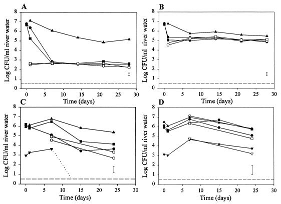
Mobilization experiments were performed with nonsterile river water which was incubated in the dark without shaking at 10°C, a typical temperature encountered in a river in a temperate region, and at 25°C, the temperature at which mobilization for this strain was found to take place most efficiently in laboratory media (Fig. 1A). B. thuringiensis subsp. israelensis(pBC16) and B. thuringiensis subsp. israelensis 658 cry Smr were coinoculated into river water at levels of 6.3 × 10⁶ and 4.8 × 10⁶ CFU ml−1, respectively, and incubated at 10°C (Fig. 2A). The number of B. thuringiensis subsp. israelensis(pBC16) cells fell slightly to 2.4 × 10⁶ CFU ml−1 after 1 day of incubation, whereas the number of B. thuringiensis subsp. israelensis 658 cry Smr cells declined to 2.3 × 10⁵ CFU ml−1 after 1 day of incubation. Subsequently, the total sizes of the donor and recipient populations declined sharply to 6.9 × 10² and 4.2 × 10² CFU ml−1, respectively, after 7 days of incubation. The proportion of the population present as spores increased from 0.0064% of the CFU detected at inoculation to nearly 88% of the total B. thuringiensis CFU detected after 7 days of incubation, due to a rapid decline in the size of the population of vegetative cells. The donor and recipient bacteria were then maintained in the river water at levels around 4 × 10² CFU ml−1, predominantly as spores. During this experiment, the size of the background culturable population was monitored and was found to increase from an ambient level of 8.7 × 10³ to 1.3 × 10⁷ CFU ml−1 1 day after inoculation of the donor and recipient populations. However, once this initial high level was attained, the population size declined gradually during the experiment and was 1.5 × 10⁵ CFU ml−1 after 28 days. No transconjugants were detected. The background culturable population during the experiment was predominantly composed of bacteria that did not display the colony morphology of B. cereus and B. thuringiensis. When B. thuringiensis subsp. israelensis(pBC16) and B. thuringiensis subsp. israelensis 658 cry Smr were inoculated individually into river water, a similar pattern of survival was observed when the results were compared to the results of the coinoculation experiments (data not shown).
FIG. 2.
Survival of B. thuringiensis strains and transfer of plasmids pBC16 and pXO16::Tn5401 in river water at 10°C (A and C) and 25°C (B and D). (A and B) B. thuringiensis subsp. israelensis IPS82(pBC16) was used as the donor strain, and B. thuringiensis subsp. israelensis 658 cry Smr was used as the recipient. (C and D) B. thuringiensis subsp. israelensis IPS78 (pXO16::Tn5401) was used as the donor, and B. thuringiensis subsp. israelensis GBJ002 was used as the recipient strain. Symbols: ▾, transconjugant cells; ▿, transconjugant spores; ●, donor cells; ○, donor spores; ■, recipient cells; □, recipient spores; ▴, CFU on nutrient agar. The dashed lines indicate the detection limit; the dotted line illustrates a decline to the detection limit. The error bars indicate the least significant differences of the means (n = 3; P < 0.05).
In water kept at 25°C the sizes of the B. thuringiensis subsp. israelensis(pBC16) and B. thuringiensis subsp. israelensis 658 cry Smr populations declined from 6.3 × 10⁶ and 4.8 × 10⁶ CFU ml−1, respectively, immediately after inoculation to 1.2 × 10⁵ and 2.2 × 10⁵ CFU ml−1, respectively, after 24 h of incubation in river water (Fig. 2B). The numbers of spores of B. thuringiensis subsp. israelensis (pBC16) and B. thuringiensis subsp. israelensis 658 cry Smr increased slightly from 3.4 × 10⁴ and 6.1 × 10⁴ CFU ml of river water−1, respectively, after 1 day to 1.5 × 10⁵ and 1.8 × 10⁵ CFU ml of river water−1, respectively, after 7 days of incubation. Plasmid mobilization was not detected when the donor and recipient strains were incubated at 25°C. The sizes of the populations of donor and recipient bacteria remained approximately 1 × 10⁵ CFU ml−1 during the experiment (28 days). The size of the culturable bacterial population present in the river water increased from 8.6 × 10³ to 6.0 × 10⁶ CFU ml−1 after 24 h following inoculation of the donor and recipient strains, and the organisms in this and subsequent samples collected during the experiment were found to predominantly display the morphology of B. cereus and B. thuringiensis strains. Subsequently, the numbers of culturable bacteria present in the river water declined to approximately 5 × 10⁵ CFU ml−1 during the experiment. When B. thuringiensis subsp. israelensis(pBC16) and B. thuringiensis subsp. israelensis 658 cry Smr were inoculated individually into river water, the patterns of survival were similar to that observed when the donor and recipient strains were coinoculated (data not shown).
Survival of B. thuringiensis and transfer of pXO16::Tn5401 in river water.
B. thuringiensis subsp. israelensis IPS78(pXO16::Tn5401) and B. thuringiensis subsp. israelensis GBJ002 were coinoculated into river water at levels of 6.3 × 10⁶ and 4.8 × 10⁶ CFU ml−1, respectively, and incubated at 10 and 25°C. Figures 2C and D show the survival of the donor and recipient strains. At both temperatures, the sizes of the populations of both donor and recipient bacteria exhibited slight, nonsignificant decreases after 1 day and slight, nonsignificant increases after 7 days. Subsequently, the sizes of all the bacterial populations monitored slowly decreased in the river water, and after 30 days the bacteria were detected predominantly as spores. Transfer of pXO16::Tn5401 between the donor and recipient strains was detected at both temperatures initially after entry into water and at later sampling times. At 10°C the transfer ratio was initially 7.7 × 10⁻⁴ transconjugant per donor and rose to 3.5 × 10⁻² transconjugant per donor on day 7. However, the number of transconjugants declined to undetectable levels by 15 days after inoculation (Fig. 2C). At 25°C, initially the size of the transconjugant population was 1.3 × 10³ CFU ml−1, and the size of this population increased significantly to 3.0 × 10⁴ CFU ml−1 after 7 days. After this the transconjugant population was found to be composed predominantly of spores. The transfer ratio increased during the experiment from 9.7 × 10⁻⁴ to 4.4 × 10⁻² transconjugant per donor.
Mobilization of pBC16 in A. aegypti larvae.
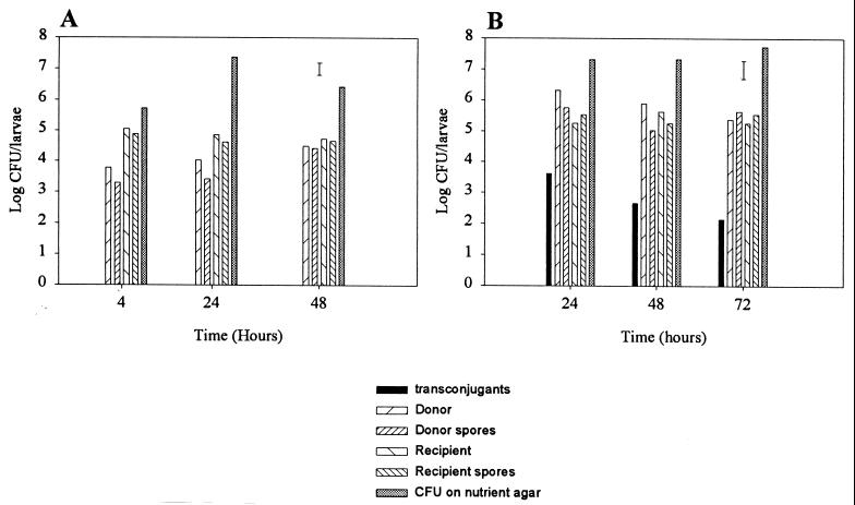
Third- or fourth-instar larvae of A. aegypti were fed the donor B. thuringiensis subsp. israelensis IPS82(pBC16) along with the recipient B. thuringiensis subsp. israelensis 658 cry Smr. After 4 h all the A. aegypti larvae were dead. Altogether, the total number of donor bacteria per larva increased from 6.1 × 10³ CFU larva−1 after 4 h to 3.2 × 10⁵ CFU larva−1 after 48 h of incubation (Fig. 3A). The number of recipient bacteria remained approximately 8 × 10⁴ CFU larva−1 throughout the experiment. There was not a significant difference in the number of donor or recipient spores at any time point. Mobilization of pBC16 between B. thuringiensis subsp. israelensis IPS82(pBC16) and B. thuringiensis subsp. israelensis 658 cry Smr was not detected. Microscopic examination of the dead larvae indicated that bacteria other than B. thuringiensis dominated the cadavers. This was reflected in the higher number of background bacteria detected, which increased from 5.6 × 10⁵ CFU larva−1 after 4 h to 2.5 × 10⁷ CFU larva−1 after 48 h.
FIG. 3.
Growth of B. thuringiensis strains and transfer of pBC16 and pXO16::Tn5401 in A. aegypti larvae. (A) B. thuringiensis subsp. israelensis IPS82(pBC16) donor and B. thuringiensis subsp. israelensis 658 cry Smr recipient. (B) B. thuringiensis subsp. israelensis IPS78(pXO16::Tn5401) donor and B. thuringiensis subsp. israelensis GBJ002 recipient. The error bars indicate the least significant differences of the means (n = 3; P < 0.05).
The total number of bacteria in the donor control increased from 1.8 × 10³ CFU larva−1 after 24 h to 3.8 × 10⁴ CFU larva−1 after 48 h (data not shown). The levels of background bacteria were much higher in all samples, reaching a maximum of 3.4 × 10⁷ CFU larva−1 after 48 h. In the recipient control, live mosquitoes were sampled during the entire experiment, and the number of recipient cells and spores remained approximately 3 × 10⁵ CFU larva−1 during the experiment. The level of background bacteria in the recipient control increased during the experiment from 3.8 × 10⁵ CFU larva−1 after 4 h to 4.6 × 10⁷ CFU larva−1 after 48 h. The level of background bacteria increased in the water-only control to a maximum of 3.4 × 10⁵ CFU larva−1 after 48 h of incubation.
Transfer of pXO16::Tn5401 in A. aegypti larvae.
A. aegypti larvae were fed a lethal dose of the donor B. thuringiensis subsp. israelensis IPS78(pXO16::Tn5401) along with an equal dose of the recipient B. thuringiensis subsp. israelensis GBJ002. After 4 h all larvae were dead in microcosms that contained the active B. thuringiensis subsp. israelensis IPS78 (pXO16::Tn5401) strain. The total number of donor and recipient bacteria remained between 5 × 10⁵ and 5 × 10⁶ CFU larva−1 between 24 and 72 h (Fig. 3B). There was not a significant difference in the number of donor or recipient spores at any sampling point. The level of background bacteria remained approximately 3 × 10⁷ CFU larva−1. Transconjugants were detected after 24 h at a level of 4.8 × 10³ CFU larva−1; after this the level of transconjugants decreased to 1.6 × 10³ and 1.6 × 10² CFU larva−1 at 48 and 72 h, respectively. No plasmid transfer between donor and recipient in water initially inoculated with spores was detected during the 3-day study.
The donor-only control behaved in a similar fashion, with both the donor and background bacteria present at high levels (data not shown). In the recipient control experiment, in which live mosquitoes were sampled during the entire experiment, the number of recipient cells and spores remained approximately 7 × 10⁴ CFU larva−1. The level of background bacteria in the recipient control remained approximately 1 × 10⁵ CFU larva−1 throughout the experiment. The number of background bacteria in the water-only control remained approximately 5 × 10⁴ CFU larva−1 (data not shown).
DISCUSSION
The widespread occurrence of different toxin genes and plasmids in B. thuringiensis strains suggests that conjugation may be an important means for dissemination of these genes and plasmids in Bacillus populations in nature. Constructed strains were used in sensitive studies to monitor the effects of a range of physical and chemical parameters on plasmid exchange between B. thuringiensis strains (Fig. 1). Initial experiments indicated that plasmid pBC16 could not be transferred from B. thuringiensis subsp. israelensis IPS82 to B. thuringiensis subsp. kurstaki cry Smr, a strain which had previously been successfully used in studies to monitor mobilization of pBC16 under environmental conditions (30). However, these experiments did indicate that pBC16 could be mobilized to another strain, B. thuringiensis subsp. israelensis 658 cry Smr, with an average transfer ratio of 8 × 10⁻². For transfer of pXO16::Tn5401, the recipient B. thuringiensis subsp. israelensis GBJ002 has previously been shown to act as an efficient recipient with a transfer ratio of 1.0, as found here (2, 21). On occasion, the donor and recipient strains were found to have a slight antagonistic effect on each other even when the donor and recipient strains were essentially the same except for their plasmid compositions (IPS70, IPS78, IPS82). Previous studies of plasmid transfer between B. thuringiensis strains in laboratory broth have shown that B. thuringiensis subsp. israelensis donors can be antagonistic to recipients (21). This can have a significant effect on the transfer rate detected. In one study the presence of bacteriocins was shown to be responsible for antagonism (26). Since the genes for production of and resistance to antagonistic molecules could be plasmid or phage encoded, production of plasmid-free strains that can act as recipients may still provide strains that are sensitive to the donor strain. This could explain the results seen in our experiments. However, the antagonistic interaction observed between B. thuringiensis strains is also likely to occur in the environment and to hinder gene exchange.
Plasmid transfer was detected quickly, in some cases after as little as 2 h. When different temperatures were used, pBC16 and pXO16::Tn5401 were found to transfer better at lower temperatures (≤30°C). This behavior is similar to that of B. thuringiensis subsp. kurstaki, whereas B. thuringiensis subsp. tenebrionis mobilized pBC16 better at higher temperatures (between 30 and 37°C) (30). The experiments described in this paper were conducted in laboratory broth or under nonselective conditions under which growth of the donor, recipient, and transconjugant could take place. This may have affected interpretation of the genuine transfer frequency, and hence the term transfer ratio was used instead (30).
No clear pH optimum was observed for plasmid transfer. This result is comparable to the results of Rochelle et al. (27), who found that in Pseudomonas species, plasmid pQM1 exhibited a clear peak transfer ratio related to pH only at a suboptimal temperature, 37°C. Greater variation between replicates was observed at pH values at the limits at which the donor and recipient strains were able to grow. Although the present experiments were designed to allow for growth of donor and recipient cells together, some results, such as those obtained at the pH extremes, showed that an increase in the total population size was not required for transfer to take place. In the case of transfer of pXO16::Tn5401, even decreases in the numbers of donor and recipients could be tolerated. However, at high salt levels both growth and plasmid transfer decreased. Growth of donor and recipient cells together has been implicated in a higher transfer ratio (20). In the present study, conditions that allowed optimal growth produced the greatest transfer ratios and transconjugant numbers. However, even under relatively severe pH and water activity conditions that did not allow growth of the donor or recipient bacteria, pXO16::Tn5401 was shown to transfer at relatively high levels.
The results obtained for plasmid transfer between strains of B. thuringiensis subsp. israelensis in river water demonstrated that at lower temperatures, the number of cells able to form spores was significantly decreased and, hence, the surviving population was smaller than that found at a higher temperature. This was clearly the case when mobilization of pBC16 was studied. This was unexpected, as other authors have reported that in the presence of a natural microbiota (as in the present study), survival is reduced at higher temperatures due to increased predation by protozoans (4). In the soil environment, in contrast to their effect on Pseudomonas species, protozoans do not affect B. thuringiensis spore populations, possibly due to toxicity of the spores or crystals (8). However, in the aquatic environment B. thuringiensis subsp. israelensis has been shown to actively grow in the food vacuoles of the protozoan Tetrahymena pyriformis (23). B. thuringiensis subsp. israelensis has been shown to form spores efficiently after a nutritional and osmotic downshift at 32°C under sterile conditions (12). In the present study the best spore survival was seen at 25°C in the presence of an indigenous population. Although no transfer of pBC16 was detected in water, transfer of pXO16::Tn5401 was detected at both temperatures examined. Since transfer was detected on day 0, transfer in freshly cultured cells that were in an active state could have been the only transfer that took place. This transfer would still have had to occur in water, as both strains were kept separate until they were mixed in the final water sample. However, the transfer ratio at this point was only 9.7 × 10⁻⁴ transconjugant per donor, compared to the transfer ratio of 1.0 transconjugant per donor that was commonly obtained for pXO16::Tn5401 in laboratory broth. Certainly, Andrup et al. (2) found that pXO16::Tn5401 was able to transfer in as little as 3.5 to 4 min in laboratory broth. However, the increase in the number of transconjugants over the first 7 days was significant, and no significant increase in the size of the donor or recipient population was detected. This indicates that plasmid transfer between cells occurred over a longer period in water or (less likely) that the transconjugant population grew under conditions under which the parental strains did not grow. The increase in the transfer ratio on day 7 coincided with the start of sporulation for the donor and recipient strains and may indicate that stationary-phase cells or cells in the early stages of sporulation are better able to transfer plasmids.
In A. aegypti larvae, as B. thuringiensis subsp. israelensis utilized the cadavers the sizes of the populations increased. However, the size of the background population increased more dramatically, and this population dominated the resources available. Mobilization of plasmid pBC16 between B. thuringiensis strains in killed insects was not detected in the present study. Although movement of pXO16::Tn5401 was detected in A. aegypti larvae, the transfer ratio was significantly lower (10−3 transconjugant per donor) than that observed in broth cultures. Transfer between donors and recipients in water was not detected in these experiments; this was probably due to the fact that spores were added and did not germinate until they were within the killed larvae. The lower transfer ratio of the normally highly potent plasmid and the lack of transfer of pBC16 may have been due to the poor environmental or microbiological conditions present in the larval gut. However, it may have been within the insect larvae that only a fraction of the engulfed spores were able to germinate and that the plasmid did indeed transfer to all of the available recipient cells, as it may do in broth matings. It was not possible to determine the numbers of donor and recipient cells in the samples without also counting spores. In killed larvae, the levels of background bacteria present in the larval cadavers may have affected the plasmid transfer rate. It is likely that a high level of background bacteria decreased the plasmid transfer rate, probably due to competition for resources. However, if the transfer rate is lower than the total number of donors and recipients that can be supported by an individual larva, transfer may not be detected unless a large sample of insects is studied. In the case of the mosquito A. aegypti, the larvae usually congregate at the surface of water when they are killed, before the cadavers eventually disintegrate and fall to the bottom and produce, as a mass, a unit larger than an individual insect. Therefore, during this process, a larger number of cells may come into contact with each other near the sediment surface, and this may be an area where increased gene transfer occurs.
Due to the high rates of transfer seen in this work, it is not surprising that gene transfer between B. thuringiensis and non-B. thuringiensis species seems to have taken place during evolution. The high numbers of background bacteria found in some experiments indicate that such transfer may be more likely than transfer to B. thuringiensis strains. It was only comparatively recently that strains of the anaerobic bacterium Clostridium bifermentans subsp. malaysia that contain cry genes were discovered, indicating that gene exchange between two very different species has taken place (5). Coincidentally, both B. thuringiensis and C. bifermentans spores are activated by the alkaline pH values (15, 33) which are normally encountered in the dipteran midgut environment (14).
In a similar study with Bacillus sphaericus, no transfer of plasmid pAMβ1 between strains infecting Culex quinquefasciatus was detected (11). The lack of plasmid transfer is surprising since it was shown that B. sphaericus spores were able to germinate and cells were able to grow vegetatively in the presence of large numbers of bacteria indigenous to the larvae (10). Since plasmid transfer between B. sphaericus strains takes place better on membranes than in broth, the lack of transfer in insects was attributed to conditions in the decomposing larvae that resembled the conditions in a broth culture. In this respect B. thuringiensis strains may have an advantage as they are in general thought to transfer plasmids better in broth rather than on surfaces, such as membranes (9). In our experiments, donor and recipient B. thuringiensis strains were introduced together, and both colonized killed insects. A key question is whether two B. thuringiensis strains are found together in the insect environment. Damgaard et al. (13) have found that strains displaying different serotypes and activities can be isolated from the same location. Therefore, dual infection is a likely event, particularly in environments that select for high populations of B. thuringiensis, such as stored grain (25).
ACKNOWLEDGMENTS
We thank Paul Jarrett (Horticulture Research International, Wellesbourne, United Kingdom) and Lars Andrup (National Institute of Occupational Health, Copenhagen, Denmark) for providing a number of strains used in this study and Julie Jones (Horticulture Research International, Wellesbourne) for undertaking the statistical analysis of our results.
We also acknowledge the financial support of the BBSRC.
REFERENCES
- 1.Andrup L, Jørgensen O, Wilcks A, Smit L, Jensen G B. Mobilization of “non-mobilizable” plasmids by the aggregation-mediated conjugation system of Bacillus thuringiensis. Plasmid. 1996;36:75–85. doi: 10.1006/plas.1996.0035. [DOI] [PubMed] [Google Scholar]
- 2.Andrup L, Smidt L, Andersen K, Boe L. Kinetics of conjugative transfer: a study of the plasmid pXO16 from Bacillus thuringiensis subsp. israelensis. Plasmid. 1998;40:30–43. doi: 10.1006/plas.1998.1346. [DOI] [PubMed] [Google Scholar]
- 3.Aronson A I, Beckman W. Transfer of chromosomal genes and plasmids in Bacillus thuringiensis. Appl Environ Microbiol. 1987;53:1525–1530. doi: 10.1128/aem.53.7.1525-1530.1987. [DOI] [PMC free article] [PubMed] [Google Scholar]
- 4.Barcina I, Arana I, Iriberri J, Egea L. Influence of light and natural microbiota of the Butron River on Escherichia coli survival. Antonie Leeuwenhoek J Microbiol. 1986;52:555–566. doi: 10.1007/BF00423416. [DOI] [PubMed] [Google Scholar]
- 5.Barloy F, Delecluse A, Nicolas L, Lecadet M M. Cloning and expression of the first anaerobic toxin gene from Clostridium bifermentans subsp. malaysia, encoding a new mosquitocidal protein with homologies to Bacillus thuringiensis delta endotoxins. J Bacteriol. 1996;178:3099–3105. doi: 10.1128/jb.178.11.3099-3105.1996. [DOI] [PMC free article] [PubMed] [Google Scholar]
- 6.Battisti L, Green B D, Thorne C B. Mating transfer of plasmids among Bacillus anthracis, Bacillus cereus, and Bacillus thuringiensis. J Bacteriol. 1985;162:543–550. doi: 10.1128/jb.162.2.543-550.1985. [DOI] [PMC free article] [PubMed] [Google Scholar]
- 7.Bernhard K, Jarrett P, Meadows M, Butt J, Ellis D J, Roberts G M, Pauli S, Rodgers P, Burgess D. Natural isolates of Bacillus thuringiensis. Worldwide distribution, characterisation, and activity against insect pests. J Invert Pathol. 1997;70:59–68. [Google Scholar]
- 8.Casida L E. Protozoan response to the addition of bacterial predators and other bacteria to soil. Appl Environ Microbiol. 1989;55:1857–1859. doi: 10.1128/aem.55.8.1857-1859.1989. [DOI] [PMC free article] [PubMed] [Google Scholar]
- 9.Chapman J S, Carlton B C. Conjugal transfer in Bacillus thuringiensis. In: Helinski D R, Cohen S N, Clewell D B, Jackson D A, Hollaender A, editors. Plasmids in bacteria. New York, N.Y: Plenum Press; 1985. pp. 453–467. [Google Scholar]
- 10.Correa M, Yousten A A. Bacillus sphaericus spore germination and recycling in mosquito larval cadavers. J Insect Pathol. 1995;66:76–81. [Google Scholar]
- 11.Correa M, Yousten A A. Conjugation by mosquito pathogenic strain of Bacillus sphaericus. Mem Inst Oswaldo Cruz. 1997;92:415–419. doi: 10.1590/s0074-02761997000300020. [DOI] [PubMed] [Google Scholar]
- 12.Cucchi A, Rivas S C. Sensitivity of spores and growing cells of Bacillus thuringiensis var. israelensis and Bacillus sphaericus to osmotic variations. Curr Microbiol. 1994;28:123–127. [Google Scholar]
- 13.Damgaard P H, Abdel-Hameed A, Eillenberg J, Smits P H. Natural occurrence of Bacillus thuringiensis on grass foliage. World J Microbiol Bio/Technology. 1998;14:239–242. [Google Scholar]
- 14.Dow J A T. Insect midgut function. Adv Insect Physiol. 1986;19:187–328. [Google Scholar]
- 15.Gibbs P A. The activation of spores of Clostridium bifermentans. J Gen Microbiol. 1967;46:285–291. doi: 10.1099/00221287-46-2-285. [DOI] [PubMed] [Google Scholar]
- 16.Goldberg J M J, Margalit J. A bacterial spore demonstrating rapid larvicidal activity against Anopheles sergentii, Uranotaenia unguiculata, Culex univitattus, Aedes aegypti and Culex pipiens. Mosq News. 1977;37:355–358. [Google Scholar]
- 17.Green B D, Battisti L, Thorne C B. Involvement of Tn4430 in transfer of Bacillus anthracis plasmids mediated by Bacillus thuringiensis plasmid pXO12. J Bacteriol. 1989;171:104–113. doi: 10.1128/jb.171.1.104-113.1989. [DOI] [PMC free article] [PubMed] [Google Scholar]
- 18.Haack B J, Andrews R E, Loynachan T E. Tn916-mediated genetic exchange in soil. Soil Biol Biochem. 1996;28:765–771. [Google Scholar]
- 19.Ishiwata S. On a kind of severe flachereie (sotto) disease. Dainihon Sanshi Kaiho. 1901;9:1–5. [Google Scholar]
- 20.Jarrett P, Stephenson M. Plasmid transfer between strains of Bacillus thuringiensis infecting Galleria mellonella and Spodoptera littoralis. Appl Environ Microbiol. 1990;56:1608–1614. doi: 10.1128/aem.56.6.1608-1614.1990. [DOI] [PMC free article] [PubMed] [Google Scholar]
- 21.Jensen G B, Andrup L, Wicks A, Smidt L, Poulsen O M. The aggregation-mediated conjugation system of B. thuringiensis subsp. israelensis: host range and kinetics of transfer. Curr Microbiol. 1996;33:228–236. doi: 10.1007/s002849900105. [DOI] [PubMed] [Google Scholar]
- 22.MacKenzie D. Friend or foe? New Sci. 1999;164:22–23. [Google Scholar]
- 23.Manasherob R, Ben-Dov E, Zaritsky A, Barak Z. Germination, growth, and sporulation of Bacillus thuringiensis subsp. israelensis in excreted food vacuoles of the protozoan Tetrahymena pyriformis. Appl Environ Microbiol. 1998;64:1750–1758. doi: 10.1128/aem.64.5.1750-1758.1998. [DOI] [PMC free article] [PubMed] [Google Scholar]
- 24.Martin P A W, Travers R S. Worldwide abundance and distribution of Bacillus thuringiensis isolates. Appl Environ Microbiol. 1989;55:2437–2422. doi: 10.1128/aem.55.10.2437-2442.1989. [DOI] [PMC free article] [PubMed] [Google Scholar]
- 25.Meadows M P, Ellis D J, Butt J, Jarrett P J, Burges H D. Distribution, frequency, and diversity of Bacillus thuringiensis in an animal feed mill. Appl Environ Microbiol. 1992;58:1344–1350. doi: 10.1128/aem.58.4.1344-1350.1992. [DOI] [PMC free article] [PubMed] [Google Scholar]
- 26.Reddy A, Battisti L, Thorne C B. Identification of self-transmissible plasmids in four Bacillus thuringiensis subspecies. J Bacteriol. 1987;169:5263–5270. doi: 10.1128/jb.169.11.5263-5270.1987. [DOI] [PMC free article] [PubMed] [Google Scholar]
- 27.Rochelle P A, Fry J C, Day M. Factors affecting conjugal transfer of plasmids encoding mercury resistance from pure cultures and mixed natural suspensions of epilithic bacteria. J Gen Microbiol. 1989;135:409–424. doi: 10.1099/00221287-135-2-409. [DOI] [PubMed] [Google Scholar]
- 28.Smith R A, Couche G A. The phylloplane as a source of Bacillus thuringiensis variants. Appl Environ Microbiol. 1991;57:311–315. doi: 10.1128/aem.57.1.311-315.1991. [DOI] [PMC free article] [PubMed] [Google Scholar]
- 29.Snedecor G W, Cochran W G. Statistical methods. 8th ed. Ames: Iowa University Press; 1989. One way and two way analysis of variance; pp. 217–268. [Google Scholar]
- 30.Thomas D J I, Morgan J A W, Whipps J M, Saunders J R. Plasmid transfer between the Bacillus thuringiensis subspecies kurstaki and tenebrionis in laboratory culture and soil and in lepidopteran and coleopteran larvae. Appl Environ Microbiol. 2000;66:118–124. doi: 10.1128/aem.66.1.118-124.2000. [DOI] [PMC free article] [PubMed] [Google Scholar]
- 31.Van Frankenhuyzen K. The challenge of Bacillus thuringiensis. In: Entwistle P F, Cory J S, Bailey M J, Higgs S, editors. Bacillus thuringiensis, an environmental biopesticide: theory and practice. London, United Kingdom: John Wiley and Sons; 1993. pp. 1–35. [Google Scholar]
- 32.Wilcks A, Jayasawal N, Lereclus D, Andrup L. Characterisation of plasmid pAW63, a second self-transmissible plasmid in B. thuringiensis subsp. kurstaki HD73. Microbiology. 1998;144:1263–1270. doi: 10.1099/00221287-144-5-1263. [DOI] [PubMed] [Google Scholar]
- 33.Wilson G R, Benoit T G. Alkaline pH activates Bacillus thuringiensis spores. J Invert Pathol. 1993;62:87–89. [Google Scholar]


