Abstract
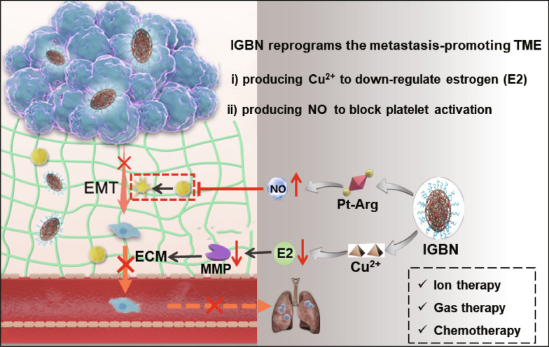
Metastasis is one of the main causes of failure in the treatment of triple-negative breast cancer (TNBC). Abnormally estrogen level and activated platelets are the key driving forces for TNBC metastasis. Herein, an “ion/gas” bioactive nanogenerator (termed as IGBN), comprising a copper-based MOF and loaded cisplatin-arginine (Pt-Arg) prodrug is developed for metastasis-promoting tumor microenvironment reprogramming and TNBC therapy. The copper-based MOF not only serves as a drug carrier, but also specifically produces Cu2+ in tumors, which catalytic oxidizing estrogen to reduce estrogen levels in situ. Meanwhile, the rationally designed Pt-Arg prodrug reduced into cisplatin to significantly promote the generation of H2O2 in the tumor, then permitting self-augmented cascade NO gas generation by oxidizing Arg through a H2O2 self-supplied way, thus blocking platelet activation in tumor. We clarified that IGBN inhibited TNBC metastasis through local estrogen deprivation and platelets blockade, affording 88.4% inhibition of pulmonary metastasis in a 4T1 mammary adenocarcinoma model. Notably, the locally copper ion interference, NO gas therapy and cisplatin chemotherapy together resulted in an enhanced therapeutic efficacy in primary tumor ablation without significant toxicity. This “ion/gas” bioactive nanogenerator offers a robust and safe strategy for TNBC therapy.
Graphical Abstract
Supplementary Information
The online version contains supplementary material available at 10.1186/s12951-022-01520-8.
Keywords: Metastasis-promoting microenvironment, Triple-negative breast cancer, Estrogen regulation, Platelets blockade, Ion/gas therapy
Background
Triple negative breast cancer (TNBC) is the most malignant subtype of breast cancer that seriously threatens women’s health and lives, and its associated morbidity and mortality have been increasing in recent years [1, 2]. Metastasis is one of the main causes of failure in the treatment of TNBC [3, 4]. Substantial studies evidenced that platelets support tumor metastatic progression by inducing epithelial-mesenchymal transition (EMT) of cancer cells and by shielding circulating tumor cells from immune-mediated elimination [5, 6]. More importantly, TNBC as a common tumor in women, estrogen may play a role in the occurrence and development of TNBC. Here we discovered that despite TNBC being an estrogen receptor (ER) -negative breast cancer, estrogens could also induce TNBC invasion and migration. According to our discoveries, apparent increased expression of calcium-activated neutral protease (Canp) and matrix metalloproteinase (MMP) could be observed along with the addition of estrogens (E2). We speculated that such E2-induced TNBC metastasis was derived from abnormally expressed MMP-mediated tumor extracellular matrix (ECM) loosening. Taken together, abnormal estrogen levels and activated platelets constitute a distinctive metastasis-promoting microenvironment of TNBC. Therefore, exploring the strategy that shifting the metastasis-promoting tumor microenvironment of TNBC toward a metastasis-inhibited status may receive significant therapeutic benefits.
Estrogen performs an important role in the metastasis of TNBC via a non-ER receptor dependent way [7–10]. However, patients with TNBC do not benefit from conventional endocrine therapy [11, 12]. Aromatase (CYP19, also called estrogen synthase) is a rate-limiting enzyme that catalyzes the production of estrogen in organisms [13]. At present, aromatase inhibitors, such as exemestane, letrozole, and anastrozole have been proved to reduce the risk of breast cancer metastasis and improve the survival time by decreasing levels of serum estrogens [14]. Unfortunately, many patients are nonadherent due to hormonal disorders mediated adverse side effects [15, 16]. Localized regulation of estrogen levels in the tumor in situ remains a challenge. Interestingly, estrogens are discovered to affect DNA stability through their downstream metabolites in many pathological processes, which requires the participation of the intracellular copper ions (Cu2+) [17]. Inspired by the role of Cu2+ in the estrogen metabolism, we assume that specific Cu2+ interference is expected to be promising in local estrogen regulation in the tumor, thus facilitating the management of TNBC metastasis.
Tumors are known to establish several mechanisms to obtain metastasis cascade, targeting a single factor is often unsatisfactory [18, 19]. Another key factor that composes the metastasis-promoting microenvironment is abnormally activated platelets in tumor tissues [20], for which diverse strategies for blocking platelet activation have been proposed, including blocking platelet receptors, inhibiting platelet-specific antibodies, and consuming circulating platelets [21–23]. But life-threatening side effects such as bleeding complications have been reported due to disruption of platelet coagulation function [24]. As an endogenous gas molecule, nitric oxide (NO) is a potent inhibitor of platelet activation [25–27]. Several significant breakthroughs have been achieved in investigation by using NO as an antiplatelet drug to prevent thrombosis [6, 28–30], which makes NO a promising candidate to reduce activated platelet mediated metastasis.
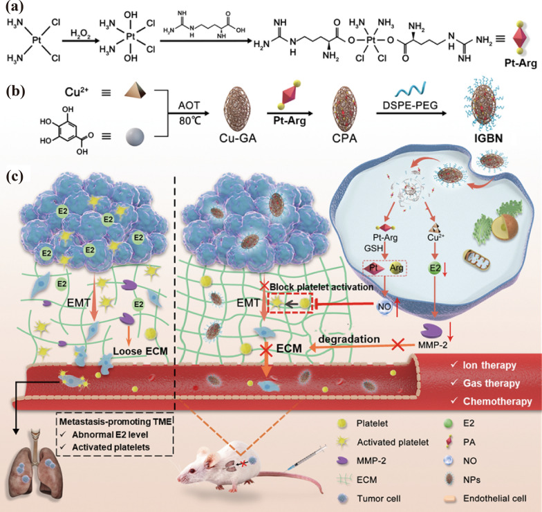
The past few decades have witnessed the revolutionary impact of nanotherapeutics. In particular, nanomaterials with more biological activities and enhanced therapeutics have attracted increasing interest [31, 32]. Herein, a multifunctional “ion/gas” bioactive nanogenerator (termed as IGBN) was constructed for highly metastatic TNBC treatment (Fig. 1). It contains copper-based MOF core with stimuli-responsive, biodegradable, as well as biosafety due to synthetic materials gallic acids (GA) and Cu2+ (limited dosage) are nearly harmless, and loads a cisplatin/l-arginine coupling prodrug (Pt-Arg) with NO producing ability and a hydrophilic shell (DSPE-PEG2000). The designed IGBN can simultaneously regulate copper ions/NO gas specific in the tumor tissue in situ to reprogram the metastasis-promoting tumor microenvironment composed of estrogen and activated platelets for TNBC metastasis inhibition. IGBN exhibits multifunctional characteristics, including (i) The pH/glutathione (GSH) cascade response design ensures the tumor specificity of controlled drug release. MOF skeleton of IGBN disintegrates in the acidic lysosome, leading to the site-specific release of Cu2+ and Pt-Arg prodrug; then Pt-Arg prodrug dissociates in response to GSH in the cytoplasm, releasing active cisplatin and l-Arg within tumor cells. (ii) Cisplatin can not only directly exert an anti-tumor effect, but also activate the highly expressed NADPH oxidases (NOX) enzymes in tumor cells and promote the production of H2O2, which can further react with the released l-Arg to produce excess NO, inhibiting the growth of primary tumor synergistically. (iii) More importantly, released Cu2+ depletes tumor estrogen, inhibiting the production of MMP. Meanwhile, the produced NO blocks tumor-induced platelet activation in situ, thus inhibiting platelet-induced EMT, both of which work together to the reprogram metastasis-promoting tumor microenvironment. In vivo experiments in a mouse orthotopic breast cancer model showed that IGBN significantly down-regulated the level of estrogen and blocked platelet activation in tumor tissue. Besides inhibiting the primary tumor, the lung metastasis efficiency of TNBC was significantly reduced. Taken together, this rational and convenient strategy led to improved antitumor and antimetastatic efficacies with marginal side effects.
Fig. 1.
a Synthetic route of cisplatin/l-arginine coupling prodrug; b Preparation of the “ion/gas” bioactive nanogenerator (IGBN); c Schematic illustration of IGBN reprograms the metastasis-promoting tumor microenvironment for metastasis-inhibited TNBC therapy. After internalized by tumor cells, IGBN could specifically produce Cu2+ in acidic lysosome, which catalytic oxidizing E2 to reduce E2 levels in tumor in situ. Meanwhile, the released Pt-Arg prodrug reduced into cisplatin to significantly promote the generation of H2O2 in the tumor, thus permitting self-augmented cascade NO gas generation by oxidizing Arg. Such a metastasis-promoting tumor microenvironment reprogramming (Cu2+ mediated E2 down-regulation and NO mediated platelet activation blockade) affords effective lung metastasis suppression. Notably, ion therapy, NO gas therapy and cisplatin chemotherapy together result in an enhanced therapeutic efficacy in primary TNBC ablation
Results and discussion
Preparation and characterization of IGBN
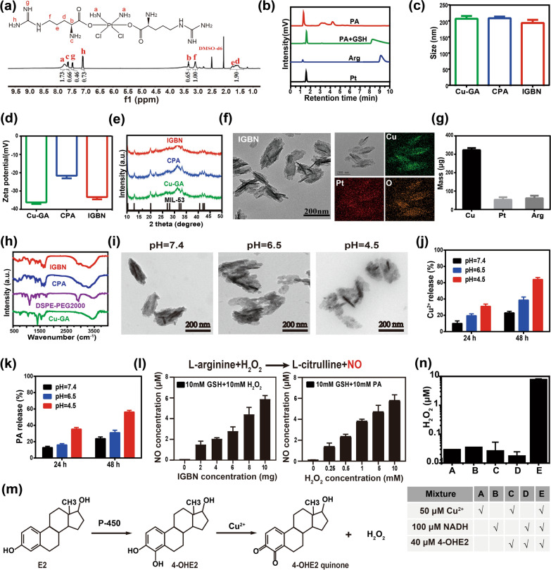
The IGBN was formed by loading cisplatin/l-arginine coupling prodrug into copper-based metal–organic frameworks (MOF) and sealing up by a hydrophilic DSPE-PEG2000. Firstly, the reduction-sensitive Pt(IV) prodrugs (Pt-Arg, termed as PA) was prepared, in which l-Arg was attached to a Pt(IV) moiety derived from cisplatin via a succinate linker. The obtained Pt(IV) prodrug was characterized by proton nuclear magnetic Resonance (1H NMR) spectra and Fourier transform infrared (FTIR) spectroscopy analysis (Fig. 2a; Additional file 1: Fig. S1). The reduction sensitivity of PA was measured by high performance liquid chromatography (HPLC), which suggested that PA have the potential of releasing cisplatin and l-Arg under reducing intracellular environment within tumor cells for their respective biological actions (Fig. 2b). Then the Cu-based MOF was synthesized by the reaction of GA and copper acetate according to a previous literature with slight changes [33]. The prepared MOF was spindle-like in shape with ~ 60 nm in width and ~ 160 nm in length under transmission electron microscope (TEM) (Additional file 1: Fig. S2a). An averaged hydrodynamic diameter of 209.0 ± 5.7 nm and negative charge surface (− 36.0 ± 0.9 mV) as determined by dynamic light scattering (DLS) (Fig. 2c, d). The successful synthesis of Cu-GA MOF was further confirmed by the X-ray diffraction (XRD) pattern (Fig. 2e). The as-prepared Cu-GA MOF showed polycrystallinity of the material, whose pattern matches well with the simulated pattern of MIL-53 frameworks (Cambridge Crystallographic Data no. CCDC-220475) [33–35]. The X-ray photoelectron spectroscopy (XPS) was performed to confirm the chemical states of elements (Additional file 1: Fig. S3). The survey spectrum in which copper (Cu), oxygen (O) and carbon (C) elements were in their respective states. After PA loading, there is no obvious change in the morphology and particle size of Cu-GA @PA (CPA) (Fig. 2c and Additional file 1: Fig. S2b), but the zeta potential of CPA increased to − 21.4 mV from − 36.0 mV for Cu-GA (Fig. 2d), which confirmed the successful loading of positively charged PA prodrug. After DSPE-PEG2000 modification, the final product Cu-GA@PA@DSPE-PEG2000 (“ion/gas” bioactive nanogenerator, IGBN) was obtained. The particle size and potential of Cu-GA@PA with different dosages of DSPE-PEG2000 surface-functionalization were then assessed (Additional file 1: Fig. S4a, b). The results showed that when the ratio of DSPE-PEG2000 and Cu-GA@PA was 0.5:1, the particle size increased slightly after 6 h, and other ratios almost had no change in particle size. Meanwhile, compared with 0.5:1, the zeta potential of IGBN prepared in other ratios (1:1, 2:1, 4:1) were lower, and there was no noticeable difference, which may be due to the saturation of modification amount at 1:1. The results indicated that the optimal modification ratio for DSPE-PEG2000 and Cu-GA@PA was 1:1. In addition, the stability of IGBN was further studied, as reflected in Additional file 1: Fig. S4c, there was no obvious change in the hydrodynamic diameter or zeta potential of IGBN in one week, laying the foundation for in vivo applications. The morphology of IGBN still showed a spindle shape with an average hydrodynamic diameter of ~ 190 nm (Fig. 2f), and the zeta potential of IGBN dropped back to − 33.1 mV (Fig. 2d). In addition, the homogeneous distributions of Cu, Pt, and O in IGBN were observed by the EDS elemental mapping (Fig. 2f), further indicating that PA prodrugs were successfully loaded into Cu-GA MOF. And the Pt and l-Arg loading amounts were about 49.8 µg and 58.9 µg in 1 mg of IGBN, respectively (Fig. 2g). The drug loading content of Pt and l-Arg was ~ 4.7% and ~ 5.6%, and the encapsulation efficiency was ~ 9.9% and 11.8%, respectively. Both the characteristic peaks of Cu-GA were also appeared in the X-ray diffraction (XRD) pattern of IGBN (Fig. 2e), proving that drug loading and surface modification did not affect the crystalline structure of MOF. Moreover, successful synthesis of IGBN was also confirmed by the FTIR spectroscopy (Fig. 2h).
Fig. 2.
Preparation and characterization of IGBN. a 1H NMR spectra of Pt-Arg prodrug. b HPLC examination of GSH-induced activation of the Pt-Arg prodrug. c Hydrodynamic size distribution of Cu-GA, CPA, and IGBN (n = 3). d Zeta potentials of Cu-GA, CPA, and IGBN (n = 3). e XRD patterns of Cu-GA, CPA, and IGBN. f Representative TEM images of IGBN and EDS elemental mapping images of IGBN with Cu, Pt and O elements. Scale bar: 200 nm. g The content of Cu, Pt and l-Arg in 1 mg of IGBN by ICP-MS (n = 3). h FTIR spectra of Cu-GA, DSPE-PEG2000, CPA, and IGBN. i Representative TEM images of IGBN in pH 7.4, pH 6.8 and pH 4.5. Scale bar: 200 nm. j Cu2+ release from IGBN at pH 7.4, pH 6.8 and pH 4.5 (n = 3). k The PA release from IGBN at pH 7.4, pH 6.8 and pH 4.5 (n = 3). l IGBN and H2O2 concentration dependent NO release in the present of 10 mM GSH (n = 3). m The mechanism for Cu mediated estrogen consumption. n Panel of five different mixtures for the generation of H2O2. A, B, C, D, and E represent Cu2+, nicotinamide adenine dinucleotide (NADH), Cu2+ + 4-OHE2, NADH + 4-OHE2, and Cu2+ + NADH + 4-OHE2, respectively. And quantification of H2O2 generated in A–E based on the absorbance from hydrogen peroxide kit (n = 3)
Drug release behaviour of IGBN
The site-specific release of drugs in tumor tissues is a prerequisite for reducing its side effects. Here the pH-responsive drug release ability of IGBN is studied. First, morphological changes of IGBN at different pH values were detected by TEM (Fig. 2i). Compared with in neutral medium, an obvious degradation of IGBN was observed under acidic medium, especially at pH 4.5, the underlying mechanism is that the protonation of the oxygen group in GA at acidic pH leads to weaker copper-oxygen bond in the framework, and then facilitates its decomposition. Then the pH-responsive Cu2+ and PA synchronous release behaviours of IGBN were investigated (Fig. 2j, k; Additional file 1: Fig. S5). The release of both Cu2+ and PA from IGBN was time-dependent and pH-dependent, and the release of Cu2+ and PA were slow at neutral pH (pH 7.4). However, at acidic pH (pH 4.5), the release rate increased significantly, about 64.7% of Cu2+ and 56.32% of PA released from IGBN was observed. Overall, the pH-dependent release of these active agents (Cu2+ and PA) is beneficial for drug-delivery purposes to desired cells.
NO production and estrogen consumption in solutions
As verified by above experiments, the released PA prodrug can generate active cisplatin and l-Arg monomers under the action of GSH (Fig. 2b). Studies have found that l-Arg can directly react with H2O2 to produce NO without relying on nitric oxide synthase [36–38]. To simulate the in vivo process, exogenous H2O2 was added, and then we tested the NO producing ability of PA. As showed in Additional file 1: Fig. S6a, without GSH treatment, negligible NO production was observed, but after treated with GSH, large amount of NO was produced from PA. As expected, NO production showed the concentration dependence of IGBN and H2O2 (Fig. 2l; Additional file 1: Fig. S6b). Taken together, PA could produce NO in the present of GSH, and the production of NO relies on the H2O2/l-Arg concentration, which was beneficial to the tumor selectivity of NO production.
In view of the key role of estrogen in cancer metastasis, the feasibility of Cu2+-mediated estrogen depletion was studied. As reported, 17β-estradiol (E2), the predominant estrogen species, undergoes oxidation by cytochrome P450 to form 4-hydroxy estradiol (4-OHE2). Then Cu2+ could oxidize the 4-OHE2 into a 4-OHE2 semiquinone radical, which subsequently transfer an electron to tissue oxygen to afford a quinone derivative and the O2−• radical, thus down-regulating the E2 level. The short-lived O2−• may then be reduced by superoxide dismutase (SOD) or nicotinamide adenine dinucleotide (NADH) to form stable H2O2 (Fig. 2m). Therefore, H2O2 was detected as a signal of Cu2+-mediated estrogen consumption (Fig. 2n). H2O2 sensor solution was added to a panel of five solutions, with A, B, C, D, and E representing Cu2+, NADH, Cu2+ + 4-OHE2, NADH + 4-OHE2, and Cu2+ + NADH + 4-OHE2, respectively. As shown in Fig. 2n, the large amount of H2O2 production was detected only in the presence of Cu2+, NADH and 4-OHE2, indicating that Cu2+ could consume estrogen under the action of intracellular enzymes according to the physiological process described in Fig. 2m. In addition, it also supplements H2O2 in tumor tissues to a certain extent.
Cellular uptake and intracellular distribution
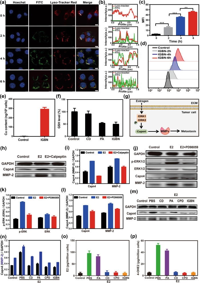
The high cellular uptake level of IGBN is major prerequisite for subsequent anti-tumor metastasis effects. Here, the fluorescent dye FITC was used as a model drug to be loaded into IGBN to mark the cell uptake and intracellular distribution of particles. As displayed in Fig. 3a, c, green fluorescence of FITC inside 4T1 cells treated with IGBN markedly enhanced along with the incubation time, indicating that IGBN were efficiently internalized into cells. These results were also verified by flow cytometer (Fig. 3d). After successful internalization, IGBN could decompose under endo/lysosomes acidic environment, and then release loaded FITC into the cytoplasm. The phenomenon was observed by CLSM (Fig. 3a, b). After incubation for 2 h, the preparation almost completely overlaped the lysosome, which proved that the nanoparticles entered the lysosome after being taken up by 4T1 cells. While after 6 h of incubation, the colocalization efficiency between the green fluorescence (FITC) and red fluorescence (lysosome) was significantly reduced, implying that FITC (model drug) could escape from lysosomes into the cytoplasm.
Fig. 3.
The estrogen regulating property of IGBN. a Representative CLSM images of biodistribution of FITC-labeled IGBN for different time treatment to 4T1 cells. Cell nuclei: blue, NPs: green, lysosome: red, scale bar: 7.5 μm. b The intensity profile along the white line in the merged image. c Mean fluorescence intensity (MFI) of FITC in IGBN treated cells for different times (n = 3). d Flow cytometer analysis of FITC-labeled IGBN treated cells for different incubation times. e Cu content in 4T1 cells after treated with IGBN for 8 h (n = 3). f Intracellular GSH levels in 4T1 cells treated with CD (DSPE-PEG2000 modified Cu-GA MOF), PA (Cisplatin-arginine) and IGBN for 24 h, respectively (n = 3). g The potential mechanism of estrogen-driven TNBC metastasis. h Western blot assay and i semi-quantitative analysis of Capn-4 and MMP-2 in 4T1 cells with estradiol and calpeptin (Capn4 inhibitor) treatments (n = 3). j Western blot assay, k and l semi-quantitative analysis of p-ERK, ERK, Capn-4 and MMP-2 in 4T1 cells with estradiol and PD98059 (ERK inhibitor) treatments. m Capn-4 and n MMP-2 levels in 4T1 cells treated with different formulations (n = 3). CPD (CD loaded with cisplatin) o E2 and p 4-OHE2 level after 4T1 cells treated with different formulations (n = 3). Results are presented as means ± s.d. Statistical significance was calculated by Student’s t-test. **P < 0.01, ***P < 0.001
Estrogen regulation in 4T1 cells
As mentioned above, Cu2+ could down-regulate estrogen by interfering with the intracellular metabolic pathway of estrogen. First, we tested the release of intracellular Cu2+ in 4T1 cells after IGBN treatment, the generation of intracellular Cu2+ were measured via ICP-MS (Fig. 3e). In comparison with untreated group, 4T1 cells treated with IGBN displayed higher Cu2+ content. The release of Cu2+ was accompanied by the collapse of MOF and the release of PA, and then PA could release active drugs in response to intracellular GSH, and at the same time down-regulating GSH. Here, the influence of IGBN on the intracellular GSH was tested to reflect the activation of PA. As shown in the Fig. 3f, both the PA and PA loaded nano particles showed a significant GSH down-regulation, while no obvious change in the other groups, indicating that the activation of PA could consume GSH.
Previous studies have confirmed that 17β-estradiol (E2) can participate in hormone-dependent tumor proliferation and metastasis through a variety of mechanisms [39, 40]. The biological role of E2, in addition to directly binding to ER to mediate genomic effects, can also rapidly induce extracellular signal-regulated kinase 1/2 (ERK1/2) phosphorylation to trigger intracellular non-genomic effect [41, 42]. Canp as the key downstream signaling molecules of ERK is involved in cancer metastasis [43], because many key mediators in the metastatic cascade are considered to be the proteolytic substrates of Canp [44]. Intriguingly, Capn4, the common small subunit of Capn, has been detected as an invasive biomarker in different cancer types. For instance, Capn4 contributes to the metastasis of non-small-cell lung cancer and cholangiocarcinoma through upregulating MMP-2 [45, 46]. However, the mechanism of E2 in promoting TNBC metastasis remains unclear. Here we propose a hypothesis that E2 may trigger the phosphorylation of ERK1/2 to induce intracellular non-gene effects, and successively promote the expression of Capn4 and MMP-2, thus improving the key link of tumor metastatic cascade—extracellular matrix (ECM) degradation for TNBC metastasis (Fig. 3g).
To verify the hypothesis, we firstly tested the effect of E2 on cell proliferation. As shown in the Additional file 1: Fig. S7a, E2 could increase the proliferation ability of 4T1 cells, and displayed a typical concentration-dependent trend. Then the effect of E2 on the expression of Capn4 and MMP-2 in 4T1 cells were examined by Western blot. The results showed that the expression of Capn4 and MMP-2 showed a concentration-dependent increase in the 10–7–10–10 M concentration range of E2. This result indicated that a certain concentration of E2 could promote the MMP-2 expression of 4T1 cells (Additional file 1: Fig. S8). To prove whether the up-regulation of MMP-2 induced by E2 was related to Capn, Capn -specific inhibitor calpeptin was used to pretreat 4T1 cells, and then stimulated with E2. It was found that calpeptin inhibited the expression of Capn4, and MMP-2 was also down-regulated accordingly (Fig. 3h, i). After that, the expression levels of phosphorylated-ERK1/2 (p-ERK1/2), total ERK1/2, Capn4 and MMP-2 proteins were examined by Western blot, and total ERK1/2 blocker PD98059 was used to pretreat 4T1 cells. The results showed that E2 could induce the up-regulation of p-ERK1/2 expression in 4T1 cells, but had no significant effect on total ERK1/2 expression, resulting in a significant increase in the ratio of p-ERK/ERK (Fig. 3j, k), while PD98059 could significantly inhibit the expression of p-ERK1/2 induced by E2 (P < 0.01). Meanwhile, PD98059 could attenuate the expression of Capn4 and MMP-2 induced by E2 (Fig. 3j, l). The above results indicate that E2 could promote the up-regulation of MMP-2 through ERK/Capn signaling pathway in a non-genomic way.
Then we investigated the effect of IGBN on the expression of Capn4 and MMP-2 in 4T1 cells. Here 4T1 cells were pre-treated with a concentration of 10–9 M E2. As shown in Fig. 3m, n, IGBN treatment resulted in a significant decrease of Capn4 and MMP-2 content. Then the intracellular E2 and 4-OHE2 were also investigated (Fig. 3o, p). The results showed that the concentrations of E2 and 4-OHE2 decreased slightly after PA treatment compared with PBS group, which might be caused by PA-mediated apoptosis of 4T1 cells. As expected, IGBN treatment also effectively reduced E2, which was consistent with the trend of 4-OHE2 level. These results indicated that the IGBN could down-regulate 4-OHE2 by releasing Cu2+ within tumor cells, thereby inhibiting the expression of E2.
Self-augmented cascade NO generation in tumor cells
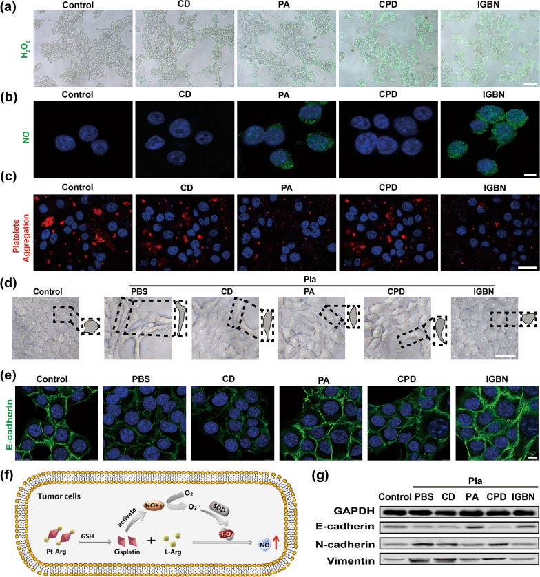
Increasing evidence have evidenced that platelet plays a vital role in phenotype transition of tumor cell, and the most accepted role is epithelial-mesenchymal transition (EMT), which is an important step for tumor metastasis [47, 48]. Blocking platelet functions may be an effective method to disrupt the interactions of tumor cells with platelets, thereby destroying the contribution of platelets to tumor metastasis. NO has been proved to be a key molecule of endogenous antiplatelet function [26]. If NO can be produced in situ in tumor tissues, it is expected to realize safe antiplatelet without affecting the function of platelets in the system. However, as a precursor of NO, it is well known that the amount of H2O2 in tumor tissues is limited. It is necessary to provide sufficient H2O2 to the tumor tissue. Studies have found that cisplatin can promote the production of H2O2 by activating overexpressed NOX enzyme within tumor cells [49, 50]. Here we first detected the IGBN-mediated H2O2 production in 4T1 cells by the specific H2O2 probe. As shown in the Fig. 4a and Additional file 1: Fig. S9a, compared with the control group, the intracellular H2O2 content in the cisplatin-containing treatment groups (PA, CPD, IGBN) was significantly increased. This was consistent with previous reports, confirming the ability of cisplatin to induce H2O2 production in tumor cells [51, 52]. The level of H2O2 induced by IGBN was higher than that of the free PA prodrug group, which may be attributed to the nanoformulation promoting the cell uptake on drug. Then specific NO probe DAF-FM DA was used to detect the production of intracellular NO. As shown in Fig. 4b and Additional file 1: Fig. S9b, the cells treated with PA and IGBN exhibited stronger green fluorescence than the control group, indicating that a large amount of NO was produced in 4T1 cells. The production of H2O2 and NO catalyzed by the Pt-Arg prodrug was summarized in Fig. 4f.
Fig. 4.
Inhibition of the platelet-induced EMT process by IGBN. a H2O2 level in 4T1 cells with different treatments. Scale bar: 200 µm. b The generation of NO in 4T1 cells after different treatments. Scale bar: 7.5 µm. c Confocal fluorescence images of Dil-labeled platelet aggregation with different treatments. Scale bar: 25 µm. d Morphology of 4T1 tumor cells treated with different formulations. Typical cell morphologies were pointed out by dotted squares. Pla: platelets. Scale bar: 50 µm. e Confocal immunofluorescence images of 4T1 cells after treated with different formulations. Green: E-cadherin; blue: nuclei. Scale bar:10 µm. f Schematic illustration of Pt-Arg cascade catalyzes the production of NO within tumor cells. g Western blot analysis of E-cadherin, N-cadherin, and Vimentin expression in 4T1 cells with different treatments (n = 3)
Inhibition of platelet activation and EMT process
Then we tested whether NO induced by IGBN could affect platelet function. The influence of IGBN on the platelet aggregation induced by tumor cell was evaluated by labelling platelets with the fluorescent dye Dil. Compared with the control group, IGBN significantly inhibited tumor cell induced platelet aggregation (Fig. 4c). After that, the effect of IGBN on platelets triggered EMT in tumor cells was tested. To avoid the interference of cytotoxicity on the assess of tumor cell migration and invasion, the cytotoxicity of cisplatin with series concentrations were measured. The result showed that less than 1 μg mL−1 of Pt had negligible influence on the growth ability of 4T1 tumor cells (Additional file 1: Fig. S7b). Thus, 1 μg mL−1 of Pt (equivalent amount of Pt in nanoformulation) was selected for subsequent experiments to evaluate the effect of IGBN on EMT. Platelets and tumor cells were co-cultured, and then the morphological changes of tumor cells with different treatments were observed using an optical microscope (Fig. 4d; Additional file 1: Fig. S10). Platelet treatment could induce tumor cells to change from a pebble shape to a spindle shape, which was a typical feature of EMT progression. There were significant morphological changes of tumor cells in the CD and CPD treated groups. However, IGBN significantly reduced the morphological changes of tumor cells, indicating that IGBN could inhibit platelet-induced EMT through NO production.
E-cadherin is a main protein that maintains cell-to-cell connections, its down-regulation is the main feature of EMT [53, 54]. As shown in the Fig. 4e, platelet treatment resulted in a significant down-regulation of the E-cadherin protein of tumor cells. PA and IGBN treatment could effectively reverse the platelet-induced down-regulation of E-cadherin expression. The down-regulation of E-cadherin expression is often accompanied by up-regulation of N-cadherin. Therefore, the key proteins of EMT (E-cadherin, N-cadherin, and vimentin) were further tested by Western blot. As shown in the Fig. 4g and Additional file 1: Fig. S11, platelets down-regulate E-cadherin levels and up-regulate N-cadherin and vimentin levels, while IGBN treatment significantly reversed the levels of the three proteins. These results illustrated that IGBN could effectively inhibit platelet-induced EMT of tumor cells.
Inhibition of cell migration and invasion
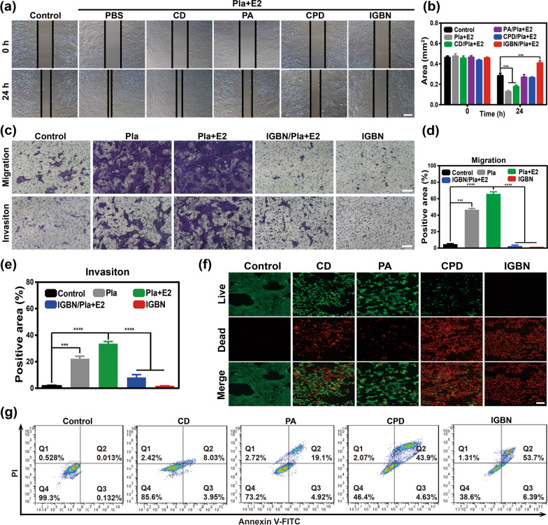
As we know, the metastasis-promoting microenvironment composed of abnormally estrogen level and activated platelets are the key driving forces of TNBC metastasis, and the prepared IGBN in this study has been proven to effectively consume estrogen and inhibit platelet activation. Here we investigated the effect of IGBN on the migration and invasive ability of breast cancer cells. Briefly, 4T1 cells grown in a six-well plate are scraped to a fixed width, and then estrogen and platelets were added to pretreat the cells. After that, different preparations were added and incubated for 24 h. As shown in Fig. 5a, b, compared with the control group, E2 and platelet treatment significantly promoted cell migration, and 4T1 cells almost covered the scratched area. However, the scratched area in IGBN treated group still maintained a clear gap. The migration ability of tumor cells was also evaluated using the transwell experiment. As shown in the Fig. 5c, d 4T1 cells were seeded to the upper chambers, then platelets and estrogen were added in the present or absent of IGBN for 24 h, tumor cells that migrated in the lower chamber are tested. Compared with the saline group, platelets significantly promoted the migration of 4T1 cells, and the addition of E2 further enhanced their migration ability. In contrast, the IGBN treatment effectively reversed cell migration induced by E2 and platelets.
Fig. 5.
In vitro anti-metastasis and anti-tumor effects of IGBN. a Representative wound-healing assay to track cell migration at 0 h and 24 h. Pla: platelets. E2: 17β-estradiol. Scale bar: 200 µm. b Quantification of cell migration by Image J (n = 6). c Representative images of migration and invasion in 4T1 tumor cells treated with different formulations in the Transwell assay. Scale bar: 200 μm. d and e Corresponding data analysis of the transwell assay by Image J (n = 6). f Fluorescence images of calcein AM (green, live cells) and PI (red, dead cells) co-stained 4T1 cells after incubation with different treatments for 24 h. Scale bar: 200 μm. g Flow cytometer analysis of apoptosis in 4T1 cells after incubation with different treatments for 24 h. Results are presented as means ± s.d. Statistical significance was calculated by Student’s t-test. ***P < 0.001, ****P < 0.0001
Next, the effect of IGBN on the invasive ability of tumor cells was studied using Matrigel precoated Transwells (Fig. 5c, e). Platelets significantly promoted the invasion of tumor cells. After adding E2, its invasion ability was further enhanced, which may be attributed to the metastasis-promoting tumor microenvironment (platelet-induced tumor EMT and E2 promoted the ECM degradation), thus synergistically promoted tumor invasion. But the IGBN treatment effectively reversed tumor cell invasion, implying the excellent anti-metastatic ability of IGBN by reprogramming the metastasis-promoting tumor microenvironment.
In vitro antitumor effect
Simultaneously block TNBC metastasis and primary tumor growth is an ideal strategy for tumor treatment. Here we studied the in vitro anti-tumor effect of IGBN by CCK-8 and apoptosis/necrosis assay. First, we investigated the effect of IGBN on the proliferation ability of 4T1 cells (Additional file 1: Fig. S12). IGBN exhibited a concentration-dependent cytotoxicity against 4T1 cells. When the concentration was 30 μg mL−1, IGBN had greater than 50% inhibition on 4T1 cells. Then we investigated the apoptosis and necrosis inducing ability of IGBN by flow cytometer and fluorescence microscope, respectively. IGBN showed strongest capability to induce apoptosis and necrosis of 4T1 cells than PA and CPD under the same concentration of Pt (Fig. 5f, g), which was consistent with the result of cell viability assay. These results confirm that IGBN could not only inhibit the invasion and metastasis of breast cancer, but also hold a strong anti-tumor effect in vitro.
In vivo antitumor and antimetastatic effect
Inspired by the prominent performance in vitro, the therapeutic efficiency of IGBN in vivo was further explored. The behaviour of IGBN in vivo was first investigated. NIR fluorescent dye IR783 was used instead of prodrug to indicate the position of nanoparticles. As shown in Additional file 1: Fig. S13, a strong fluorescence signal in the tumor site at 6 h post-injection was observed in IGBN treated group, and there was still strong fluorescence at 24 h post-injection. While the free IR783 group had a weak fluorescence signal in the tumor site at 6 h post-injection, and then the fluorescence decreased rapidly. These results indicated that nano-scale materials contribute to the enrichment of tumor tissues, attributing to size of the prepared IGBN was about ∼ 200 nm, which was able to passively target the tumor site through the EPR effect.
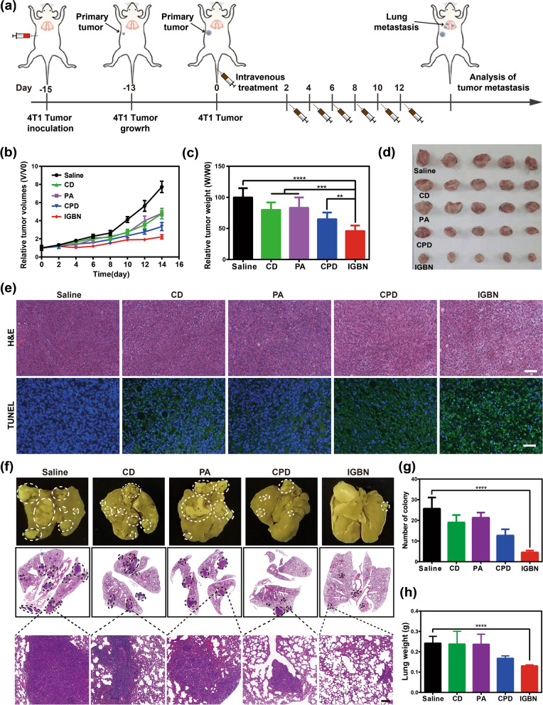
To evaluate the antitumor effect of IGBN, the 4T1 breast cancer orthotopic tumor model was constructed by subcutaneously injecting 4T1 cells into the right mammary gland of Balb/c mice [55]. 4T1 tumor-bearing mice were administered according to the set schedule (Fig. 6a). The tumor sizes and body weight of mice were recorded every second day. As shown in Fig. 6b, the tumor volume after treatment of IGBN was significantly decreased contrast to those of control groups, which was attributed to the increased EPR effect, the synergistic anti-tumor effect of copper ion therapy, NO gas therapy and cisplatin chemotherapy. The tumor weight and photos presented in Fig. 6c, d further confirmed the anti-tumor effect of IGBN. Then the hematoxylin and eosin (H&E) staining and terminal deoxynucleotidyl transferase dUTP nick end labelling (TUNEL) assay were performed to assess apoptosis of tumor after different therapies (Fig. 6e). Large amounts of karyopyknosis and the severer structural deformation was observed in IGBN treated group, indicated that tumor cells in the tissue section with IGBN treatment were severely damaged. Similarly, more apoptotic cells were detected in the IGBN group by TUNEL assay, which was obviously more than that in other groups (Additional file 1: Fig. S14). These results demonstrated the excellent ability of IGBN (ion therapy, gas therapy and chemotherapy) to suppress the primary tumor.
Fig. 6.
In vivo antitumor and antimetastatic evaluation of IGBN in 4T1 tumor-bearing mice. a Schematic illustrating the establishment of 4T1 tumor-bearing mice mode and administration schedule. b Relatively primary tumor growth profiles of 4T1 tumor-bearing mice treated with Saline, CD, PA, CPD and IGBN (n = 5). c The relative weight of tumor tissue on day 14 after treatments (n = 5). d Photographs of tumor tissue on day 14 after treatments (n = 5). e Representative images of H&E (upper panel) and TUNEL (lower panel) staining of tumors sections on day 14 after treatments. Scale bar: 100 μm. f Representative lung photos (upper panel) and H&E-stained lung slices (lower panel) on day 14 after treatments. Metastatic nodules are represented by white circles. Scale bar (upper panel): 2 mm. Scale bar of H&E images in bottom is 200 µm. g Number of lung tumor nodules (n = 5). h The weight of lung in 4T1-bearing mice on day 14 after treatments. Error bars are based on SD (n = 5). Results are presented as means ± s.d. Statistical significance was calculated by Student’s t-test. **p < 0.01, ***p < 0.001, ****p < 0.0001
Next, the anti-metastatic effect of IGBN was evaluated. The 4T1 tumor model we used in this study was a highly aggressive TNBC tumor model, which is potent for distant lung metastasis. The lungs of mice after therapy were extracted for metastatic nodule counting and histological analysis. As shown in Fig. 6f, g, approximate 26 metastatic nodules were observed in each lung of the saline-injected mice, and CPD-mediated cancer therapy slightly reduced the metastatic nodule number to 13, while the number of pulmonary metastasis nodules in IGBN-treated mice was 3. At the same time the weight of the lung is significantly reduced in IGBN-treated mice (Fig. 6h). As shown in H&E staining images, nearly no tumor metastasis was observed in the lungs of mice after IGBN treated group, while metastatic tumors distributed in the lungs of mice after the other treatments (Fig. 6f). These results indicated that IGBN-mediated cancer therapy could not only effectively inhibit the primary tumor, but also block lung metastasis.
The metastasis-promoting tumor microenvironment reprogramming
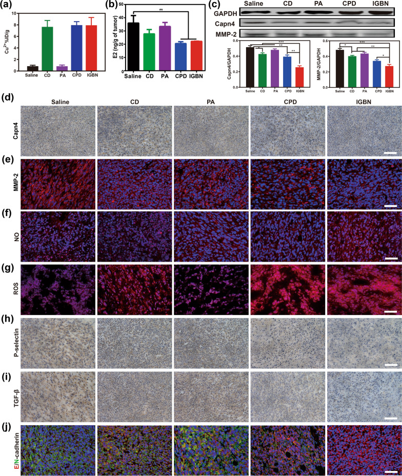
To confirm the anti-metastasis mechanism in vivo, tumor tissues from different treatment groups were sliced. Firstly, the release of Cu2+ in tumor site was measured by ICP-MS. As shown in Fig. 7a, a large amount of Cu2+ were detected in the CD, CPD and IGBN treated groups, indicating the superior Cu2+ regulation ability of Cu-based MOF in tumor tissues. Then, we investigated the effect of the ICBN on the level of estrogen in tumor tissues (Fig. 7b), which was consistent with the result of Cu2+ content testing, confirming the estrogen regulation ability of Cu2+. As mentioned earlier, estrogen can regulate the expression of Capn4 by activating the ERK pathway, thereby up-regulating MMP-2 to accelerate tumor metastasis. Herein, the expression of Capn4 and MMP-2 in tumor tissue was analyzed by immunohistochemistry/immunofluorescence assay and Western blot assay (Fig. 7c–e). The expression levels of Capn4 and MMP-2 showed varying degrees of down-regulation in the CD, CPD and IGBN treated groups, confirmed that Cu2+ could inhibit the expression of MMP-2 by down-regulating estrogen. Compared with other groups, IGBN induced the most significant down-regulation of estrogen in tumor tissues, the possible reason was the synergistic effect mediated by the cisplatin and NO in addition to the role of Cu2+.
Fig. 7.
In vivo antimetastatic mechanism of IGBN. a Cu2+ and b E2 content in tumor tissue of 4T1 tumor-bearing mice on day 14 after different treatments (n = 5). c Qualitative Western blot data of GAPDH, Capn4 and MMP-2 in tumor tissue from 4T1 tumor-bearing mice on day 14 after treatment (upper panel), (n = 3). Corresponding gray analysis of Capn4 and MMP-2 from Western blot by Image J (lower panel), (n = 3). d Immunohistochemistry images of Capn4 in tumor tissue from 4T1 tumor-bearing mice on day 14 after treatment. Scale bars: 100 μm. e Immunofluorescence staining images of MMP-2 in tumor tissue from 4T1 tumor-bearing mice on day 14 after treatment. Scale bars: 50 μm. f Immunofluorescence staining images of NO. Scale bars: 50 μm. g Immunofluorescence staining images of ROS. Scale bars: 50 μm. h Immunohistochemistry staining images of P-selectin and i TGF-β. Scale bars: 50 μm. j Immunofluorescence staining images of E-cadherin and N-cadherin. Scale bars: 50 μm. Results are presented as means ± s.d. Statistical significance was calculated by Student’s t-test. *p < 0.05, **p < 0.01 and ***p < 0.001
As another important component of the metastasis-promoting tumor microenvironment, platelet activation could lead to EMT of tumor cells, thereby promoting tumor cell invasion and metastasis.5. 6 NO is a well-known anti-platelet molecule, and we have demonstrated that it could inhibit platelet activation in vitro. Here NO production in tumor tissues induced by IGBN was detected. Compared with other groups, the NO level of tumor tissue with IGBN treatment was significantly increased (Fig. 7f). As a key raw material for NO production, the content of ROS in tumor tissues after different treatments was also tested (Fig. 7g), both the CPD and IGBN led to a significant increase of ROS, confirming that cisplatin could up-regulate ROS in tumor cells. While the free PA prodrug resulted to a slight increase of ROS, which may be due to its lower blood half-life.
Then we tested whether IGBN-mediated NO production could block platelet activation. P-selectin (CD62P), a marker of platelet activation, was first tested (Fig. 7h). The results of immunohistochemistry showed that compared with the control group, the amount of P-selectin in the tumors with IGBN treatment was greatly reduced. Activated platelets could secrete TGF-β, which promoted the EMT pathway of tumor cells. The results of immunohistochemistry confirm that IGBN also inhibited the secretion of TGF-β (Fig. 7i). These results indicated that IGBN could inhibit tumor-induced platelet activation by producing NO. After that, the EMT process was monitored by detecting the expression of EMT-related proteins in tumor tissues. As shown in Fig. 7j, compared with other treatments, IGBN treatment significantly reduced the expression of N-cadherin and at the same time up-regulated the expression of E-cadherin, indicating that IGBN inhibited the EMT process of tumors. These results confirmed that IGBN could synergistically block TNBC metastasis by reprogramming the metastasis-promoting tumor microenvironment.
Finally, in vivo biosafety of IGBN was evaluated. The body weight of mice from IGBN group displayed a reasonable weight change in the normal range, indicating negligible systemic toxicity of IGBN (Additional file 1: Fig. S15). Besides, the major organs (heart, liver, spleen, and kidney) of the mice with different treatments were assessed by H&E staining. No observable pathological abnormalities were found in major organs (Additional file 1: Fig. S16). After systemic treatment, serum biochemical indicators and whole blood were also tested (Additional file 1: Fig. S17). There was no obvious abnormality in the IGBN-treated mouse group. Meanwhile, IGBN treatment did not alter the platelet counts in whole blood of mice, the underlying mechanism was that NO produced in situ in tumor by IGBN and had negligible effect on platelets in the blood. Correspondingly, IGBN only consumed E2 in the tumor in situ, and would not affect the hormone balance in vivo. These results indicated that IGBN had good biocompatibility and safety for anti-tumor therapy in vivo.
Conclusion
In summary, we proposed a metastasis-promoting tumor microenvironment reprogramming strategy for metastatic TNBC treatment. The rationally designed “ion/gas” bioactive nanogenerator (IGBN) in this study, on one hand, produced Cu2+ in tumors and reduce estrogen levels in situ; on the other hand, performed tumor-specific prodrug activation and self-augmented cascade NO gas generation. We clarified that IGBN inhibits TNBC metastasis through ERK/CAPN pathway activation and tumor epithelial-mesenchymal transition (EMT) pathway blockade, and such a metastasis-promoting tumor microenvironment reprogramming afforded a 3.6-fold inhibition of pulmonary metastasis in a metastatic 4T1 mammary adenocarcinoma model as compared to sole cisplatin chemotherapy. More importantly, the Cu2+ interference, NO gas therapy and cisplatin chemotherapy together eventually resulted in the significant regression of the primary tumors without significant toxicity. This “ion/gas” bioactive nanogenerator offered a robust and safe strategy for metastatic TNBC therapy.
Experimental methods
Materials
Copper acetate was obtained from Macklin. Gallic acid (GA) was purchased from Kermel. Surfactant Aerosol OT was obtained from Alfa Aesar. Cisplatin was purchased from Shanghaiyuanye Bio-Technology Co., Ltd. l-arginine (l-Arg), NADH, glutathione (GSH) and H2O2 Assay Kit were obtained from Beijing Solarbio Science & Technology Co., Ltd. DSPE-PEG2000 was purchased from Top-Peptide Co., Ltd. BCA Kit, Lyso-Tracker Red, Hoechst 33342, DAPI, GSH Assay Kit, Calcein-AM/PI staining kit and Annexin V-FITC/PI were obtained from Beyotime Biotechnology. Charcoal-stripped fetal bovine serum (CS-FBS) was supplied by Hyclony. Nitric Oxide Assay Kit was obtained from Nanjing Jiancheng Bioengineering Institute. NO fluorescence probe (DAF-FM DA) was purchased from meilunbio. 4-hydroxyestradiol (4-OHE2) and 17β-estradiol (E2) were obtained from Cayman Chemical. PD98059 and Calpeptin were purchased from MCE. All other chemicals were provided by Sigma Aldrich (St. Louis, MO, USA) unless mentioned otherwise.
Cell culture
The murine breast cancer 4T1 cells were purchased from American Type Culture Collection (ATCC) and cultured in RPMI-1640 medium with 10% FBS supplemented with 1% antibiotics (penicillin and streptomycin). The cells were incubated in the cell incubator with 37 °C under a humidified atmosphere containing 5% CO2.
Animals
Female BALB/c mice (6–8 weeks old, average body weight 17–19 g) were purchased from Henan Laboratory Animal Center and raised in the specific pathogenfree (SPF) laboratory. All animal experiment procedures were performed following the guidelines of the Institutional Animal Care and Treatment Committee of Zhengzhou University. 4T1 cells (2 × 10⁷ cells per milliliter, 50 µL) were subcutaneous injected into the third pair of breast fat pads in the mice to generate the metastatic 4T1 mammary adenocarcinoma model.
Synthesis of Cu-GA MOF
Copper-gallate metal–organic framework (Cu-GA MOF) was synthesized according to a previous study [33]. In brief, 0.22 g of surfactant AOT and 400 μL of n-butanol were solubilized in double-distilled water at room temperature, followed by the addition 200 μL each of 0.1 M copper acetate and gallic acid prepared in DMF. The reaction was stirred at 80℃ for 12 h, then adding ethanol to precipitate the particles and centrifuged at 12,000 rpm for 10 min to obtain Cu-GA MOF.
Synthesis of Pt-Arg prodrug
Appropriate amount of cisplatin (Pt) was dissolved in 30% H2O2 solution, stirred at 50℃ for 1 h, then stood at room temperature for 24 h. After centrifugation and vacuum drying, the hydroxylated cisplatin compound (Pt-OH) was obtained. Then Pt-OH (10 g) and succinic anhydride (7 g) were dissolved in DMF solution, stirring at 75 ℃ for 24 h, then the DMF in the reaction solution was removed by rotary steam at 90 ℃ to obtain the carboxylated cisplatin compound (Pt-COOH). Pt-COOH was dissolved in EDC/NHS solution, and stirred at room temperature in the dark for 2 h, and then l-Arg was added. The l-Arg to Pt-COOH mass ratio was 5:6. After stirring for 24 h at room temperature, GSH-sensitive Pt-Arg (PA) prodrug were collected.
Synthesis of CPA
Cu-GA (10 mg) and Pt-Arg (5 mg) were added to a round bottom flask with 30 mL of saline. The reaction was stirred at room temperature in the dark for 12 h. CPA (Cu-GA@Pt-Arg) was collected by centrifugation and washed three times with saline. Then, drug-loaded content and the drug-loading efficiency were indirectly calculated using the following equations:
Synthesis of IGBN: The CPA (10 mg) nanoparticles were dispersed in DSPE-PEG2000 (5 mg) solution, after stirred at room temperature for 4 h, IGBN nanoparticles (Cu-GA@Pt-Arg@DSPE-PEG2000) were obtained by centrifuged. CD (Cu-GA@DSPE-PEG2000) nanoparticles were synthesized as described above.
Statistical analysis
All results were reported as the mean ± standard deviation (SD) of at least three samples. All statistical analysis was evaluated by Student’s t-test. The *p < 0.05 was defined as statistically significant, whereas, the threshold for highly and extremely significance were *p < 0.05, **p < 0.01, ***p < 0.001 and ****p < 0.0001.
Supplementary Information
Additional file 1: Fig. S1 FTIR spectra of Pt, Arg and PA. Fig. S2 (a) Transmission electron micrographs (TEM) of Cu-GA and (b) CPA nanoparticles. Fig. S3 XPS spectra of Cu-GA. Fig. S4 Size (a) and zeta potential (b) of IGBN with different dosage of DSPE-PEG2000 (n = 3). (c) The long-term stability of IGBN in FBS at 4℃ during 7 days measured by dynamic light scattering (n = 3). Fig. S5 (a) Cumulative Cu2+ and (b) PA release from IGBN with different pH values (n = 3). Fig. S6 (a) NO producing ability of PA in the present or absent of GSH (n = 3). (b) PA and H2O2 concentration dependent NO release in the present of 10 mM GSH (n = 3). Fig. S7 (a) The cell viability of 4T1 cells with exposed to E2 and (b) Pt at different concentrations. Data are presented as the mean ± SD (n = 6). Fig. S8 (a) Qualitative Western blot data of Capn4 and MMP-2 in 4T1 cells after different concentrations of E2 treated for 24 h. (b) Corresponding quantification of Capn-4 and MMP-2 calculated by Image J (n = 3). Fig. S9 (a) The corresponding mean fluorescence intensity of H2O2 and (b) NO in 4T1 cells with different treatments (n = 3). Fig S10. Representative morphology of 4T1 tumor cells with different treatments. Fig. S11 The corresponding gray analysis of E-cadherin, N-cadherin, and Vimentin by ImageJ (n = 3). Fig. S12 Cell viability of 4T1 cells after treated with CD, CPD, IGBN, respectively (n = 6). Fig. S13 (a) The fluorescent imaging of 4T1 tumor-bearing mice were administered a single intravenous injection of free IR783 (upper panel) or CIR783D (lower panel) at 1, 3, 6, 9, 12, 24, and 48 h post-injection. White circle: tumor site. (b) Ex vivo images of the tumor and major organs after injection for 48 h. Fig. S14 Corresponding mean fluorescence intensity analysis of TUNEL staining in tumors on day 14 after treatments (n = 5). Fig. S15 Changes in body weights of 4T1 tumor-bearing mice treated with Saline, CD, PA, CPD and IGBN (n = 5). Fig. S16 Major organs (heart, liver, spleen, and kidney) harvested from the mice after the last treatment were subjected to H&E staining pathological analysis. Fig. S17 Blood routine examination and blood biochemistry analysis.
Acknowledgements
All animal experiments were performed in compliance with the Institutional Animal Care and Use Committee of Zhengzhou University (ZZU-19211-1-6). S.W. and N.Y. contributed equally to this work.
Abbreviations
- TNBC
Triple-negative breast cancer
- ER
Estrogen receptor
- PR
Progesterone receptor
- HER-2
Human epidermal growth factor receptor-2
- MOF
Metal–organic framework
- IGBN
“Ion/gas” bioactive nanogenerator
- PA
Cisplatin/arginine prodrug
- Pt
Cisplatin
- NO
Nitric oxide
- GSH
Glutathione
- H2O2
Hydrogen peroxide
- E2
17β-Estradiol
- 4-OHE2
4-Hydroxy estradiol
- ERK
Extracellular-signal regulated protein kinase
- Canp
Calcium-activated neutral protease
- capn-4
Calpain-s1
- MMP-2
Matrix metalloproteinase-2
- Pla
Platelets
- EMT
Epithelial–mesenchymal transition (EMT)
- TGF-β
Transforming growth factor-β
- ECM
Extracellular matrix
- EPR
Enhanced permeability and retention
Author contributions
Conceptualization: JJS, JJL, SJW and NY Data curation, methodology, investigation and writing–original draft: SJW, NY, TTX, WXJ, YJL and XZ. Project administration and writing–review and editing: ZZZ, WL and KXZ. All authors analyzed and interpreted the data, contributed to the writing of the manuscript, discussed the results and implications, and edited the manuscript at all stages. SJW and NY contributed equally to this work. All authors read and approved the final manuscript.
Funding
This work was supported by the National Natural Science Foundation of China [Nos. 31900991, 82172762, 21904119, 82073395, 82102937]; Innovation Talent Support Program of Henan Province [No. 21HASTIT043]; Postdoctoral Science Foundation of China [Nos. 2020TQ0288, 2021M690140]; Postdoctoral Innovative Talent Support Program of Henan Province [No. ZYYCYU202012179]; Youth Talent Promotion Foundation of Henan Province [No. 2021HYTP047]. Health Commission of Henan Province (YXKC2021026; LHGJ20190580), Science and Technology Department of Henan Province (202102310035), Henan association for science and technology (2020HYTP035). Authors thank Modern Analysis and Computing Center of Zhengzhou University for technical assistance.
Availability of data and materials
All data needed to evaluate the conclusions in the paper are present in the paper and/or the Additional files. Additional data related to this paper may be requested from the authors.
Declarations
Ethics approval and consent to participate
All animal experiments were performed in compliance with the Institutional Animal Care and Use Committee of Zhengzhou University (ZZU-19211-1-6).
Consent for publication
All authors agree to be published.
Competing interests
The authors declare no competing interests.
Footnotes
This article has been retracted. Please see the retraction notice for more detail: https://doi.org/10.1186/s12951-024-02589-z
Publisher's Note
Springer Nature remains neutral with regard to jurisdictional claims in published maps and institutional affiliations.
Sijie Wang and Na Yin contributed equally to this work
Change history
5/31/2024
This article has been retracted. Please see the Retraction Notice for more detail: 10.1186/s12951-024-02589-z
Contributor Information
Xingming Guo, Email: guoxm@cqu.edu.cn.
Pilei Si, Email: siplei2013@pku.edu.cn.
Junjie Liu, Email: liujunjie@zzu.edu.cn.
References
- 1.Harbeck N, Gnant M. Breast cancer. The Lancet. 2017;389:1134–1150. doi: 10.1016/S0140-6736(16)31891-8. [DOI] [PubMed] [Google Scholar]
- 2.Guo P, Yang J, Liu D, Huang L, Fell G, Huang J, Moses MA, Auguste DT. Dual complementary liposomes inhibit triple-negative breast tumor progression and metastasis. Sci Adv. 2019;5:eaav5010. doi: 10.1126/sciadv.aav5010. [DOI] [PMC free article] [PubMed] [Google Scholar]
- 3.Li X, Chen M, Lu W, Tang J, Deng L, Wen Q, Huang M, Deng R, Ye G, Ye W, et al. Targeting FAPα-expressing tumor-associated mesenchymal stromal cells inhibits triple-negative breast cancer pulmonary metastasis. Cancer Lett. 2021;503:32–42. doi: 10.1016/j.canlet.2021.01.013. [DOI] [PubMed] [Google Scholar]
- 4.Zhao L, Gu C, Gan Y, Shao L, Chen H, Zhu H. Exosome-mediated siRNA delivery to suppress postoperative breast cancer metastasis. J Control Release. 2020;318:1–15. doi: 10.1016/j.jconrel.2019.12.005. [DOI] [PubMed] [Google Scholar]
- 5.Zhou Z, Zhang B, Zai W, Kang L, Yuan A, Hu Y, Wu J. Perfluorocarbon nanoparticle-mediated platelet inhibition promotes intratumoral infiltration of T cells and boosts immunotherapy. Proc Natl Acad Sci USA. 2019;116:11972–11977. doi: 10.1073/pnas.1901987116. [DOI] [PMC free article] [PubMed] [Google Scholar]
- 6.Xu Y, Liu J, Liu Z, Ren H, Yong J, Li W, Wang H, Yang Z, Wang Y, Chen G, et al. Blockade of platelets using tumor-specific NO-releasing nanoparticles prevents tumor metastasis and reverses tumor immunosuppression. ACS Nano. 2020;14:9780–9795. doi: 10.1021/acsnano.0c01687. [DOI] [PubMed] [Google Scholar]
- 7.Jiang QF, Wu TT, Yang JY, Dong CR, Wang N, Liu XH, Liu ZM. 17β-estradiol promotes the invasion and migration of nuclear estrogen receptor-negative breast cancer cells through cross-talk between GPER1 and CXCR1. J Steroid Biochem. 2013;138:314–324. doi: 10.1016/j.jsbmb.2013.07.011. [DOI] [PubMed] [Google Scholar]
- 8.Nair S, Sachdeva G. Estrogen matters in metastasis. Steroids. 2018;138:108–116. doi: 10.1016/j.steroids.2018.07.006. [DOI] [PubMed] [Google Scholar]
- 9.Wang R, Li J, Yin C, Zhao D, Zhao Y, Li Y, Yin L. Role of β-estradiol in MCF-7 breast cancer cell line based on the bioinformatics analysis. Gynecol Obstet Inves. 2019;84:268–276. doi: 10.1159/000487945. [DOI] [PubMed] [Google Scholar]
- 10.Shang D, Li Z, Zhu Z, Chen H, Zhao L, Wang X, Chen Y. Baicalein suppresses 17-β-estradiol-induced migration, adhesion and invasion of breast cancer cells via the G protein-coupled receptor 30 signaling pathway. Oncol Rep. 2015;33:2077–2085. doi: 10.3892/or.2015.3786. [DOI] [PubMed] [Google Scholar]
- 11.Volovat SR, Volovat C, Hordila I, Hordila DA, Mirestean CC, Miron OT, Lungulescu C, Scripcariu DV, Stolniceanu CR, Konsoulova-Kirova AA, et al. MiRNA and lncRNA as potential biomarkers in triple-negative breast cancer: a review. Front Oncol. 2020;10:526850. doi: 10.3389/fonc.2020.526850. [DOI] [PMC free article] [PubMed] [Google Scholar]
- 12.Lyons TG. Targeted therapies for triple-negative breast cancer. Curr Treat Options Oncol. 2019;20:82. doi: 10.1007/s11864-019-0682-x. [DOI] [PubMed] [Google Scholar]
- 13.Blakemore J, Naftolin F. Aromatase: contributions to physiology and disease in women and men. Physiology. 2016;31:258–269. doi: 10.1152/physiol.00054.2015. [DOI] [PubMed] [Google Scholar]
- 14.Carmocan C, Drăgănescu M. Hormone therapy in breast cancer. Chirurgia. 2017;112:413. doi: 10.21614/chirurgia.112.4.413. [DOI] [PubMed] [Google Scholar]
- 15.Khosrow-Khavar F, Filion KB, Bouganim N, Suissa S, Azoulay L. Aromatase inhibitors and the risk of cardiovascular outcomes in women with breast cancer: a population-based cohort study. Circulation. 2020;141:549–559. doi: 10.1161/CIRCULATIONAHA.119.044750. [DOI] [PubMed] [Google Scholar]
- 16.Foglietta J, Inno A, de Iuliis F, Sini V, Duranti S, Turazza M, Tarantini L, Gori S. Cardiotoxicity of aromatase inhibitors in breast cancer patients. Clin Breast Cancer. 2017;17:11–17. doi: 10.1016/j.clbc.2016.07.003. [DOI] [PubMed] [Google Scholar]
- 17.Rehmani N, Zafar A, Arif H, Hadi SM, Wani AA. Copper-mediated DNA damage by the neurotransmitter dopamine and L-DOPA: a pro-oxidant mechanism. Toxicol in Vitro. 2017;40:336–346. doi: 10.1016/j.tiv.2017.01.020. [DOI] [PMC free article] [PubMed] [Google Scholar]
- 18.Ganesh K, Massagué J. Targeting metastatic cancer. Nat Med. 2021;27:34–44. doi: 10.1038/s41591-020-01195-4. [DOI] [PMC free article] [PubMed] [Google Scholar]
- 19.Lambert AW, Pattabiraman DR, Weinberg RA. Emerging biological principles of metastasis. Cell. 2017;168:670–691. doi: 10.1016/j.cell.2016.11.037. [DOI] [PMC free article] [PubMed] [Google Scholar]
- 20.Labelle M, Begum S, Hynes RO. Direct signaling between platelets and cancer cells induces an epithelial-mesenchymal-like transition and promotes metastasis. Cancer Cell. 2011;20:576–590. doi: 10.1016/j.ccr.2011.09.009. [DOI] [PMC free article] [PubMed] [Google Scholar]
- 21.Zhou Z, Zhang B, Wang S, Zai W, Yuan A, Hu Y, Wu J. Perfluorocarbon nanoparticles mediated platelet blocking disrupt vascular barriers to improve the efficacy of oxygen-sensitive antitumor drugs. Small. 2018;14:e1801694. doi: 10.1002/smll.201801694. [DOI] [PubMed] [Google Scholar]
- 22.McFadyen JD, Schaff M, Peter K. Current and future antiplatelet therapies: emphasis on preserving haemostasis. Nat Rev Cardiol. 2018;15:181–191. doi: 10.1038/nrcardio.2017.206. [DOI] [PubMed] [Google Scholar]
- 23.Gareau AJ, Brien C, Gebremeskel S, Liwski RS, Johnston B, Bezuhly M. Ticagrelor inhibits platelet-tumor cell interactions and metastasis in human and murine breast cancer. Clin Exp Metastas. 2018;35:25–35. doi: 10.1007/s10585-018-9874-1. [DOI] [PubMed] [Google Scholar]
- 24.Scharf RE. Drugs that affect platelet function. Semin Thromb Hemost. 2012;38:865–883. doi: 10.1055/s-0032-1328881. [DOI] [PubMed] [Google Scholar]
- 25.Zhao Y, Vanhoutte PM, Leung SW. Vascular nitric oxide: beyond eNOS. J Pharmacol Sci. 2015;129:83–94. doi: 10.1016/j.jphs.2015.09.002. [DOI] [PubMed] [Google Scholar]
- 26.Radomski MW, Palmer RM, Moncada S. Endogenous nitric oxide inhibits human platelet adhesion to vascular endothelium. Lancet. 1987;2:1057–1058. doi: 10.1016/S0140-6736(87)91481-4. [DOI] [PubMed] [Google Scholar]
- 27.Jin RC, Voetsch B, Loscalzo J. Endogenous mechanisms of inhibition of platelet function. Microcirculation. 2005;12:247–258. doi: 10.1080/10739680590925493. [DOI] [PubMed] [Google Scholar]
- 28.Gresele P, Momi S, Guglielmini G. Nitric oxide-enhancing or-releasing agents as antithrombotic drugs. Biochem Pharmacol. 2019;166:300–312. doi: 10.1016/j.bcp.2019.05.030. [DOI] [PubMed] [Google Scholar]
- 29.Gromotowicz-Poplawska A, Kloza M, Aleksiejczuk M, Marcinczyk N, Szemraj J, Kozlowska H, Chabielska E. Nitric oxide as a modulator in platelet-and endothelium-dependent antithrombotic effect of eplerenone in diabetic rats. J Physiol Pharmacol. 2019;70:187–198. doi: 10.26402/jpp.2019.2.02. [DOI] [PubMed] [Google Scholar]
- 30.Ma Z, Liu S, Ke Y, Wang H, Chen R, Xiang Z, Xie Z, Shi Q, Yin J. Biomimetic nano-NOS mediated local NO release for inhibiting cancer-associated platelet activation and disrupting tumor vascular barriers. Biomaterials. 2020;255:120141. doi: 10.1016/j.biomaterials.2020.120141. [DOI] [PubMed] [Google Scholar]
- 31.Liu JJ, Jin YJ, Song Z, Xu LH, Yang Y, Zhao X, Wang BH, Liu W, Zhang KX, Zhang ZZ, Shi JJ. Boosting tumor treatment by dredging the hurdles of chemodynamic therapy synergistic ion therapy. Chem Eng J. 2021;411:128440. doi: 10.1016/j.cej.2021.128440. [DOI] [Google Scholar]
- 32.Liu JJ, Zhu CY, Xu LH, Wang DY, Liu W, Zhang KX, Zhang ZZ, Shi JJ. Nanoenabled intracellular calcium bursting for safe and efficient reversal of drug resistance in tumor cells. Nano Lett. 2020;20:8102–8111. doi: 10.1021/acs.nanolett.0c03042. [DOI] [PubMed] [Google Scholar]
- 33.Sharma S, Mittal D, Verma AK, Roy I. Copper-gallic acid nanoscale metal-organic framework for combined drug delivery and photodynamic therapy. ACS Appl Bio Mater. 2019;2:2092–2101. doi: 10.1021/acsabm.9b00116. [DOI] [PubMed] [Google Scholar]
- 34.Loiseau T, Serre C, Huguenard C, Fink G, Taulelle F, Henry M, Bataille T, Ferey G. A rationale for the large breathing of the porous aluminum terephthalate (MIL-53) upon hydration. Chem Eur J. 2004;10:1373–1382. doi: 10.1002/chem.200305413. [DOI] [PubMed] [Google Scholar]
- 35.Sanchez-Sanchez M, Getachew N, Diaz K, Diaz-Garcia M, Chebude Y, Diaz I. Synthesis of metal–organic frameworks in water at room temperature: salts as linker sources. Green Chem. 2015;17:1500–1509. doi: 10.1039/C4GC01861C. [DOI] [Google Scholar]
- 36.Fu LH, Qi C, Hu YR, Lin J, Huang P. Glucose oxidase-instructed multimodal synergistic cancer therapy. Adv Mater. 2020;32:1808325. doi: 10.1002/adma.202003130. [DOI] [PubMed] [Google Scholar]
- 37.Yuan Z, Lin C, He Y, Tao B, Chen M, Zhang J, Liu P, Cai K. Near-infrared light-triggered nitric-oxide-enhanced photodynamic therapy and low-temperature photothermal therapy for biofilm elimination. ACS Nano. 2020;14:3546–3562. doi: 10.1021/acsnano.9b09871. [DOI] [PubMed] [Google Scholar]
- 38.Fan W, Lu N, Huang P, Liu Y, Yang Z, Wang S, Yu G, Liu Y, Hu J, He Q, et al. Glucose-responsive sequential generation of hydrogen peroxide and nitric oxide for synergistic cancer starving-like/gas therapy. Angew Chem Int Edit. 2017;56:1229–1233. doi: 10.1002/anie.201610682. [DOI] [PubMed] [Google Scholar]
- 39.Gupta PB, Kuperwasser C. Contributions of estrogen to ER-negative breast tumor growth. J Steroid Biochem. 2006;102:71–78. doi: 10.1016/j.jsbmb.2006.09.025. [DOI] [PubMed] [Google Scholar]
- 40.Zhuo Y, Li X, Zheng Q, Fan X, Ma W, Chen J, Zhao X, Zhao P, Liu X, Tang F, et al. Estrogen enhances tumor growth and angiogenesis indirectly via mediation of bone marrow-derived cells as well as directly through stimulation of tumor and endothelial cells. Oncol Rep. 2018;40:2147–2156. doi: 10.3892/or.2018.6631. [DOI] [PubMed] [Google Scholar]
- 41.Visram H, Greer PA. 17beta-estradiol and tamoxifen stimulate rapid and transient ERK activationin MCF-7 cells via distinct signaling mechanisms. Cancer Biol Ther. 2006;5:1677–1682. doi: 10.4161/cbt.5.12.3378. [DOI] [PubMed] [Google Scholar]
- 42.Thomas W, Coen N, Faherty S, Flatharta CO, Harvey BJ. Estrogen induces phospholipase A2 activation through ERK1/2 to mobilize intracellular calcium in MCF-7 cells. Steroids. 2006;71:256–265. doi: 10.1016/j.steroids.2005.10.010. [DOI] [PubMed] [Google Scholar]
- 43.Dai Z, Zhou S-L, Zhou Z-J, Bai D-S, Xu X-Y, Fu X-T, Chen Q, Zhao Y-M, Zhu K, Yu L, et al. Capn4 contributes to tumour growth and metastasis of hepatocellular carcinoma by activation of the FAK-Src signalling pathways. J Pathol. 2014;234:316–328. doi: 10.1002/path.4395. [DOI] [PubMed] [Google Scholar]
- 44.Goll DE, Thompson VF, Li H, Wei W, Cong J. The calpain system. Physiol Rev. 2003;83:731–801. doi: 10.1152/physrev.00029.2002. [DOI] [PubMed] [Google Scholar]
- 45.Gu J, Xu FK, Zhao GY, Lu CL, Lin ZW, Ding JY, Ge D. Capn4 promotes non-small cell lung cancer progression via upregulation of matrix metalloproteinase 2. Med Oncol. 2015;32:51. doi: 10.1007/s12032-015-0500-7. [DOI] [PubMed] [Google Scholar]
- 46.Zhang C, Bai D-S, Huang X-Y, Shi G-M, Ke A-W, Yang L-X, Yang X-R, Zhou J, Fan J. Prognostic significance of capn4 overexpression in intrahepatic cholangiocarcinoma. PLoS ONE. 2013;8(1):e54619. doi: 10.1371/journal.pone.0054619. [DOI] [PMC free article] [PubMed] [Google Scholar]
- 47.Haemmerle M, Stone RL, Menter DG, Afshar-Kharghan V, Sood AK. The platelet lifeline to cancer: challenges and opportunities. Cancer Cell. 2018;33:965–983. doi: 10.1016/j.ccell.2018.03.002. [DOI] [PMC free article] [PubMed] [Google Scholar]
- 48.Schumacher D, Strilic B, Sivaraj KK, Wettschureck N, Offermanns S. Platelet-derived nucleotides promote tumor-cell transendothelial migration and metastasis via P2Y2 receptor. Cancer Cell. 2013;24:130–137. doi: 10.1016/j.ccr.2013.05.008. [DOI] [PubMed] [Google Scholar]
- 49.Chu C, Lyu X, Wang Z, Jin H, Lu S, Xing D, Hu X. Cocktail polyprodrug nanoparticles concurrently release cisplatin and peroxynitrite-generating nitric oxide in cisplatin-resistant cancers. Chem Eng J. 2020;402:126125. doi: 10.1016/j.cej.2020.126125. [DOI] [Google Scholar]
- 50.Nerush AS, Shchukina KM, Balalaeva IV, Orlova AG. Hydrogen peroxide in the reactions of cancer cells to cisplatin. BBA-Gen Subjects. 2019;1863:692–702. doi: 10.1016/j.bbagen.2019.01.013. [DOI] [PubMed] [Google Scholar]
- 51.Chen Y, Yao Y. Cascade-reaction-based nanodrug for combined chemo/starvation/chemodynamic therapy against multidrug-resistant tumors. ACS Appl Mater Inter. 2019;11:46112–46123. doi: 10.1021/acsami.9b15848. [DOI] [PubMed] [Google Scholar]
- 52.Ren Z, Sun S, Sun R, Cui G, Hong L, Rao B, Li A, Yu Z, Kan Q, Mao Z. A metal-polyphenol-coordinated nanomedicine for synergistic cascade cancer chemotherapy and chemodynamic therapy. Adv Mater. 2020;32:e1906024. doi: 10.1002/adma.201906024. [DOI] [PubMed] [Google Scholar]
- 53.Paolillo M, Schinelli S. Extracellular matrix alterations in metastatic processes. Int J Mol Sci. 2019;20:4947. doi: 10.3390/ijms20194947. [DOI] [PMC free article] [PubMed] [Google Scholar]
- 54.Serrano-Gomez SJ, Maziveyi M, Alahari SK. Regulation of epithelial–mesenchymal transition through epigenetic and post-translational modifications. Mol Cancer. 2016;15:18. doi: 10.1186/s12943-016-0502-x. [DOI] [PMC free article] [PubMed] [Google Scholar]
- 55.Zhang Y, Wei J, Liu S, Wang J, Han X, Qin H, Lang J, Cheng K, Li Y, Qi Y, et al. Inhibition of platelet function using liposomal nanoparticles blocks tumor metastasis. Theranostics. 2017;7:1062–1071. doi: 10.7150/thno.17908. [DOI] [PMC free article] [PubMed] [Google Scholar]
Associated Data
This section collects any data citations, data availability statements, or supplementary materials included in this article.
Supplementary Materials
Additional file 1: Fig. S1 FTIR spectra of Pt, Arg and PA. Fig. S2 (a) Transmission electron micrographs (TEM) of Cu-GA and (b) CPA nanoparticles. Fig. S3 XPS spectra of Cu-GA. Fig. S4 Size (a) and zeta potential (b) of IGBN with different dosage of DSPE-PEG2000 (n = 3). (c) The long-term stability of IGBN in FBS at 4℃ during 7 days measured by dynamic light scattering (n = 3). Fig. S5 (a) Cumulative Cu2+ and (b) PA release from IGBN with different pH values (n = 3). Fig. S6 (a) NO producing ability of PA in the present or absent of GSH (n = 3). (b) PA and H2O2 concentration dependent NO release in the present of 10 mM GSH (n = 3). Fig. S7 (a) The cell viability of 4T1 cells with exposed to E2 and (b) Pt at different concentrations. Data are presented as the mean ± SD (n = 6). Fig. S8 (a) Qualitative Western blot data of Capn4 and MMP-2 in 4T1 cells after different concentrations of E2 treated for 24 h. (b) Corresponding quantification of Capn-4 and MMP-2 calculated by Image J (n = 3). Fig. S9 (a) The corresponding mean fluorescence intensity of H2O2 and (b) NO in 4T1 cells with different treatments (n = 3). Fig S10. Representative morphology of 4T1 tumor cells with different treatments. Fig. S11 The corresponding gray analysis of E-cadherin, N-cadherin, and Vimentin by ImageJ (n = 3). Fig. S12 Cell viability of 4T1 cells after treated with CD, CPD, IGBN, respectively (n = 6). Fig. S13 (a) The fluorescent imaging of 4T1 tumor-bearing mice were administered a single intravenous injection of free IR783 (upper panel) or CIR783D (lower panel) at 1, 3, 6, 9, 12, 24, and 48 h post-injection. White circle: tumor site. (b) Ex vivo images of the tumor and major organs after injection for 48 h. Fig. S14 Corresponding mean fluorescence intensity analysis of TUNEL staining in tumors on day 14 after treatments (n = 5). Fig. S15 Changes in body weights of 4T1 tumor-bearing mice treated with Saline, CD, PA, CPD and IGBN (n = 5). Fig. S16 Major organs (heart, liver, spleen, and kidney) harvested from the mice after the last treatment were subjected to H&E staining pathological analysis. Fig. S17 Blood routine examination and blood biochemistry analysis.
Data Availability Statement
All data needed to evaluate the conclusions in the paper are present in the paper and/or the Additional files. Additional data related to this paper may be requested from the authors.


