Abstract
Minichromosome Maintenance Complex Component 3 Associated Protein Antisense 1 (MCM3AP-AS1) is an RNA gene located on 21q22.3. The sense transcript from this locus has dual roles in the pathogenesis of solid tumors and hematological malignancies. MCM3AP-AS1 has been shown to sequester miR-194-5p, miR-876-5p, miR-543-3p, miR-28-5p, miR-93, miR-545, miR-599, miR‐193a‐5p, miR-363-5p, miR-204-5p, miR-211-5p, miR-15a, miR-708-5p, miR-138, miR-138-5p, miR-34a, miR-211, miR‐340‐5p, miR-148a, miR-195-5p and miR-126. Some cancer-related signaling pathway, namely PTEN/AKT, PI3K/AKT and ERK1/2 are influenced by this lncRNA. Cell line studies, animal studies and clinical studies have consistently reported oncogenic role of MCM3AP-AS1 in different tissues except for cervical cancer in which this lncRNA has tumor suppressor role. In the current manuscript, we collected evidence from these three sources of evidence to review the impact of MCM3AP-AS1 in the carcinogenesis.
Keywords: MCM3AP-AS1, lncRNA, Cancer
Introduction
Long non-coding RNAs (lncRNAs) have been vastly evaluated for their functions in the carcinogenesis [1]. These transcripts have more than 200 nucleotides and are mostly located in the nucleus [2, 3]. From a functional point of view, they can enhance or suppress establishment of transcription loops, and recruit or inhibit function of expression regulators, thus modulating transcriptional events [4–6]. In addition, they have fundamental roles in the regulation of mRNA splicing and can function as precursors for making other non-coding RNAs, particularly miRNAs [7]. The subcellular location of lncRNAs affects their functions. Within the nucleus, lncRNAs can regulate gene expression at epigenetic and transcriptional levels. In the cytoplasm, lncRNAs interact with proteins and regulate metabolism of mRNAs. Growing evidence shows that lncRNAs are imperative modulators of diverse biological processes. Through regulation of several signaling pathways, lncRNAs can act as tumor suppressive or oncogenic transcripts [8]. There are several examples of cancer-related lncRNAs in the literature. For instance, the oncofetal transcript H19 is an lncRNA which is normally expressed exclusively from the maternal allele. Yet, due to abnormalities in the imprinting, it can be up-regulated in numerous malignant tissues. Up-regulation of H19 in tumoral tissues or circulation of patients has potentiated this lncRNA as a cancer marker [9]. On the other hand, MEG3 is an lncRNA being repeatedly down-regulated in human cancers through hypermethylation of the promoter region. This lncRNA regulates expression of p53, RB, MYC and TGF-β. Moreover, it can affect epithelial-mesenchymal transition and response to chemotherapy [10]. NEAT1 is another lncRNA with fundamental roles in the establishment of paraspeckles. These nuclear sub-structures can regulate gene expression via diverse mechanisms. NEAT1 has been shown to be up-regulated in most types of cancer except for leukemia and multiple myeloma. Thus, it has been speculated that NEAT1 has distinctive roles in solid tumors and hematological malignancies [11].
Minichromosome Maintenance Complex Component 3 Associated Protein Antisense 1 (MCM3AP-AS1) is an RNA gene located on chr21:46,228,977–46,259,390 (GRCh38/hg38), plus strand. The corresponding cytogenetic band is 21q22.3. The lncRNA encoded by this gene has 15 splice variants with sizes ranging from 572 bp (MCM3AP-AS1-209) to 3213 bp (MCM3AP-AS1-213) (http://asia.ensembl.org/Homo_sapiens/Gene/).
MCM3 is an important regulator in the process of DNA replication. MCM3AP is an acetyltransferase that acetylates MCM3 [12]. Up-regulation of MCM3AP suppresses DNA replication through blocking S phase entry [13]. MCM3AP contributes in the modulation of carcinogenesis process in numerous human malignant tumors [14]. In fact, MCM3AP serves as a tumor suppressive protein in breast cancer, glioma and other solid tumors [14, 15]. On the other hand, expression of MCM3AP is elevated in B-cell lymphomas and hematological malignancies [14, 16]. The antisense transcript from this locus is also involved in the pathetiology of human cancers. In the current manuscript, we collected evidence from cell line studies, investigations in xenograft models of cancers and clinical studies to review the impact of MCM3AP-AS1 in the carcinogenesis.
Cell line studies
Cell line studies have important roles in determination of functions of lncRNAs. In these studies, the effects of up-regulation or down-regulation of a transcript on cellular functions can be assessed using apoptosis assays and migration assays. Moreover, RNA binding protein immunoprecipitation and RNA pull-down assays can be used to identify interaction between these transcripts and protein [17]. MCM3AP-AS1 silencing has blocked proliferation, colony forming ability and cell cycle progression of hepatocellular carcinoma cells, and stimulated their apoptosis. MCM3AP-AS1 has been found to interact with miR-194-5p and decrease its bioavailability, thus enhancing expression of target gene of this miRNA i.e. forkhead box A1 (FOXA1). Consistently, FOXA1 up-regulation has reversed the effect of MCM3AP-AS1 silencing on proliferation, cell cycle arrest and apoptosis [18].
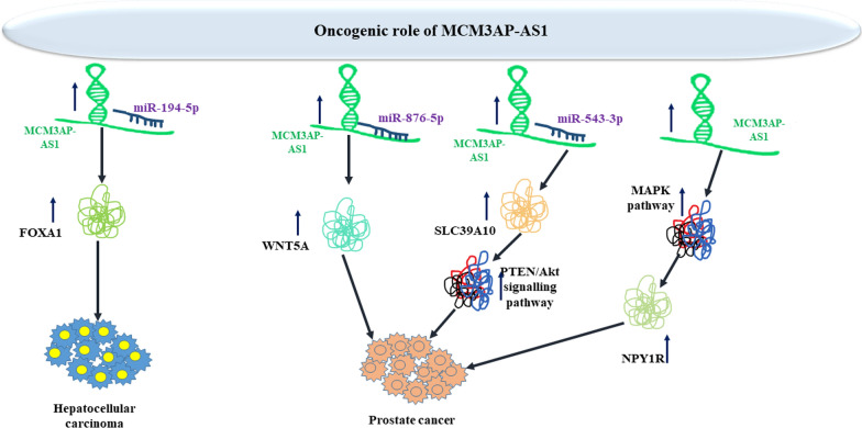
Short hairpin-mediated silencing of MCM3AP-AS1 has suppressed the proliferative capacity of prostate cancer cells and prompted their apoptosis. Mechanistically, MCM3AP-AS1 acts as a sponge of miR-876-5p to up-regulate levels of the target of this miRNA i.e. WNT5A [19]. To discover the function of MCM3AP-AS1 in prostate cancer, Wu et al. have knocked down this lncRNA in LNCaP and PC-3 cells. Based on the results of CCK-8 and EdU assays, authors have reported that proliferation of these cells has been remarkably reduced after MCM3AP-AS1 silencing. Moreover, flow cytometry technique has shown that MCM3AP-AS1 silencing increases apoptosis of these cells. Notably, expression of Bax has been increased in MCM3AP-AS1 knockdown cells, while Bcl-2 levels have been decreased, proposing that this lncRNA participates in the apoptotic processes in prostate cancer cells. These findings can also indicate that MCM3AP-AS1 is involved in the malignant features of prostate cancer [19]. Moreover, up-regulation of MCM3AP-AS1 in prostate cancer cells has been found to increase proliferation and invasive capacity through sponging miR-543-3p and influencing the SLC39A10/PTEN/Akt axis [20]. Another study in prostate cancer cells has revealed up-regulation of MCM3AP-AS1 in association with down-regulation of NPY1R. Lentivirus-mediated up-regulation of this lncRNA has increased proliferation, invasiveness, and migratory potential of prostate cancer cells while decreasing their apoptosis. Suppression of MAPK pathway has exerted opposite effects. Functionally, MCM3AP-AS1 enhances methylation of NPY1R promoter through recruiting of DNMT1/DNMT3 (A/B). Subsequent downregulation of NPY1R expression increases activity of the MAPK pathway [21]. Figure 1 depicts the oncogenic impact of MCM3AP-AS1 in liver and prostate cancers.
Fig. 1.
Oncogenic impact of MCM3AP-AS1 in hepatocellular carcinoma and prostate cancer
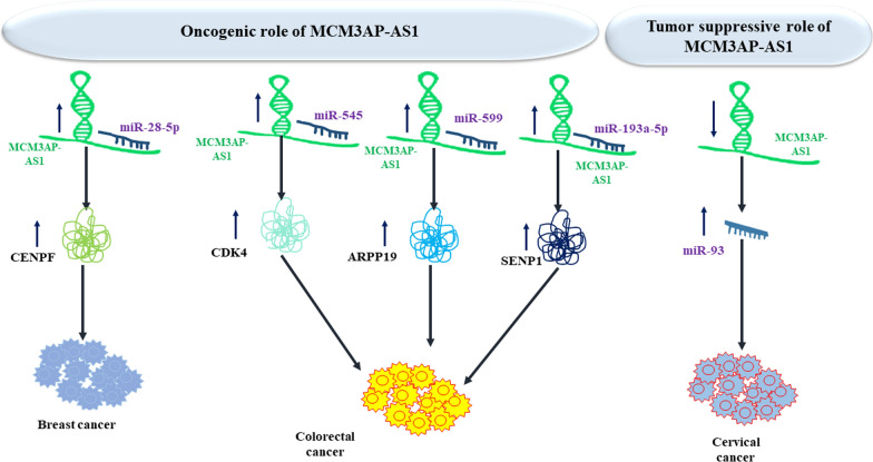
MCM3AP-AS1 has been found to be down-regulated in cervical squamous cell carcinoma cells. Enhancement of MCM3AP-AS1 expression has resulted in down-regulation of miR-93 and reduction of cell proliferation. Methylation-specific PCR has shown that MCM3AP-AS1 enhances methylation of miR-93 gene. Cumulatively, MCM3AP-AS1 can act as a tumor suppressor transcript in cervical cancer through decreasing miR-93 levels [22]
In colorectal cancer cells, up-regulation of MCM3AP-AS1 has enhanced expression of CDK4, which is directly targeted by miR-545. Up-regulation of MCM3AP-AS1 and CDK4 has inhibited G1 arrest caused by miR-545 over-expression. Furthermore, up-regulation of MCM3AP-AS1 has decreased the facilitating impact of miR-545 up-regulation on cell cycle progression. Thus, MCM3AP-AS1 has been suggested to increase CDK4 expression through sequestering miR-545 to arrest colorectal cancer cells at G1 phase [23]. Another study in colorectal cancer cells has recognized MCM3AP-AS1 as a molecular sponge of miR-599. MCM3AP-AS1 silencing has reduced ARPP19 transcript levels and enhanced expression of miR-599. Taken together, MCM3AP-AS1 facilitates progression of colorectal cancer through affecting the miR-599/ARPP19 axis [24]. MCM3AP‐AS1 also stimulates proliferation and metastatic potential of colorectal cancer cells through regulating miR‐193a‐5p/SENP1 axis [25]. In addition, MCM3AP-AS1 enhances progression of breast cancer through affecting the activity of miR-28-5p/CENPF axis [26]. Figure 2 shows the oncogenic impact of MCM3AP-AS1 in breast and colorectal cancers and its tumor suppressor role in cervical cancer.
Fig. 2.
Oncogenic effect of MCM3AP-AS1 in breast and colorectal cancers and its tumor suppressor role in cervical cancer
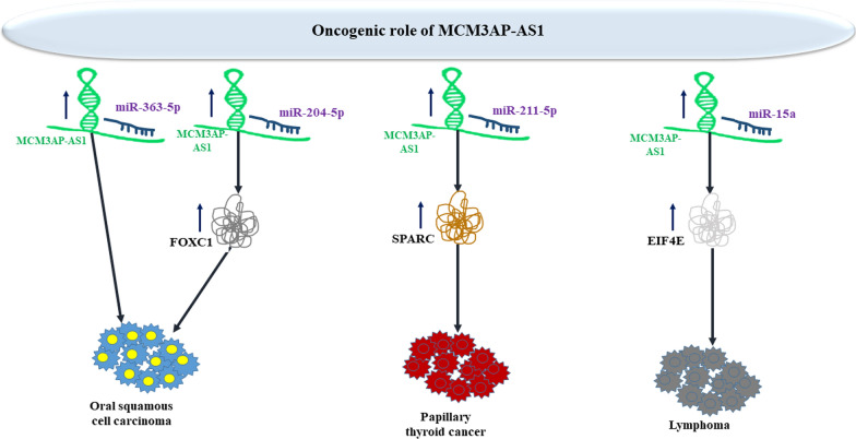
Over-expression of MCM3AP-AS1 has been correlated with poor prognosis in these patients. Small interfering RNA-mediated MCM3AP‑AS1 silencing has decreased optical density value and migratory potential of oral squamous cell carcinoma cell lines. MCM3AP-AS1 has been shown to inhibit miR‑363‑5p expression [27]. miR-204-5p/FOXC1 has been identified as another molecular axis through which MCM3AP-AS1 contributes in the pathoetiology of oral squamous cell carcinoma [28]. In papillary thyroid carcinoma, MCM3AP-AS1 enhances proliferation and invasiveness via modulating miR-211-5p/SPARC axis [29]. Moreover, in lymphoma cells, MCM3AP-AS1 silencing has been shown to attenuate resistance to doxorubicin via affecting the miR-15a/EIF4E axis [30]. Figure 3 shows the oncogenic impact of MCM3AP-AS1 in oral squamous cell cancer, papillary thyroid cancer and lymphoma.
Fig. 3.
Oncogenic impact of MCM3AP-AS1 in oral squamous cell carcinoma, papillary thyroid cancer and lymphoma
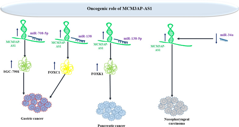
MCM3AP-AS1 has been revealed to regulate proliferation and apoptosis of gastric cancer cells through regulating miR-708-5p levels [31]. Levels of MCM3AP-AS1 have been found to be higher cisplatin-resistant gastric cancer cells. MCM3AP-AS1 silencing has reduced cisplatin resistance in these cells. Mechanistically, MCM3AP-AS1 up-regulates FOXC1 levels through sequestering miR-138. Up-regulation of FOXC1 has stopped the effects of MCM3AP-AS1 silencing or miR-138 mimic on resistance to cisplatin. Taken together, MCM3AP-AS1 confers resistance to cisplatin through sponging miR-138 and up-regulating FOXC1 levels [32]. miR-138-5p/FOXK1 has been found as the molecular axis mediating the pro-proliferative effects of MCM3AP-AS1 in pancreatic cancer cells [33]. Moreover, MCM3AP-AS1 affects proliferastion and apoptosis of nasopharyngeal cancer cells through regulating expression of miR-34a [34]. Figure 4 shows oncogenic impact of MCM3AP-AS1 in gastric, pancreatic and nasopharyngeal cancers.
Fig. 4.
Oncogenic impact of MCM3AP-AS1 in gastric, pancreatic and nasopharyngeal cancers
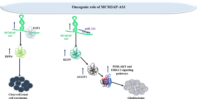
Assessment of methylation status of MCM3AP-AS1 in clear cell renal cell carcinoma cells has revealed demethylation of its promoter. Up-regulation of MCM3AP-AS1 has increased proliferation, production of proinflammatory cytokines, and the tube establishment of endothelial cells. MCM3AP-AS1 increases E2F1 recruitment at the DPP4 promoter to facilitate DPP4 expression. DPP4 silencing has abolished pro-angiogenic and pro-inflammatory effects of MCM3AP-AS1 in renal cancer cells [35]. In glioblastoma cells, MCM3AP-AS1 enhances angiogenesis through modulation of miR-211/KLF5/AGGF1 route [36]. Figure 5 shows oncogenic impact of MCM3AP-AS1 in renal cell carcinoma and glioblastoma.
Fig. 5.
Oncogenic impact of MCM3AP-AS1 in renal cell carcinoma and glioblastoma
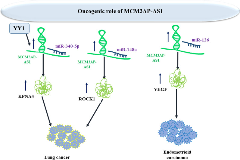
In lung cancer, transcription of MCM3AP‐AS1 is induced by YY1. Enhancement of MCM3AP‐AS1 levels enhances angiogenic process and cancer progression through influencing miR‐340‐5p/KPNA4 molecular axis [37]. Moreover, it sequesters miR-148a to increase invasiveness and migratory potential [38]. In endometrioid carcinoma, miR-126/VEGF axis mediates cancer cell invasiveness and migration [39]. Figure 6 shows oncogenic impact of MCM3AP-AS1 in lung and endometroid cancers.
Fig. 6.
Oncogenic impact of MCM3AP-AS1 in lung and endometroid cancers
Table 1 shows expression levels of MCM3AP-AS1 in cell lines.
Table 1.
Summary of research papers appraised expression of MCM3AP-AS1 in cell lines
| Tumor type | Target/ Regulator/ Signaling | Cell line | Function | References |
|---|---|---|---|---|
| Hepatocellular carcinoma | miR-194-5p, FOXA1 | LO2, HepG2, Hep3B, Huh7, SMMC-7721 | ∆ MCM3AP-AS1: ↓ proliferation, ↑ cell cycle arrest, ↑ apoptosis | [18] |
| Prostate cancer | miR-876-5p, WNT5A | C4-2, PC-3, LNCaP, DU145, 22Rv1 | ∆ MCM3AP-AS1: ↓ proliferation, ↑ apoptosis | [19] |
| miR-543-3p, SLC39A10, PTEN/Akt signaling pathway | PC-3, DU145, 22RV1, LNCaP, WPMY-1 | ∆ MCM3AP-AS1: ↓ proliferation, ↓ migration, ↓ invasion, ↑ cell cycle arrest, did not affect apoptosis | [20] | |
| NPY1R, MAPK pathway | RWPE1, 22RV1, LNCaP, and DU145 | ∆ MCM3AP-AS1: ↓ proliferation, ↓ migration, ↓ invasion, ↑ apoptosis | [21] | |
| Breast cancer | miR-28-5p, CENPF | MCF-10A, MCF-7, BT-549, MDA-MB-468, HS578 T | ∆ MCM3AP-AS1: ↓ proliferation, ↓ migration, ↓ invasion | [26] |
| Cervical cancer | miR-93 | C-33A, SiHa | ↑ MCM3AP-AS1: ↓ proliferation | [22] |
| Colorectal cancer | miR-545, CDK4 | CR4 | ↑ MCM3AP-AS1: ↑ proliferation, ↓ G1 arrest | [23] |
| miR-599, ARPP19 | HCT-116, SW620, SW480, LoVo, NCM460, 293 T | ↑ MCM3AP-AS1: ↑ proliferation, ↑ migration | [24] | |
| miR‐193a‐5p, SENP1 | HCT‐8, HCT116, LoVo, HT29, and SW620 | ∆ MCM3AP-AS1: ↓ proliferation, ↓ migration, ↓ invasion | [25] | |
| Oral squamous cell carcinoma | miR-363-5p | HOK, SCC-9, SCC-15, CAL-27 | ∆ MCM3AP-AS1: ↓ proliferation, ↓ migration, ↓ invasion | [27] |
| miR-204-5p, FOXC1 | HN-6, SCC-9 | ∆ MCM3AP-AS1: ↓ proliferation, ↓ migration, ↓ invasion | [28] | |
| Papillary thyroid cancer | miR-211-5p, SPARC | TPC-1, HTH83, 8505C, SW1736, BCPAP | ∆ MCM3AP-AS1: ↓ proliferation, ↓ migration, ↓ invasion | [29] |
| Lymphoma | miR-15a, EIF4E | Daudi, Namalwa | ∆ MCM3AP-AS1: ↓ proliferation, ↑ apoptosis, ↑ sensitivity to doxorubicin | [30] |
| Gastric cancer | miR-708-5p, SGC-7901 | GES-1, MGc-803, SGC-7901 | ∆ MCM3AP-AS1: ↓ proliferation, ↑ apoptosis | [31] |
| miR-138, FOXC1 | AGS, MKN45, NCI-N87, SNU638, GES-1, 293 T | ∆ MCM3AP-AS1: ↑ CDDP sensitivity | [32] | |
| Pancreatic cancer | miR-138-5p, FOXK1 | HPDE6-C7, PANC-1, BxPC-3, MIA PaCa-2, Capan-2, AsPC-1, HEK-293 | ↑ MCM3AP-AS1: ↑ proliferation, ↑ migration, ↑ invasion | [33] |
| Nasopharyngeal carcinoma | miR-34a | FNA, C666-1, 13-9B | ↑ MCM3AP-AS1: ↑ proliferation, ↓ apoptosis | [34] |
| Clear cell renal cell carcinoma | DPP4, E2F1 | 786-O, Caki-1, UT14, UT48 HK-2 | ↑ MCM3AP-AS1: ↑ proliferation, ↑ angiogenesis, ↑ inflammatory responses | [35] |
| Glioblastoma | miR-211, KLF5, AGGF1, PI3K/AKT and ERK1/2 signaling pathways | hCMEC/D3, HEK293T | ∆ MCM3AP-AS1: ↓ viability, ↓ migration, ↓ tube formation, ↓ angiogenesis | [36] |
| Lung cancer | miR‐340‐5p, KPNA4, YY1 | A549, H1299, H596, H520 | ∆ MCM3AP-AS1: ↓ angiogenesis, ↓ migration | [37] |
| miR-148a, ROCK1 | SHP-77 | ∆ MCM3AP-AS1: ↓ migration, ↓ invasion | [38] | |
| miR-195-5p, E2F3 | A549, H358, H1299, H460, H226, BEAS-2B | ↑ MCM3AP-AS1: ↑ proliferation, ↑ migration, ↑ invasion ↓ apoptosis | [40] | |
| Endometrioid carcinoma | miR-126, VEGF | HEC-1 | ↑ MCM3AP-AS1: ↑ migration, ↑ invasion | [39] |
∆: knock-down or deletion, CDDP: cisplatin
Animal studies
Animal studies have important positions in cancer research. Animal models have been used for simulation of human body. Several animal models have been established and their application has been evaluated in cancer research. Chemical induction, xenotransplanted models and gene programmed models have been used in this field [41]. Xenotransplantation has been the most widely used model for assessment of function of MCM3AP-AS1. In this method, cancer cell lines have been manipulated to over-express or down-regulate MCM3AP-AS1. Then, these cell lines have been transplanted into animals.
To appraise the impact of MCM3AP-AS1 silencing on tumorigenesis of hepatocellular carcinoma in vivo, Wang et al. have implanted MCM3AP-AS1-silence Hep3B cells into nude mice through subcutaneous injection. They have reported that MCM3AP-AS1 silencing significantly reduces tumor growth in animals. Moreover, subcutaneous lesions produced by MCM3AP-AS1-silenced Hep3B had lower proportion of Ki-67 expressing cells compared to tumor produced by control cells [18]. In vivo experiments in xenograft models of prostate cancer has also shown that MCM3AP-AS1 silencing considerably reduces tumor volume, and decreases the ratio of Ki67-expressing cells and expression levels of SLC39A10 in lesions [20]. Another study in prostate cancer has shown that MCM3AP-AS1 over-expression facilitates cancer development in vivo, which can be inverted by up-regulation of NPY1R [21]. Additional experiments in xenograft model of renal cell cancer has confirmed pro-angiogenic and pro-inflammatory impact of MCM3AP-AS1 [35]. Table 2 shows the results of animal studies investigating the impact of MCM3AP-AS1 in tumorigenesis.
Table 2.
Appraisal of function of MCM3AP-AS1 in animal models
| Tumor Type | Animal models | Results | (type of cells injected to the mice) (type of deletion and control) | References |
|---|---|---|---|---|
| Hepatocellular carcinoma |
12 male BALB/c nude mice (divided into two groups (n = 6 per group)) |
∆ MCM3AP-AS1: ↓ tumor growth, ↓ tumor weight |
Hep3B cells were infected with sh-MCM3AP-AS1 and sh-control |
[18] |
| Prostate cancer | 5 BALB/c mice | ∆ MCM3AP-AS1: ↓ tumor volumes | Cells were transfected with LV-si-MCM3AP-AS1 or LV-si-NC | [20] |
| Breast cancer | BALB/c nude mice | ∆ MCM3AP-AS1: ↓ tumor volumes, ↓ tumor weight |
MCF-7 cells were transfected with sh-NC, sh-MCM3AP-AS1#1 or sh-MCM3AP-AS1#1 + pcDNA3.1/CENPF |
[26] |
| Colorectal cancer |
10 BALB/c athymic nude mice (divided into two groups (n = 5 per group)) |
↑ MCM3AP-AS1: ↑ tumor volumes, ↑ tumor weigh | CR4 cells with overexpression of MCM3AP-AS1 and of C (mock cells) | [23] |
|
10 male BALB/c nude mice (divided into two groups (n = 5 per group)) |
∆ MCM3AP-AS1: ↓ tumor growth, ↓ tumor weight | LoVo cells were transfected with sh‐MCM3AP‐AS1 or sh‐Ctrl lentiviral vector | [25] | |
| Papillary thyroid cancer |
10 BALB/c nude mice (divided into two groups (n = 5 per group)) |
↑ MCM3AP-AS1: ↑ tumor volume |
TPC-1 cells were transfected with lentivirus mediated sh- MCM3AP-AS1 and sh-NC |
[29] |
| Lymphoma |
24 male BALB/c nude mice (divided into four groups n = 6 per group: (Daudi-siNC, Daudi-siMCM, Namalwa-siNC and Namalwa-siMCM)) |
∆ MCM3AP-AS1: ↓ tumor growth | Daudi and Namalwa cells were transfected with siNC or siMCM3AP-AS1 | [30] |
| Pancreatic cancer |
24 BALB/c nude mice (divided into two groups (n = 12 per group)) |
∆ MCM3AP-AS1: ↓ tumor volumes, ↓ tumor weight, ↓ tumor growth | BxPC-3 cells were transfected with shRNA-NC and MCM3AP-AS1 shRNA-2# | [33] |
| Clear cell renal cell carcinoma |
20 BALB/C nude mice (divided into two groups (n = 10 per group)) |
∆ MCM3AP-AS1: ↓ tumor volumes, ↓ tumor weight, ↓ angiogenesis | ccRCC cells expressing sh-MCM3AP-AS1 expression or the relevant NC | [35] |
| Glioblastoma | male BALB/c athymic nude mice | ∆ MCM3AP-AS1 + ↑ miR-211: ↓ angiogenesis | GECs were transfected with sh-MCM3AP-AS1, pre-miR-211, sh-MCM3AP-AS1 + pre-miR-211 and control | [36] |
| Lung cancer |
10 male BALB/c nude mice (divided into two groups (n = 5 per group)) |
↑ MCM3AP-AS1: ↑ metastasis | A549 cells with overexpression of MCM3AP-AS1 and NC | [40] |
∆: knock-down or deletion
Clinical studies
MCM3AP-AS1 has been identified as an up-regulated lncRNA in liver carcinoma samples compared to normal liver samples according to a microarray data (GSE65485). Moreover, its expression has been reported to be increased in hepatocellular carcinoma patients in another cohort in correlation with large tumor bulk, higher grade and stage of tumors and poor prognosis of cancer [18]. Analysis of TCGA data has shown higher expression of MCM3AP-AS1 in hepatocellular carcinoma samples that have higher tumor grades compared with those with low grade tumors. Besides, MCM3AP-AS1 has been shown to be over-expressed in tumors of more advanced stages compared with early stages. Using the median of expression level of MCM3AP-AS1 in tumor samples of the mentioned cohort as a cut-off value, patients have been divided into low and high MCM3AP-AS1 subgroups. Notably, high levels of this lncRNA have been correlated with larger tumor dimension, higher grade and more advanced TNM stage. Based on the results of Kaplan–Meier survival analysis, over-expression of MCM3AP-AS1 has been correlated with poor overall survival [18]. Another study on GSE65485 dataset has revealed correlation between up-regulation of MCM3AP-AS1 in liver cancer samples and shorted survival [42].
Expression of MCM3AP-AS1 has also been demonstrated to be up-regulated in prostate cancer samples in correlation with levels of WNT5A [19]. Consistent with this finding, assessment of TCGA data has confirmed up-regulation of MCM3AP-AS1 in prostate cancer tissues compared with normal tissues. Kaplan–Meier survival analysis of TCGA data has shown that over-expression of MCM3AP-AS1 is associated with shorter disease-free survival of patients, indicating that the abnormal expression of MCM3AP-AS1 can participate in the progression of prostate cancer [19]. Another study in prostate cancer has shown up-regulation of MCM3AP-AS1 cancer samples compared with healthy tissues. Long-term follow-up of these patients has shown that over-expression of this lncRNA is associated with decreased long-term survival rate of patients. Moreover, Gleason scores for N staging has been significantly different between low and high expression group [20]. An in silico analysis of GSE3868 dataset as well as TCGA data has confirmed up-regulation of this lncRNA in prostate cancer [21].
Similarly, MCM3AP-AS1 has been upregulated in colorectal cancer tissues and its over-expression has been correlated with poor survival of these patients [23]. Moreover, levels of MCM3AP-AS1 have been shown to be higher in oral squamous cell carcinoma specimens versus normal tissues [27]. MCM3AP-AS1 has been shown to be highly expressed in clear cell renal carcinoma tissues in association with poor patient survival [35].
In breast cancer samples, expression of this lncRNA has been associated with estrogen or progesterone receptor expression [43]. In endometrioid carcinoma, up-regulation of this lncRNA has been associated with poor overall and progression-free survival [39].
Contrary to these studies, MCM3AP-AS1 has been found to be down-regulated in cervical squamous cell carcinoma in correlation with poor survival [22]. Table 3 shows the levels of MCM3AP-AS1 in different cancer types and their relevance with clinical outcomes.
Table 3.
Abnormal levels of MCM3AP-AS1 in clinical samples
| Tumor type | Samples | Expression (tumor vs. Normal) | Kaplan–Meier analysis (impact of MCM3AP-AS1 up-regulation) | Univariate/multivariate cox regression | Association of MCM3AP-AS1expression with clinicopathologic characteristics | Quantification method | Normalization method | References |
|---|---|---|---|---|---|---|---|---|
| Hepatocellular carcinoma (HCC) | 80 pairs of HCC and ANCTs (80 patients) | Up | Poorer OS | _ | Advanced tumor stages, tumor size, high tumor grade, and advanced TNM stages | qRT-PCR | 2−ΔΔCt Method (normalized by GAPDH and U6) | [18] |
| GSE65485 (50 HCC tissues and 5 ANCTs) and GSE54236 | Up | _ | _ | _ | _ | _ | ||
|
GEO databases: daGSE29721, |
Up | Shorter OS | _ | _ | _ | _ | [42] | |
| Prostate cancer (PCa) |
30 pairs of PCa and ANCTs (30 patients) |
Up | _ | _ | _ | qRT-PCR | 2−ΔΔCt method (normalized by GAPDH) | [19] |
| TCGA analysis: _ | Up | Shorter DFS | _ | _ | _ | _ | ||
|
64 pairs of PCa and ANCTs (64 patients) |
Up | Poorer OS | _ | _ | qRT-PCR | 2−ΔΔCt method (normalized by GAPDH) | [20] | |
| GEO analysis: GSE3868 | Up | _ | _ | _ | _ | _ | [21] | |
| TCGA analysis: | Up | _ | _ | _ | _ | _ | ||
|
46 pairs of PCa and ANCTs (46 patients) |
Up | _ | _ | _ | qRT-PCR | 2−ΔΔCt method (normalized by GAPDH) | ||
| GEO analysis: GSE32269, GSE26964 | Up | _ | _ | PCa bone metastasis by regulating immune microenvironment, especially M2 macrophages | _ | _ | [44] | |
| TCGA analysis: 490 PRAD patients | Up | _ | _ | _ | _ | |||
| Breast cancer (BC) | 102 pairs of BC and ANCTs (102 patients) | Up | _ | _ | Estrogen or progesterone receptor expression | qRT-PCR | 2−ΔΔCt method | [43] |
| Cervical cancer | 64 pairs of CSCC and ANCTs (64 patients) | Down | Higher OS | _ | _ | qRT-PCR | 2−ΔΔCt method (normalized by GAPDH) | [22] |
| Colorectal cancer (CRC) | 60 pairs of CRC and ANCTs (60 patients) | Up | Lower OS | _ | _ | qRT-PCR | 2−ΔΔCt method (normalized by GAPDH) | [23] |
| 30 pairs of CRC and ANCTs (30 patients) | Up | _ | _ | _ | qRT-PCR | 2−ΔΔCt method (normalized by GAPDH) | [24] | |
| 131 pairs of CRC and ANCTs (131 patients) | Up | _ | _ | Larger tumor size | qRT-PCR | 2−ΔΔCt method (normalized by β‐actin) | [25] | |
| TCGA database and GEO GSE21510 database | Up | _ | _ | _ | _ | _ | ||
| Oral squamous cell carcinoma (OSCC) | 36 pairs of OSCC and ANCTs (36 patients) | Up | _ | _ | Clinical stage and lymph node metastasis | qRT-PCR |
2-ΔΔCt method (normalized by U6) |
[27] |
| Papillary thyroid cancer (PTC) | 68 pairs of papillary thyroid cancer samples and ANCTs (68 patients) | Up | Lower OS | _ | _ | qRT-PCR | 2−ΔΔCt method (normalized by GAPDH and U6) | [29] |
| Lymphoma | 41 pairs of papillary thyroid cancer tissues and ANCTs (41 patients) | Up | Lower OS | _ | Tumor size, Tumor stage | qRT-PCR | _ | [30] |
| Pancreatic cancer (PC) | 86 pairs of PC and ANCTs (86 patients) | Up | Shorter OS | _ | _ | qRT-PCR | 2−ΔΔCt method (normalized by GAPDH and U6) | [33] |
| Nasopharyngeal carcinoma (NPC) | 55 pairs of NPC and ANCTs (55 patients) | Up | Lower OS | _ | _ | qRT-PCR | triplicate and 2−ΔΔCT method (normalized by GAPDH) | [34] |
| Clear cell renal cell carcinoma (ccRCC) | GEO GSE15641 database | Up | _ | _ | _ | _ | _ | (35) |
| 78 pairs of ccRCC and ANCTs (78 patients) | Up | Shorter OS | Expression of MCM3AP-AS1 was an independent prognostic factor for ccRCC patients | Tumors size > 7 cm | qRT-PCR | Triplicate and 2−ΔΔCT method (normalized by β-actin) | ||
| Lung cancer | 60 pairs of SCLC and ANCTs (60 patients) | Up | Lower OS | _ | _ | qRT-PCR | Triplicate and 2−ΔΔCT method (normalized by GAPDH) | [38] |
| 63 pairs of NSCLC and ANCTs (63 patients) | Up | Lower OS | _ | _ | qRT-PCR | 2−ΔΔCT method (normalized by GAPDH) | (40) | |
| Endometrioid carcinoma (EC) | 60 pairs of EC and ANCTs (60 patients) | Up | Poor OS and PFS | _ | _ | qRT-PCR | 2−ΔΔCT method (normalized by GAPDH) | [39] |
ANCTs adjacent non-cancerous tissues, OS overall survival, DFS disease-free survival, TNM tumor‐node‐metastasis, PRAD prostate adenocarcinoma, SCLC small cell lung cancer, NSCLC non-small cell lung cancer, PFS progression-free survival
Discussion
During recent years, transcriptome analyses have revealed thousands of lncRNAs. Growing numbers of these transcripts have been found to be associated with carcinogenesis process. These cancer-related lncRNAs have been found to affect development and progression of different cancers. Several of them have been found to participate in the pathetiology of diverse cancers, thus being proposed as biomarkers for these conditions. The functional roles of several lncRNAs in the carcinogenesis have been extensively reviewed during recent years [45–50]. MCM3AP-AS1 is a tumor-promoting lncRNA in almost all tissues except for cervical tissue. Although it is transcribed from the antisense of MCM3AP, its roles in oncogenesis seems to be independent from MCM3AP. This lncRNA has been shown to sequester miR-194-5p, miR-876-5p, miR-543-3p, miR-28-5p, miR-93, miR-545, miR-599, miR‐193a‐5p, miR-363-5p, miR-204-5p, miR-211-5p, miR-15a, miR-708-5p, miR-138, miR-138-5p, miR-34a, miR-211, miR‐340‐5p, miR-148a, miR-195-5p and miR-126. Thus, the sponging role of MCM3AP-AS1 on miRNAs is the most probable mechanism of contribution of this transcript in the carcinogenesis. A number of cancer-related signaling pathways such as PTEN/AKT, PI3K/AKT and ERK1/2 are influenced by this lncRNA.
Notably, miR-138 has been found to be sponged by MCM3AP-AS1 in gastric and pancreatic cancers. Moreover, expression of FOXC1 has been shown to be regulated by MCM3AP-AS1 in oral squamous cell carcinoma and gastric cancer through modulation of expressions of miR-204-5p and miR-138, respectively. Apart from these two examples, there is no other verified example of the same miRNA targeted by MCM3AP-AS1 in different tumors or certain mRNAs being targeted by this lncRNA through different miRNAs. In addition, AKT signaling has been shown to be regulated by MCM3AP-AS1 in prostate cancer and glioblastoma. These examples provide clues for design of specific therapies for each type of cancer.
In addition to regulation of cell apoptosis, migration and invasiveness, MCM3AP-AS1 affects response of neoplastic cells to chemotherapeutic agents, namely cisplatin and doxorubicin.
MCM3AP-AS1 can also affect tumor microenvironment, since it can influence expression of VEGF, thus affecting angiogenesis. Moreover, studies in renal cell carcinoma have suggested involvement of this lncRNA in the regulation of inflammatory responses in tumor microenvironment [35]. However, the exact role of this lncRNA on immune cell cells should be investigated in future studies.
Up-regulation of MCM3AP-AS1 in cancer patients has been correlated with poor survival of these patients. Moreover, its levels have been associated with several parameters related to aggressiveness of tumors such as greater tumor size and involvement of local lymph nodes or distant organs.
Therapeutic targeting of MCM3AP-AS1 has not been investigated in clinical settings. However, there are several putative strategies for targeting lncRNAs. For instance, Antisense oligonucleotides (ASOs), CRISPR-Cas9-based methods and small molecules have been used for manipulation of gene expression. Moreover, therapeutic manipulation of the promoter region is another strategy in this regard [51]. Other strategies to target function of MCM3AP-AS1are small molecules, nanobodies, aptamers, and RNA decoys. These strategies can possibly interrupt interaction between MCM3AP-AS1 and proteins through competition or steric blockade [52]. Although sequence-based nucleic acid treatment methods are developing rapidly, several questions principally those associated with the safety and efficiency of these methods should be answered before adaptation of these techniques in the clinical settings. The tissue-specificity of MCM3AP-AS1 targeting methods is another imperative subject which can be accomplished using specific vectors that have affinity to target tissues. Development of a number of receptor-targeted adeno-associated viral vectors is an important achievement in this regard [53].
Cumulatively, MCM3AP-AS1 is an oncogenic lncRNA which facilitates oncogenesis through different routes, thus therapeutic intervention with its expression represents a possible modality for cancer treatment. However, since it has been shown to exert tumor suppressor role in cervical cancer, a context-dependent effect might exist for this lncRNA. This note should be considered in design of anticancer modalities.
Acknowledgements
This study was financially supported by Grant from Medical School of Shahid Beheshti University of Medical Sciences.
Author contributions
SGF wrote the manuscript and revised it. MT supervised and designed the study. TK, MS and BMH collected the data and designed the figures and tables. All authors read and approved the submitted version.
Funding
Open Access funding enabled and organized by Projekt DEAL.
Availability of data and materials
The analyzed data sets generated during the study are available from the corresponding author on reasonable request.
Declarations
Ethics approval and consent to participant
All procedures performed in studies involving human participants were in accordance with the ethical standards of the institutional and/or national research committee and with the 1964 Helsinki declaration and its later amendments or comparable ethical standards. Informed consent forms were obtained from all study participants. The study protocol was approved by the ethical committee of Shahid Beheshti University of Medical Sciences. All methods were performed in accordance with the relevant guidelines and regulations.
Consent of publication
Not applicable.
Competing interests
The authors declare they have no conflict of interest.
Footnotes
Publisher's Note
Springer Nature remains neutral with regard to jurisdictional claims in published maps and institutional affiliations.
Contributor Information
Mohammad Taheri, Email: mohammad.taheri@uni-jena.de.
Mohammad Samadian, Email: mdsamadian@gmail.com.
References
- 1.Qian Y, Shi L, Luo Z. Long non-coding RNAs in cancer: implications for diagnosis, prognosis, and therapy. Front Med (Lausanne). 2020;7:612393. doi: 10.3389/fmed.2020.612393. [DOI] [PMC free article] [PubMed] [Google Scholar]
- 2.Wang KC, Chang HY. Molecular mechanisms of long noncoding RNAs. Mol Cell. 2011;43(6):904–914. doi: 10.1016/j.molcel.2011.08.018. [DOI] [PMC free article] [PubMed] [Google Scholar]
- 3.Quinn JJ, Chang HY. Unique features of long non-coding RNA biogenesis and function. Nat Rev Genet. 2016;17(1):47. doi: 10.1038/nrg.2015.10. [DOI] [PubMed] [Google Scholar]
- 4.Li W, Notani D, Ma Q, Tanasa B, Nunez E, Chen AY, et al. Functional roles of enhancer RNAs for oestrogen-dependent transcriptional activation. Nature. 2013;498(7455):516–520. doi: 10.1038/nature12210. [DOI] [PMC free article] [PubMed] [Google Scholar]
- 5.Lai F, Orom UA, Cesaroni M, Beringer M, Taatjes DJ, Blobel GA, et al. Activating RNAs associate with mediator to enhance chromatin architecture and transcription. Nature. 2013;494(7438):497–501. doi: 10.1038/nature11884. [DOI] [PMC free article] [PubMed] [Google Scholar]
- 6.Khalil AM, Guttman M, Huarte M, Garber M, Raj A, Morales DR, et al. Many human large intergenic noncoding RNAs associate with chromatin-modifying complexes and affect gene expression. Proc Natl Acad Sci. 2009;106(28):11667–11672. doi: 10.1073/pnas.0904715106. [DOI] [PMC free article] [PubMed] [Google Scholar]
- 7.Jarroux J, Morillon A, Pinskaya M. History, discovery, and classification of lncRNAs. In: Rao MRS, editor. Long non coding RNA biology. Singapore: Springer Singapore; 2017. pp. 1–46. [DOI] [PubMed] [Google Scholar]
- 8.Huarte M. The emerging role of lncRNAs in cancer. Nat Med. 2015;21(11):1253–1261. doi: 10.1038/nm.3981. [DOI] [PubMed] [Google Scholar]
- 9.Ghafouri-Fard S, Esmaeili M, Taheri M. H19 lncRNA: roles in tumorigenesis. Biomed Pharmacother. 2020;123:109774. doi: 10.1016/j.biopha.2019.109774. [DOI] [PubMed] [Google Scholar]
- 10.Ghafouri-Fard S, Taheri M. Maternally expressed gene 3 (MEG3): a tumor suppressor long non coding RNA. Biomed Pharmacother. 2019;118:109129. doi: 10.1016/j.biopha.2019.109129. [DOI] [PubMed] [Google Scholar]
- 11.Ghafouri-Fard S, Taheri M. Nuclear enriched abundant transcript 1 (NEAT1): a long non-coding RNA with diverse functions in tumorigenesis. Biomed Pharmacother. 2019;111:51–59. doi: 10.1016/j.biopha.2018.12.070. [DOI] [PubMed] [Google Scholar]
- 12.Takei Y, Swietlik M, Tanoue A, Tsujimoto G, Kouzarides T, Laskey R. MCM3AP, a novel acetyltransferase that acetylates replication protein MCM3. EMBO Rep. 2001;2(2):119–123. doi: 10.1093/embo-reports/kve026. [DOI] [PMC free article] [PubMed] [Google Scholar]
- 13.Poole E, Bain M, Teague L, Takei Y, Laskey R, Sinclair J. The cellular protein MCM3AP is required for inhibition of cellular DNA synthesis by the IE86 protein of human cytomegalovirus. PLoS ONE. 2012 doi: 10.1371/journal.pone.0045686. [DOI] [PMC free article] [PubMed] [Google Scholar]
- 14.Kuwahara K, Yamamoto-Ibusuki M, Zhang Z, Phimsen S, Gondo N, Yamashita H, et al. GANP protein encoded on human chromosome 21/mouse chromosome 10 is associated with resistance to mammary tumor development. Cancer Sci. 2016;107(4):469–477. doi: 10.1111/cas.12883. [DOI] [PMC free article] [PubMed] [Google Scholar]
- 15.Ohta K, Kuwahara K, Zhang Z, Makino K, Komohara Y, Nakamura H, et al. Decreased expression of germinal center–associated nuclear protein is involved in chromosomal instability in malignant gliomas. Cancer Sci. 2009;100(11):2069–2076. doi: 10.1111/j.1349-7006.2009.01293.x. [DOI] [PMC free article] [PubMed] [Google Scholar]
- 16.Singh SK, Maeda K, Eid MMA, Almofty SA, Ono M, Pham P, et al. GANP regulates recruitment of AID to immunoglobulin variable regions by modulating transcription and nucleosome occupancy. Nat Commun. 2013;4(1):1–12. doi: 10.1038/ncomms2823. [DOI] [PMC free article] [PubMed] [Google Scholar]
- 17.Bierhoff H. Analysis of lncRNA-protein interactions by RNA-protein pull-down assays and RNA immunoprecipitation (RIP) In: Daniel Lacorazza H, editor. Cellular quiescence: methods and protocols. New York, NY: Springer New York; 2018. pp. 241–250. [DOI] [PubMed] [Google Scholar]
- 18.Wang Y, Yang L, Chen T, Liu X, Guo Y, Zhu Q, et al. A novel lncRNA MCM3AP-AS1 promotes the growth of hepatocellular carcinoma by targeting miR-194-5p/FOXA1 axis. Mol Cancer. 2019;18(1):1–16. doi: 10.1186/s12943-019-0957-7. [DOI] [PMC free article] [PubMed] [Google Scholar]
- 19.Wu J, Lv Y, Li Y, Jiang Y, Wang L, Zhang X, et al. MCM3AP-AS1/miR-876-5p/WNT5A axis regulates the proliferation of prostate cancer cells. Cancer Cell Int. 2020;20(1):1–12. doi: 10.1186/s12935-019-1086-5. [DOI] [PMC free article] [PubMed] [Google Scholar] [Retracted]
- 20.Jia Z, Li W, Bian P, Liu H, Pan D, Dou Z. LncRNA MCM3AP-AS1 promotes cell proliferation and invasion through regulating miR-543-3p/SLC39A10/PTEN axis in prostate cancer. Onco Targets Therapy. 2020;13:9365. doi: 10.2147/OTT.S245537. [DOI] [PMC free article] [PubMed] [Google Scholar] [Retracted]
- 21.Li X, Lv J, Liu S. MCM3AP-AS1 KD inhibits proliferation, invasion, and migration of PCa cells via DNMT1/DNMT3 (A/B) methylation-mediated upregulation of NPY1R. Mol Therapy-Nucleic Acids. 2020;20:265–278. doi: 10.1016/j.omtn.2020.01.016. [DOI] [PMC free article] [PubMed] [Google Scholar] [Retracted]
- 22.Lan L, Liang Z, Zhao Y, Mo Y. LncRNA MCM3AP-AS1 inhibits cell proliferation in cervical squamous cell carcinoma by down-regulating miRNA-93. Biosci Rep. 2020;40(2):BSR20193794. doi: 10.1042/BSR20193794. [DOI] [PMC free article] [PubMed] [Google Scholar]
- 23.Ma X, Luo J, Zhang Y, Sun D, Lin Y. LncRNA MCM3AP-AS1 upregulates CDK4 by sponging miR-545 to suppress G1 Arrest in colorectal cancer. Cancer Manag Res. 2020;12:8117. doi: 10.2147/CMAR.S247330. [DOI] [PMC free article] [PubMed] [Google Scholar]
- 24.Yu Y, Lai S, Peng X. Long non-coding RNA MCM3AP-AS1 facilitates colorectal cancer progression by regulating the microRNA-599/ARPP19 axis. Oncol Lett. 2021;21(3):1. doi: 10.3892/ol.2021.12486. [DOI] [PMC free article] [PubMed] [Google Scholar]
- 25.Zhou M, Bian Z, Liu B, Zhang Y, Cao Y, Cui K, et al. Long noncoding RNA MCM3AP-AS1 enhances cell proliferation and metastasis in colorectal cancer by regulating miR-193a-5p/SENP1. Cancer Med. 2021;10(7):2470–2481. doi: 10.1002/cam4.3830. [DOI] [PMC free article] [PubMed] [Google Scholar]
- 26.Chen Q, Xu H, Zhu J, Feng K, Hu C. LncRNA MCM3AP-AS1 promotes breast cancer progression via modulating miR-28-5p/CENPF axis. Biomed Pharmacother. 2020;128:110289. doi: 10.1016/j.biopha.2020.110289. [DOI] [PubMed] [Google Scholar]
- 27.Hou C, Wang X, Du B. lncRNA MCM3AP-AS1 promotes the development of oral squamous cell carcinoma by inhibiting miR-363-5p. Exp Ther Med. 2020;20(2):978–984. doi: 10.3892/etm.2020.8738. [DOI] [PMC free article] [PubMed] [Google Scholar]
- 28.Li H, Jiang J. LncRNA MCM3AP-AS1 promotes proliferation, migration and invasion of oral squamous cell carcinoma cells via regulating miR-204-5p/FOXC1. J Investig Med. 2020;68(7):1282–1288. doi: 10.1136/jim-2020-001415. [DOI] [PubMed] [Google Scholar]
- 29.Liang M, Jia J, Chen L, Wei B, Guan Q, Ding Z, et al. LncRNA MCM3AP-AS1 promotes proliferation and invasion through regulating miR-211-5p/SPARC axis in papillary thyroid cancer. Endocrine. 2019;65(2):318–326. doi: 10.1007/s12020-019-01939-4. [DOI] [PubMed] [Google Scholar]
- 30.Guo C, Gong M, Li Z. Knockdown of lncRNA MCM3AP-AS1 attenuates chemoresistance of Burkitt lymphoma to doxorubicin treatment via targeting the miR-15a/EIF4E Axis. Cancer Manag Res. 2020;12:5845. doi: 10.2147/CMAR.S248698. [DOI] [PMC free article] [PubMed] [Google Scholar]
- 31.Wang H, Xu T, Wu L, Xu H, Liu R. Molecular mechanisms of MCM3AP-AS1 targeted the regulation of miR-708-5p on cell proliferation and apoptosis in gastric cancer cells. Eur Rev Med Pharmacol Sci. 2020;24(5):2452–2461. doi: 10.26355/eurrev_202003_20512. [DOI] [PubMed] [Google Scholar]
- 32.Sun H, Wu P, Zhang B, Wu X, Chen W. MCM3AP-AS1 promotes cisplatin resistance in gastric cancer cells via the miR-138/FOXC1 axis. Oncol Lett. 2021;21(3):1. doi: 10.3892/ol.2021.12472. [DOI] [PMC free article] [PubMed] [Google Scholar]
- 33.Yang M, Sun S, Guo Y, Qin J, Liu G. Long non-coding RNA MCM3AP-AS1 promotes growth and migration through modulating FOXK1 by sponging miR-138-5p in pancreatic cancer. Mol Med. 2019;25(1):1–10. doi: 10.1186/s10020-019-0121-2. [DOI] [PMC free article] [PubMed] [Google Scholar]
- 34.Sun P, Feng Y, Guo H, Li R, Yu P, Zhou X, et al. MiR-34a inhibits cell proliferation and induces apoptosis in human nasopharyngeal carcinoma by targeting lncRNA MCM3AP-AS1. Cancer Manag Res. 2020;12:4799. doi: 10.2147/CMAR.S245520. [DOI] [PMC free article] [PubMed] [Google Scholar]
- 35.Qiu L, Ma Y, Yang Y, Ren X, Wang D, Jia X. Pro-angiogenic and pro-inflammatory regulation by lncRNA MCM3AP-AS1-mediated upregulation of DPP4 in clear cell renal cell carcinoma. Front Oncol. 2020;10:705. doi: 10.3389/fonc.2020.00705. [DOI] [PMC free article] [PubMed] [Google Scholar]
- 36.Yang C, Zheng J, Xue Y, Yu H, Liu X, Ma J, et al. The effect of MCM3AP-AS1/miR-211/KLF5/AGGF1 axis regulating glioblastoma angiogenesis. Front Mol Neurosci. 2018;10:437. doi: 10.3389/fnmol.2017.00437. [DOI] [PMC free article] [PubMed] [Google Scholar] [Retracted]
- 37.Li X, Yu M, Yang C. YY1-mediated overexpression of long noncoding RNA MCM3AP-AS1 accelerates angiogenesis and progression in lung cancer by targeting miR-340-5p/KPNA4 axis. J Cell Biochem. 2020;121(3):2258–2267. doi: 10.1002/jcb.29448. [DOI] [PubMed] [Google Scholar]
- 38.Luo H, Zhang Y, Qin G, Jiang B, Miao L. LncRNA MCM3AP-AS1 sponges miR-148a to enhance cell invasion and migration in small cell lung cancer. BMC Cancer. 2021;21(1):1–8. doi: 10.1186/s12885-020-07763-8. [DOI] [PMC free article] [PubMed] [Google Scholar]
- 39.Yu J, Fan Q, Li L. The MCM3AP-AS1/miR-126/VEGF axis regulates cancer cell invasion and migration in endometrioid carcinoma. World J Surg Oncol. 2021;19(1):1–8. doi: 10.1186/s12957-015-0754-8. [DOI] [PMC free article] [PubMed] [Google Scholar]
- 40.Shen D, Li J, Tao K, Jiang Y. Long non-coding RNA MCM3AP antisense RNA 1 promotes non-small cell lung cancer progression through targeting microRNA-195-5p. Bioengineered. 2021;12(1):3525–3538. doi: 10.1080/21655979.2021.1950282. [DOI] [PMC free article] [PubMed] [Google Scholar]
- 41.Li Z, Zheng W, Wang H, Cheng Y, Fang Y, Wu F, et al. Application of animal models in cancer research: recent progress and future prospects. Cancer Manag Res. 2021;13:2455. doi: 10.2147/CMAR.S302565. [DOI] [PMC free article] [PubMed] [Google Scholar]
- 42.Song M, Zhong A, Yang J, He J, Cheng S, Zeng J, et al. Large-scale analyses identify a cluster of novel long noncoding RNAs as potential competitive endogenous RNAs in progression of hepatocellular carcinoma. Aging (Albany NY) 2019;11(22):10422. doi: 10.18632/aging.102468. [DOI] [PMC free article] [PubMed] [Google Scholar]
- 43.Riahi A, Hosseinpour-Feizi M, Rajabi A, Akbarzadeh M, Montazeri V, Safaralizadeh R. Overexpression of long non-coding RNA MCM3AP-AS1 in breast cancer tissues compared to adjacent non-tumour tissues. Br J Biomed Sci. 2021;78(2):53–57. doi: 10.1080/09674845.2020.1798058. [DOI] [PubMed] [Google Scholar]
- 44.Chen Y, Chen Z, Mo J, Pang M, Chen Z, Feng F, et al. Identification of HCG18 and MCM3AP-AS1 that associate with bone metastasis, poor prognosis and increased abundance of M2 macrophage infiltration in prostate cancer. Technol Cancer Res Treat. 2021;20:1533033821990064. doi: 10.1177/1533033821990064. [DOI] [PMC free article] [PubMed] [Google Scholar]
- 45.Ghafouri-Fard S, Khoshbakht T, Taheri M, Ebrahimzadeh K. A review on the carcinogenic roles of DSCAM-AS1. Front Cell Dev Biol. 2021;9:758513. doi: 10.3389/fcell.2021.758513. [DOI] [PMC free article] [PubMed] [Google Scholar]
- 46.Ghafouri-Fard S, Khoshbakht T, Taheri M, Shojaei S. A review on the role of small nucleolar RNA host gene 6 long non-coding RNAs in the carcinogenic processes. Front Cell Dev Biol. 2021;9:741684. doi: 10.3389/fcell.2021.741684. [DOI] [PMC free article] [PubMed] [Google Scholar]
- 47.Ghafouri-Fard S, Khoshbakht T, Taheri M, Khashefizadeh A. Hepatocyte nuclear factor 1A-antisense: review of its role in the carcinogenesis. Pathol Res Pract. 2021;20(227):153623. doi: 10.1016/j.prp.2021.153623. [DOI] [PubMed] [Google Scholar]
- 48.Ghafouri-Fard S, Azimi T, Hussen BM, Abak A, Taheri M, Dilmaghani NA. Non-coding RNA activated by DNA damage: review of its roles in the carcinogenesis. Front Cell Dev Biol. 2021;9:714787. doi: 10.3389/fcell.2021.714787. [DOI] [PMC free article] [PubMed] [Google Scholar]
- 49.Ghafouri-Fard S, Khoshbakht T, Taheri M, Mokhtari M. A review on the role of GAS6 and GAS6-AS1 in the carcinogenesis. Pathol Res Pract. 2021;226:153596. doi: 10.1016/j.prp.2021.153596. [DOI] [PubMed] [Google Scholar]
- 50.Hussen BM, Azimi T, Abak A, Hidayat HJ, Taheri M, Ghafouri-Fard S. Role of lncRNA BANCR in human cancers: an updated review. Front Cell Dev Biol. 2021;9:689992. doi: 10.3389/fcell.2021.689992. [DOI] [PMC free article] [PubMed] [Google Scholar]
- 51.Arun G, Diermeier SD, Spector DL. Therapeutic targeting of long non-coding RNAs in cancer. Trends Mol Med. 2018;24(3):257–277. doi: 10.1016/j.molmed.2018.01.001. [DOI] [PMC free article] [PubMed] [Google Scholar]
- 52.Gutschner T, Richtig G, Haemmerle M, Pichler M. From biomarkers to therapeutic targets—the promises and perils of long non-coding RNAs in cancer. Cancer Metastasis Rev. 2018;37(1):83–105. doi: 10.1007/s10555-017-9718-5. [DOI] [PubMed] [Google Scholar]
- 53.Münch RC, Muth A, Muik A, Friedel T, Schmatz J, Dreier B, et al. Off-target-free gene delivery by affinity-purified receptor-targeted viral vectors. Nat Commun. 2015;6(1):1–9. doi: 10.1038/ncomms7246. [DOI] [PubMed] [Google Scholar]
Associated Data
This section collects any data citations, data availability statements, or supplementary materials included in this article.
Data Availability Statement
The analyzed data sets generated during the study are available from the corresponding author on reasonable request.


