Abstract
Adenoid cystic carcinoma (ACC) is a type of malignant tumor arising from the salivary glands. The tumor is characterized by a predilection for recurrence and metastasis. At present, there is no effective treatment for ACC complicated with lung metastasis. The present study reported on a case of suboral ACC with bilateral lung metastasis and the clinical features and treatment options were discussed based on a literature review. A 55-year-old female presented with suboral ACC accompanied with bilateral lung metastases. The main symptoms were masses in the right mandible and shortness of breath. Low-dose radiotherapy (LDRT) combined with immunotherapy were administered to control lung metastases. The patient is currently in a stable condition and is receiving immunotherapy. LDRT combined with immunotherapy is a feasible treatment option for such patients. Our experience with this case and the information from the literature review provide insight into the therapeutic approach for this relatively rare entity.
Keywords: adenoid cystic carcinoma, lung metastasis, immunotherapy, low-dose radiotherapy, treatment outcome
Introduction
Adenoid cystic carcinoma (ACC) is a relatively rare low-grade malignant tumor type, which accounts for ~1% of all head and neck tumors and 10% of all malignant tumors of salivary gland origin (1). At present, surgery and postoperative radiotherapy are the standard treatment modalities for localized ACC; however, the optimal treatment for ACC with lung metastasis has remained to be established (2). Use of low-dose radiotherapy (LDRT) combined with immunotherapy is being gradually used in clinical settings (3,4). The present study reported on a case of suboral ACC with lung metastasis who was treated at our hospital. The pertinent literature on the mechanism and the advantages of LDRT combined with immunotherapy in patients with pulmonary metastases was also reviewed. The patient diagnosis and treatment are outlined in a timeline presented in Fig. 1. The present case may provide insight regarding the treatment of ACC with bilateral lung metastasis.
Figure 1.
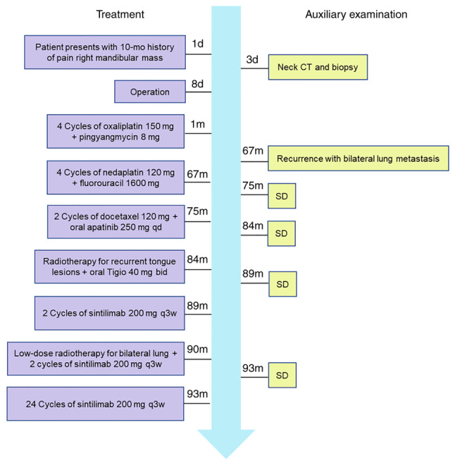
Timeline of patient diagnosis and treatment. Mo, month; SD, stable disease.
Case study
Case presentation
A 55-year-old female presented to the Department of Oncology, West China Hospital, Sichuan University (Nanchong, China) due to a painful right mandibular mass in May 2012. There was no history of chronic disease or family history of adenoid cystic carcinoma. Physical examination revealed a right mandibular mass with no signs of inflammation. The mass had a hard texture and its boundary was poorly demarcated from the surrounding tissues. The mass measured ~3×3 cm. In addition, a prominent 2-cm right cervical lymph node was noted.
Diagnostic assessment
The patient did not undergo magnetic resonance imaging (MRI) due to financial constraints. Contrast-enhanced computed tomography (CT) revealed a mass in the floor of the right mouth, denervation atrophy of the right submandibular gland and signs of lymph node involvement. Biopsy of the mass indicated adenoid cystic carcinoma (ACC) of the floor of the right side of the mouth.
Therapeutic intervention
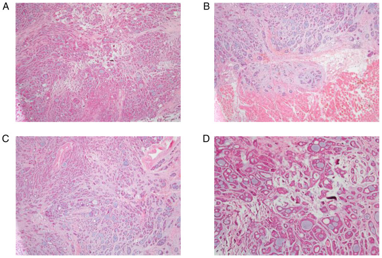
Enlarged resection of the mass plus box-like resection of the right side of mandible plus dissection of the right neck lymph node was performed. Histopathological examination of the surgical specimen confirmed the diagnosis (Fig. 2). The patient had indications for post-operative radiotherapy. However, the patient refused radiotherapy due to various reasons, such as financial constraints, and was only willing to accept chemotherapy. Post-operatively, the patient was administered chemotherapy with four cycles of oxaliplatin 150 mg plus pingyangmycin 8 mg. Subsequently, the patient underwent regular chemotherapy and follow-up evaluation.
Figure 2.
Histopathological examination of surgical specimen. (A) The tumor cells are cribriform and associated with interstitial fibrosis (H&E stain, ×4 objective); (B) tumor cells have infiltrated the surrounding skeletal muscle tissue (H&E stain, ×4 objective); (C) the tumor cells are cribriform and have invaded the nerve (H&E stain, ×4 objective); (D) the nuclei of tumor cells are small and consistent; interstitial mucus is obvious (H&E stain, ×10 objective).
In October 2018, the patient was re-admitted to the oncology department of our hospital due to shortness of breath and dyspnea. Tongue MRI indicated a mass of ~5.3×4.7×4.6 cm on the right side of the bottom of the mouth. Chest CT revealed multiple diffuse soft tissue nodules in both lungs. Considering tumor recurrence with bilateral lung metastasis, four cycles of PF (nedaplatin 40 mg D1-3 plus fluorouracil 400 mg D1-4) chemotherapy were administered at our department. On repeat tongue MRI and chest CT performed in June 2019, the primary lesions and the metastatic lesions in both lungs were slightly larger than those in October 2018 (stable disease, SD). According to the Response Evaluation Criteria in Solid Tumours guidelines (5), SD was defined as neither sufficient shrinkage to qualify for PR nor sufficient increase to qualify for PD, pertaining to the smallest sum of diameters. PD means at least a 20% increase in the sum of diameters of target lesions. Clearly, the lung metastases in the present case did not reach PD. The patient was administered two cycles of docetaxel 120 mg D1, while oral apatinib 250 mg qd was administered as antiangiogenic therapy.
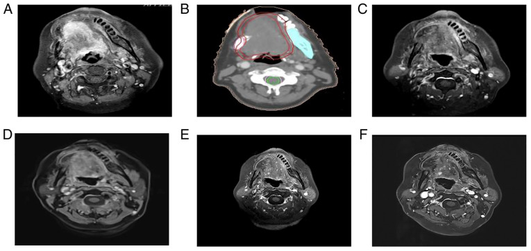
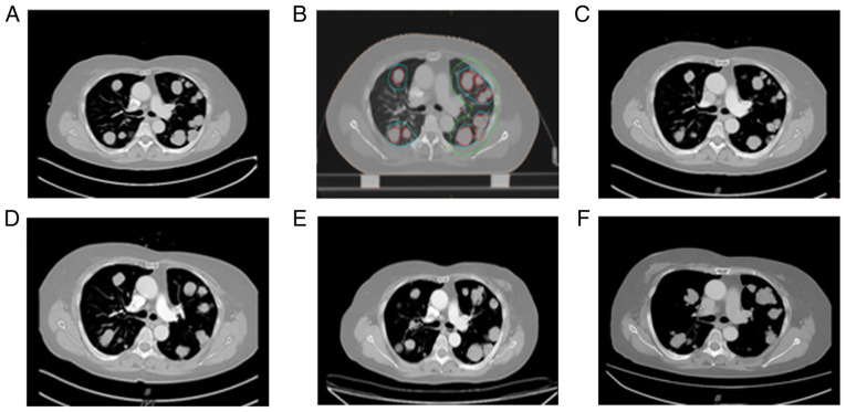
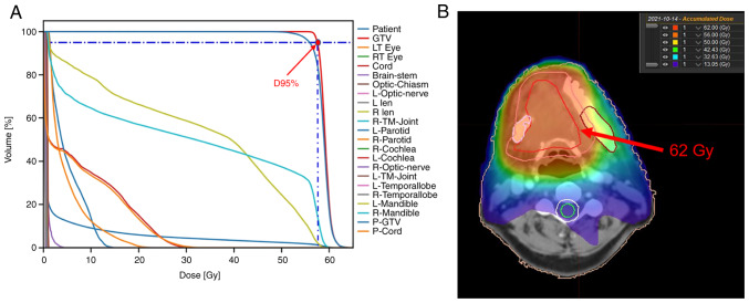
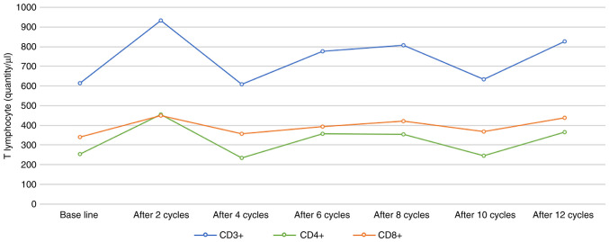
Repeat evaluation in March 2020 indicated a slight increase in lung metastases, cervical lymph nodes and lesions in the floor of the mouth (SD) (Figs. 3 and 4). Intensity-modulated radiation therapy was used for palliative radiotherapy of tongue tumors in March 2020. The patient was subjected to radiation therapy, targeting the gross tumor volume expanding 1 cm outwards at a dose of 60 Gy/2.0 Gy/30Fx (Fig. 5) and oral Tigio 40 mg twice a day was given as concurrent chemotherapy. Chest CT examination in August 2020 indicated that certain lung nodules were larger than those in March 2020 (evaluation: SD). Subsequently, two cycles of sintilimab 200 mg q3w immunotherapy were administered and lung radiotherapy was initiated in September 2020. The prescription dose was as follows: Clinical target volume (CTV1) (right lung metastasis) 2.0 Gy/1Fx; CTV2 (left lung metastasis) 2.0 Gy/1Fx; two cycles of concomitant sintilimab 200 mg q3w immunotherapy. This led to a significant alleviation of shortness of breath. After 24 cycles of sintilimab 200 mg q3w immunotherapy from November 2020 to February 2022, the lung lesions were reduced in size and the tongue lesions were significantly smaller than previously (evaluation: SD). At present (February 2022), the patient is in a stable condition and the general condition is satisfactory. The main symptom is shortness of breath induced by exercise. Laboratory examination indicated a slight decrease in thyroid function. The counts of T-cell subsets were measured in the peripheral blood of the patient after chemotherapy and the temporal trend of its change is presented in Fig. 6. The patient continued treatment with immunotherapy.
Figure 3.
Recurrence of tumor on the tongue, which significantly shrank after radiotherapy. (A) Prior to radiotherapy (March 2020). (B) Target area of tongue radiotherapy (March 2020). Red line: Gross tumor volume of tongue; blue line: OAR of mandible; green line: OAR of cord; pink line: Cord expanding 0.5 cm outwards. (C) nine months after radiotherapy (February 2021); (D) 12 months after radiotherapy (May 2021). (E) 17 months after radiotherapy (September 2021). (F) 22 months after radiotherapy (February 2022). OAR, organ at risk.
Figure 4.
CT (axial view) indicated bilateral lung metastasis and tumor size reduction after low-dose radiotherapy. (A) Prior to radiotherapy (August 2020). (B) Target area of low-dose radiotherapy of the lung (September 2020), Red line, GTV of lung; blue line, CTV (GTV expanding 0.5 cm outwards); green line, planning target volume (CTV expanding 1.5 cm outwards). (C) two months after radiotherapy (December 2020). (D) four months after radiotherapy (February 2021). (E) seven months after radiotherapy (May 2021). (F) 16 months after radiotherapy (February 2022). GTV, gross tumor volume; CTV, clinical target volume.
Figure 5.
Radiotherapy target map of recurrent lesions in the tongue. (A) Dose volume histogram graph: Maximum dose administered to P-GTV, 2.88 Gy; dose administered to 95% of target volume, 55.86 Gy. (B) Isodose line. L/LT, left; R/RT, right; TM-Joint, temporomandibular joint; P-Cord, cord expanding 0.5 cm outwards; P-GTV, gross tumor volume expanding 1 cm outwards.
Figure 6.
Changes in the counts of various subsets of T cells during the course of treatment with low-dose radiotherapy combined with anti-programmed cell death-1.
Discussion
ACC typically occurs in salivary gland tissues such as minor salivary gland, sublingual gland, parotid gland and submandibular gland (1). The typical characteristics of ACC are slow growth, no local lymph node metastasis and a tendency for recurrence and metastasis after several years (6). The ACC lesion is usually painless and the clinical course is typically insidious. Therefore, the condition is frequently ignored by patients in the early stage of the disease and most cases are typically diagnosed at an advanced stage. With the progression of the disease, it may metastasize to sites including the lung, bone, liver, brain and kidney; of these, lung metastasis accounts for ~70% of metastatic cases (7). Distant metastasis usually occurs within 5 years after treatment. Failure to prevent and control distant metastasis is one of the key determinants of the low long-term survival rate of patients with ACC (6,8). Tumor size, peripheral nerve infiltration and local recurrence are risk factors for lung metastasis (9). In a study of 125 patients with ACC by Jeong et al (10), the 10-year OS rates of patients without distant metastasis and those with distant metastasis were 81.5 and 60.2%, respectively. Due to the biological characteristics of ACC, surgery is the first-choice treatment for localized ACC. However, there is no effective treatment for patients with ACC with lung metastasis. Girelli et al (8) studied 109 patients with lung metastasis of ACC and observed that surgical treatment of lung metastasis was effective in patients who had indications for surgery; the 5- and 10-year survival rate of these patients was 66.8 and 40.5%, respectively. Systemic therapy is the main treatment for patients with multiple metastases who are not able to receive surgery or palliative radiotherapy (11).
Local radiotherapy may also have a certain effect on the unirradiated distant metastatic lesions, a phenomenon referred to as the remote effect (12). Radiotherapy directly destroys tumor DNA and endoplasmic reticulum. Tumor cells release a large quantity of tumor-associated antigens after injury or death. These antigens are presented to T lymphocytes, which stimulates their proliferation, inducing a specific immune response (13). Furthermore, radiotherapy may promote the expression of tumor antigens by upregulating the expression of major histocompatibility complex-I molecules, which improves the ability of antigen therapy to recognize tumor cells (12). In addition, radiotherapy may also induce the infiltration of T cells and neutrophils in the tumor and activate the local immune inflammatory response, resulting in inhibition of unirradiated tumor cells (14). The combination of radiotherapy and immunotherapy helps activate the immune response and transforms the tumor microenvironment from immunosuppressive to immunoreactive. Several studies have indicated that the combination of radiotherapy and immunotherapy improves the remote effect. The immune response may vary depending on the radiation doses and radiotherapy regimens. In the study by Yin et al (15), a 2Gy*1f radiotherapy regimen was indicated to induce higher infiltration of CD4+ and CD8+ T cells into tumor tissue than 2Gy*3f and 2Gy*5f regimens. Shevtsov et al (16) obtained similar results. They observed that, although LDRT (2Gy*1f) had a poor killing effect on tumor cells, it may normalize tumor blood vessels, thus promoting T-cell infiltration into the tumor and improving the curative effect. LDRT (<1Gy) may induce differentiation of macrophages into the M2 anti-inflammatory phenotype, while high-dose radiotherapy (1–10 Gy) may induce their differentiation into M1 pro-inflammatory phenotype (17). Yin et al (15) explored triple therapy of low-dose radiotherapy (LDRT) and hypofractionated radiotherapy (HFRT) combined with immunotherapy for non-small cell lung cancer. It was indicated that the LDRT of distant metastases significantly enhances the distant effect of HFRT combined with immunotherapy and that the triple regimen is well tolerated by the patients (15). The present study also suggested that LDRT upregulated the expression of genes involved in antigen presentation and genes that promote tumor T-cell invasion.
In the present case, the previous multi-line therapy programs, including surgery, chemotherapy, targeted therapy and radiotherapy for recurrent lesions, failed, and there was an increase in the number of metastatic lesions in both lungs. At six years after the treatment, the patient developed extensive metastasis in both lungs. According to the International Lung Metastasis staging system, the patient was categorized as stage IV (unable to be resected completely) with no surgical indication. Considering the size and the number of pulmonary metastases and the general condition of the patient, LDRT combined with immunotherapy was administered, which controlled pulmonary metastases and local recurrence. Studies have indicated that changes in lymphocyte subsets may be used to assess the efficacy of immunotherapy and the prognosis of patients with cancer (18). Immune markers such as natural killer cells, dendritic cells and T-regulatory cells are not routinely assessed but would be useful for monitoring the effectiveness of the treatment. In a bilateral mouse colon tumor model study (15), the number of CD8+ T cells was significantly increased after low-dose radiotherapy plus anti-programmed cell death-1 (PD-1) therapy. After 1 cycle of anti-PD-1 treatment, the number of peripheral blood T lymphocytes in the patient of the present study exhibited a marked increase (CD3+, 933/µl; CD4+, 455/µl; CD8+, 449/µl; and CD4+/CD8+ ratio, 1.01) as compared with the baseline (CD3+, 613/µl; CD4+, 254/µl; CD8+, 339/µl; CD4+/CD8+, ratio 0.75), indicating the effectiveness of the treatment. Subsequently, fluctuations in the number of immune cells were observed over time, but the levels were largely above the baseline level. To date, no consensus has been reached regarding the optimal sequence of radiotherapy and immunotherapy. Certain researchers suggested that anti-PD-1 therapy has the best effect when administered within one week after radiotherapy, but it may also be related to the selection and efficacy of immunotherapy drugs. This patient of the present study was treated with immunotherapy combined with sequential radiotherapy. Repeat examination after 4 months of radiotherapy indicated a decrease in the size of lung and tongue lesions, which may reflect the remote effect, indicating that low-dose radiotherapy may enhance T-lymphocyte infiltration in distant tumors.
In conclusion, for the present case of ACC with bilateral lung metastasis, satisfactory results were achieved with low-dose lung radiotherapy combined with immunotherapy. At present, there is no standardized treatment for bilateral lung metastasis of ACC and this approach may be used as a feasible treatment model. However, further studies are required to determine the optimal radiotherapy dose and the optimal sequence of radiotherapy and immunotherapy.
Acknowledgements
Not applicable.
Glossary
Abbreviations
- ACC
adenoid cystic carcinoma
- CT
computed tomography
- MRI
magnetic resonance imaging
- LDRT
low-dose radiotherapy
- HFRT
hypofractionated radiotherapy
- SD
stable disease
- GTV
gross tumor volume
- CTV
clinical target volume
- PD-1
programmed cell death-1
Funding Statement
Funding: No funding was received.
Availability of data and materials
The datasets used and/or analysed during the current study are available from the corresponding author on reasonable request.
Authors' contributions
DYL was responsible for the conception, design, content and writing of the manuscript. XZP made substantial contributions to acquisition, analysis and interpretation of data and revising the manuscript critically for important intellectual content. XXZ and QYS reviewed the pathology findings and made substantial contributions to conception, design, drafting and revising the manuscript. GW was responsible for the treatment and management of the patient and was responsible for acquisition, analysis and interpretation of the images. DYM made substantial contributions to conception and design, revising and proofreading the manuscript and gave final approval of the version to be published. DYL and XXZ confirm the authenticity of all the raw data. All authors were involved in writing the manuscript. All authors read and approved the final manuscript.
Ethics approval and consent to participate
This study was reviewed and approved by the Ethics Committee of Affiliated Hospital of North Sichuan Medical College (Nanchong, China).
Patient consent for publication
Written informed consent was obtained from the subject for the publication of any potentially identifiable images or data included in this article.
Competing interests
The authors declare that they have no competing interests.
References
- 1.Dodd RL, Slevin NJ. Salivary gland adenoid cystic carcinoma: A review of chemotherapy and molecular therapies. Oral Oncol. 2006;42:759–769. doi: 10.1016/j.oraloncology.2006.01.001. [DOI] [PubMed] [Google Scholar]
- 2.Bradley PJ. Adenoid cystic carcinoma evaluation and management: Progress with optimism! Curr Opin Otolaryngol Head Neck Surg. 2017;25:147–153. doi: 10.1097/MOO.0000000000000347. [DOI] [PubMed] [Google Scholar]
- 3.Patel RR, He K, Barsoumian HB, Chang JY, Tang C, Verma V, Comeaux N, Chun SG, Gandhi S, Truong MT, et al. High-dose irradiation in combination with non-ablative low-dose radiation to treat metastatic disease after progression on immunotherapy: Results of a phase II trial. Radiother Oncol. 2021;162:60–67. doi: 10.1016/j.radonc.2021.06.037. [DOI] [PMC free article] [PubMed] [Google Scholar]
- 4.Menon H, Chen D, Ramapriyan R, Verma V, Barsoumian HB, Cushman TR, Younes AI, Cortez MA, Erasmus JJ, de Groot P, et al. Influence of low-dose radiation on abscopal responses in patients receiving high-dose radiation and immunotherapy. J Immunother Cancer. 2019;7:237. doi: 10.1186/s40425-019-0718-6. [DOI] [PMC free article] [PubMed] [Google Scholar]
- 5.Eisenhauer EA, Therasse P, Bogaerts J, Schwartz LH, Sargent D, Ford R, Dancey J, Arbuck S, Gwyther S, Mooney M, et al. New response evaluation criteria in solid tumours: Revised RECIST guideline (version 1.1) Eur J Cancer. 2009;45:228–2247. doi: 10.1016/j.ejca.2008.10.026. [DOI] [PubMed] [Google Scholar]
- 6.van Weert S, Reinhard R, Bloemena E, Buter J, Witte BI, Vergeer MR, Leemans CR. Differences in patterns of survival in metastatic adenoid cystic carcinoma of the head and neck. Head Neck. 2017;39:456–463. doi: 10.1002/hed.24613. [DOI] [PubMed] [Google Scholar]
- 7.Seok J, Lee DY, Kim WS, Jeong WJ, Chung EJ, Jung YH, Kwon SK, Kwon TK, Sung MW, Ahn SH. Lung metastasis in adenoid cystic carcinoma of the head and neck. Head Neck. 2019;41:3976–3983. doi: 10.1002/hed.25942. [DOI] [PubMed] [Google Scholar]
- 8.Girelli L, Locati L, Galeone C, Scanagatta P, Duranti L, Licitra L, Pastorino U. Lung metastasectomy in adenoid cystic cancer: Is it worth it? Oral Oncol. 2017;65:114–118. doi: 10.1016/j.oraloncology.2016.10.018. [DOI] [PubMed] [Google Scholar]
- 9.Sharma VJ, Gupta A, Yaftian N, Ball D, Brown R, Barnett S, Antippa P. Low recurrence of lung adenoid cystic carcinoma with radiotherapy and resection. ANZ J Surg. 2019;89:1051–1055. doi: 10.1111/ans.15222. [DOI] [PubMed] [Google Scholar]
- 10.Jeong IS, Roh JL, Cho KJ, Choi SH, Nam SY, Kim SY. Risk factors for survival and distant metastasis in 125 patients with head and neck adenoid cystic carcinoma undergoing primary surgery. J Cancer Res Clin Oncol. 2020;146:1343–1350. doi: 10.1007/s00432-020-03170-5. [DOI] [PMC free article] [PubMed] [Google Scholar]
- 11.Coca-Pelaz A, Rodrigo JP, Bradley PJ, Vander Poorten V, Triantafyllou A, Hunt JL, Strojan P, Rinaldo A, Haigentz M, Jr, Takes RP, et al. Adenoid cystic carcinoma of the head and neck-An update. Oral Oncol. 2015;51:652–661. doi: 10.1016/j.oraloncology.2015.04.005. [DOI] [PubMed] [Google Scholar]
- 12.Locy H, de Mey S, de Mey W, De Ridder M, Thielemans K, Maenhout SK. Immunomodulation of the tumor microenvironment: Turn foe into friend. Front Immunol. 2018;9:2909. doi: 10.3389/fimmu.2018.02909. [DOI] [PMC free article] [PubMed] [Google Scholar]
- 13.Bhalla N, Brooker R, Brada M. Combining immunotherapy and radiotherapy in lung cancer. J Thorac Dis. 2018;10((Suppl 13)):S1447–S1460. doi: 10.21037/jtd.2018.05.107. [DOI] [PMC free article] [PubMed] [Google Scholar]
- 14.Theelen WS, de Jong MC, Baas P. Synergizing systemic responses by combining immunotherapy with radiotherapy in metastatic non-small cell lung cancer: The potential of the abscopal effect. Lung Cancer. 2020;142:106–113. doi: 10.1016/j.lungcan.2020.02.015. [DOI] [PubMed] [Google Scholar]
- 15.Yin L, Xue J, Li R, Zhou L, Deng L, Chen L, Zhang Y, Li Y, Zhang X, Xiu W, et al. Effect of low-dose radiation therapy on abscopal responses to hypofractionated radiation therapy and Anti-PD1 in mice and patients with non-small cell lung cancer. Int J Radiat Oncol Biol Phys. 2020;108:212–224. doi: 10.1016/j.ijrobp.2020.07.1741. [DOI] [PubMed] [Google Scholar]
- 16.Shevtsov M, Sato H, Multhoff G, Shibata A. Novel approaches to improve the efficacy of immuno-radiotherapy. Front Oncol. 2019;9:156. doi: 10.3389/fonc.2019.00156. [DOI] [PMC free article] [PubMed] [Google Scholar]
- 17.Dhawan G, Kapoor R, Dhawan R, Singh R, Monga B, Giordano J, Calabrese EJ. Low dose radiation therapy as a potential life saving treatment for COVID-19-induced acute respiratory distress syndrome (ARDS) Radiother Oncol. 2020;147:212–216. doi: 10.1016/j.radonc.2020.05.002. [DOI] [PMC free article] [PubMed] [Google Scholar]
- 18.Schalper KA, Brown J, Carvajal-Hausdorf D, McLaughlin J, Velcheti V, Syrigos KN, Herbst RS, Rimm DL. Objective measurement and clinical significance of TILs in non-small cell lung cancer. J Natl Cancer Inst. 2015;107:dju435. doi: 10.1093/jnci/dju435. [DOI] [PMC free article] [PubMed] [Google Scholar]
Associated Data
This section collects any data citations, data availability statements, or supplementary materials included in this article.
Data Availability Statement
The datasets used and/or analysed during the current study are available from the corresponding author on reasonable request.


