Figure 4.
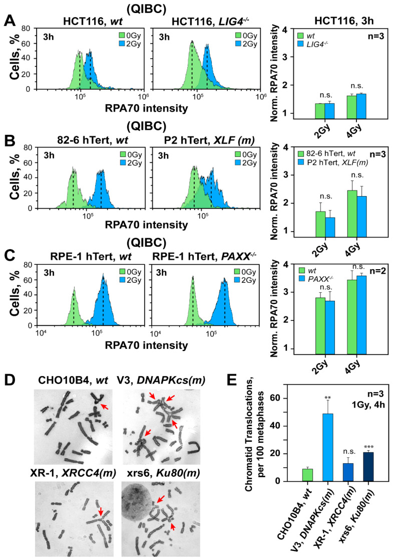
Deficiency in c-NHEJ factors other than DNA-PKcs leaves unchanged resection in G2-phase, but DNA-PKcs deficiency dramatically increases translocations. (A) QIBC analysis of RPA70 in parental and LIG4−/− HCT116 human cells in G2-phase. Bar plots on the right represent the normalized RPA70 signal intensity from three experiments. (B) As in panel (A) for 82-6 hTert and the P2 hTert fibroblast cell line with a defect in XLF. (C) As in panel (A), for RPE-1 hTert cells and a derivative PAXX−/− cell line. (D) Representative metaphase spreads of CHO10B4, V3, XR-1 and xrs6 cells depicting chromatid translocations at 4 h post 1 Gy IR. Translocations are marked with arrows (E) Frequency of IR induced translocations in wt CHO10B4 and c-NHEJ mutants (V3, XR-1 and xrs6 cell lines) at 4 h post 1 Gy IR. The mean ± SD shown represent data from three independent experiments. ** indicates p < 0.01 and *** indicates p < 0.001, while n.s. indicates non-significance.

