Abstract
Three new flavonoid derivatives, melodorones A–C (1–3), together with four known compounds, tectochrysin (4), chrysin (5), onysilin (6), and pinocembrin (7), were isolated from the stem bark of Melodorum fruticosum. Their structures were determined on the basis of extensive spectroscopic methods, including NMR and HRESIMS, and by comparison with the literature. Compounds 1–7 were evaluated for their in vitro α-glucosidase inhibition and cytotoxicity against KB, Hep G2, and MCF7 cell lines. Among them, compound 1 exhibited the best activity against α-glucosidase and was superior to the positive control with an IC50 value of 2.59 μM. On the other hand, compound 1 showed moderate cytotoxicity toward KB, Hep G2, and MCF7 cell lines with the IC50 values of 23.5, 19.8, and 23.7 μM, respectively. These findings provided new evidence that the stem bark of M. fruticosum is a source of bioactive flavonoid derivatives that are highly valuable for medicinal development.
Keywords: Annonaceae, Melodorum fruticosum, melodorones A–C, flavonoids, α-glucosidase inhibition, cytotoxicity
1. Introduction
Melodorumfruticosum Lour (Annonaceae) is a shrub with fragrant yellow flowers distributed in South East Asia, more specifically indigenous to Laos, Cambodia, Thailand, and Vietnam. This plant has been used as a mild cardiac stimulant, a tonic, and as a hematinic to resolve dizziness [1]. Previous phytochemical studies on this plant have led to the isolation of terpenoids, aromatic compounds, butenolides, heptenoids, aporphine alkaloids, and flavonoids [2,3,4,5,6,7], some of which showed significant antioxidant, anti-inflammatory, cytotoxic, antiphytopathogenic, and antifungal activities [8,9,10,11]. However, there are no reports concerning α-glucosidase inhibition from M. fruticosum.
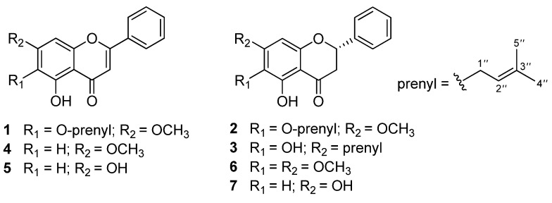
Flavonoids are a class of polyphenol secondary metabolites that are present in plants [12]. Some flavonoids, such as luteolin, kaempferol, baicalein, and apigenin, have hypoglycemic potential and have been reported as α-glucosidase inhibitors [13]. As an extension of a search for new flavonoids as α-glucosidase inhibitors from the M. fruticosum, three new flavonoid derivatives, melodorones A–C (1–3), together with four known compounds, tectochrysin (4) [14], chrysin (5) [15], onysilin (6) [16], and pinocembrin (7) [17] (Figure 1), were isolated from the dried stem bark of M. fruticosum. We describe here the isolation and structural elucidation of these compounds, together with the evaluations of their in vitro α-glucosidase inhibition and their cytotoxicity against different cancer cell lines (KB, Hep G2, and MCF7).
Figure 1.
Chemical structures of 1–7.
2. Results and Discussion
2.1. Structural Elucidation of the Isolates
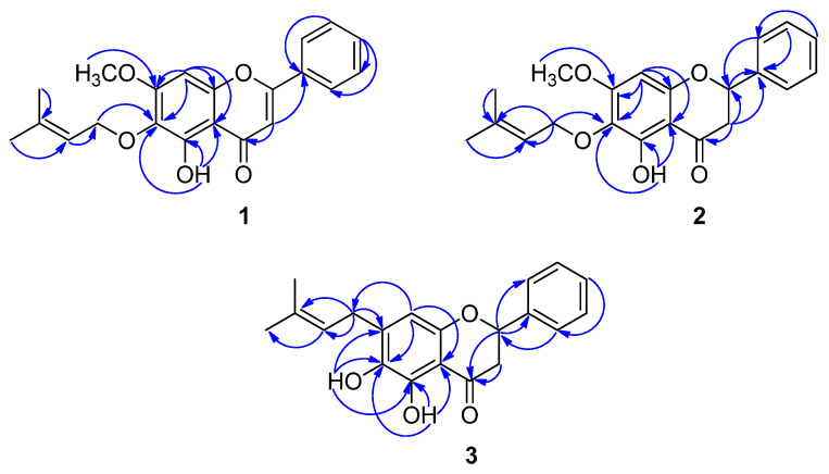
Compound 1 was isolated as a colorless gum. The molecular formula C21H20O5 was determined by its HRESIMS, which displayed a molecular ion peak at m/z 351.1178 [M-H]− (calcd. for C21H19O5 351.1232), together with NMR data (Table 1, Supplementary Materials). The characteristic resonances of a flavone core structure were evident from the 1H NMR data at δH 7.03 (s, H-3), and 13C NMR data at δC 163.4 (C-2), 104.9 (C-3), and 182.4 (C-4) [18]. The NMR data revealed the presence of one hydrogen-bonded hydroxy (δH 12.77), one methoxy (δH 3.94), and one prenyloxy (one oxymethylene protons at δH 4.46 (2H, d, J = 7.5 Hz), one olefinic proton at δH 5.45 (1H, m), two methyl protons at δH 1.71 (3H, s) and 1.63 (3H, s)) substituents. A singlet proton at δH 6.97 was assigned to aromatic proton H-8 of the ring A. The remaining proton resonances were typical of an unsubstituted ring B flavone at δH 8.11 (H-2′/6′) and 7.58–7.63 (H-3′/4′/5′). The HMBC correlation of H-1″ (δH 4.46) with C-6 (δC 130.6) allowed the placement of the prenyloxy moiety at C-6 (Figure 2). The methoxy group at δH 3.94 (3H, s) located at C-7 was proven by the HMBC correlation of the methoxy substituent protons with C-7 (δC 159.3). In turn, the HMBC correlations of 5-OH (δH 12.77) with C-5 (δC 152.8), C-6 (δC 130.6), and C-10 (δC 105.3) supported the position of the hydroxyl group at C-5. A careful comparison of the 1H and 13C NMR spectral data of 1 with baicalein [19] identified similar signals, distinguished by two hydroxyl groups at C-6 and C-7 (ring A) were replaced by the prenyloxy and methoxy substituents, respectively, in 1. This deduction was strongly confirmed by the HSQC and HMBC correlations (Figure 2). From the aforementioned results, structure 1 was assigned as melodorone A.
Table 1.
1H and 13C NMR spectroscopic data of 1–3 recorded in DMSO-d6 (δ in ppm).
| Position | 1 a | 2 b | 3 a | |||
|---|---|---|---|---|---|---|
| δH (J in Hz) | δC | δH (J in Hz) | δC | δH (J in Hz) | δC | |
| 2 | 163.4 | 5.62, dd (12.6, 3.0) | 78.7 | 5.55, dd (13.0, 3.0) | 78.4 | |
| 3ax | 7.03, s | 104.9 | 3.31, dd (17.4, 13.2) | 42.2 | 3.31, overlapped | 42.8 |
| 3eq | 2.82, dd (16.8, 3.0) | 2.83, dd (17.0, 3.0) | ||||
| 4 | 182.4 | 197.1 | 198.7 | |||
| 5 | 152.8 | 154.4 | 148.3 | |||
| 6 | 130.6 | 128.3 | 136.1 | |||
| 7 | 159.3 | 161.1 | 139.5 | |||
| 8 | 6.97, s | 91.6 | 6.30, s | 92.0 | 6.27, s | 106.4 |
| 9 | 152.3 | 158.3 | 153.0 | |||
| 10 | 105.3 | 102.5 | 106.2 | |||
| 1′ | 130.6 | 138.5 | 138.8 | |||
| 2′ | 8.11, dd (7.0, 1.5) | 126.4 | 7.54, d (7.2) | 126.6 | 7.53, dd (8.5, 1.5) | 126.5 |
| 3′ | 7.58–7.63, m | 129.1 | 7.44, dd (7.2, 7.2) | 128.5 | 7.37–7.44, m | 128.5 |
| 4′ | 132.1 | 7.40, m | 128.6 | 128.4 | ||
| 5′ | 129.1 | 7.44, dd (7.2, 7.2) | 128.5 | 128.5 | ||
| 6′ | 8.11, dd (7.0, 1.5) | 126.4 | 7.54, d (7.2) | 126.6 | 7.53, dd (8.5, 1.5) | 126.5 |
| 1″ | 4.46, d (7.5) | 68.3 | 4.35, d (7.2) | 68.4 | 3.26, overlapped | 28.7 |
| 2″ | 5.45, m | 120.5 | 5.42, m | 120.6 | 5.24, m | 121.1 |
| 3″ | 137.4 | 137.1 | 132.7 | |||
| 4″ | 1.71, s | 25.4 | 1.70, s | 25.4 | 1.69, s | 25.4 |
| 5″ | 1.63, s | 17.7 | 1.61, s | 17.7 | 1.65, s | 17.6 |
| 5-OH | 12.77, s | 11.94, s | 11.71, s | |||
| 6-OH | 8.51, s | |||||
| 7-OCH3 | 3.94, s | 56.5 | 3.83, s | 56.3 | ||
a NMR at 500/125 MHz. b NMR at 600/125 MHz.
Figure 2.
Key HMBC correlations of 1–3.
Compound 2 was obtained as a colorless gum with [α]25D −73.0 (c 0.01, CHCl3). Its molecular formula, C21H22O5, was established by its HREIMS, which showed a molecular ion peak at m/z 399.1448 [M+HCOO]− (calcd. for C22H23O7 399.1444). This was further supported by the 1H and 13C NMR spectral data, which displayed one oxymethine, one methoxy, one olefinic methine, two methylene, two methyl, six aromatic methine, and eight quaternary carbons (Table 1). The spectroscopic (1H and 13C NMR) patterns of 2 were very similar to those of 1 except for the presence of a single bond between C-2 and C-3 in the heterocyclic ring C. This flavanone moiety was proven by the 1H NMR spectrum, which showed an ABX spin system at δH 5.62 (1H, dd, J = 12.6, 3.0 Hz), 3.31 (1H, dd, J = 17.4, 13.2 Hz), and 2.82 (1H, dd, J = 16.8, 3.0 Hz), corresponding to H-2, H-3ax, and H-3eq, respectively [20]. The 13C NMR spectrum also confirmed the presence of a flavanone core structure because of the signals resonating at δC 42.2 (C-3), 78.7 (C-2), and 197.1 (C-4) of the flavanone ring C. The absolute configuration of C-2 was assigned as S-configuration through contrastive analysis of the optical rotation data of 2 ([α]25D −73.0) with the known compound (−)-butin ([α]26D −39.9) [21]. Based on the above spectral evidence, compound 2 was identified as a new flavanone and was named melodorone B.
Compound 3 was obtained as a colorless gum with [α]25D −26.8 (c 0.01, CHCl3). The molecular formula C20H20O4 was determined by its HREIMS, which displayed a molecular ion peak at m/z 369.1363 [M+HCOO]− (calcd. for C21H21O6 369.1338). The 13C NMR spectrum (Table 1) showed three carbon signals at δC 198.7 (C-4), 78.4 (C-2), and 42.8 (C-3), which are frequently observed in the flavanone series [22]. The 1H NMR spectral data also justified the assignment of a flavanone moiety of 3 (Table 1). The presence of two hydroxyl substituents at C-5 (δC 148.3) and C-6 (δC 136.1) was identified by the HMBC correlations of 5-OH with C-5, C-6, and C-10 (δC 106.2), and of 6-OH with C-5, C-6, and C-7 (δC 139.5) (Figure S1). The 1H and 13C NMR data of 3 were nearly identical to those of (2S)-dihydrobaicalein [22], differing only in the resonance of the hydroxyl group was replaced by the resonance of a prenyl moiety at C-7 of ring A. This prenyl group was substituted at C-7, which was proven by the HMBC correlation of H-1″ (δH 3.26) with C-7. On the basis of these data, the structure of 3 was unambiguously established and named melodorone C.
2.2. α-Glucosidase Inhibitory Activity
Compounds 1–7 were assessed for their α-glucosidase inhibition, with acarbose as a positive control. The resulting IC50 values (Table 2) showed that all isolates, except 4, 6, and 7, displayed stronger inhibitory effects on α-glucosidase than acarbose (IC50 179 μM). Especially, compounds 1–3 and 5 exhibited α-glucosidase inhibition with IC50 values in the range of 2.59 to 4.00 μM, which were more strongly than acarbose. Among isolates, compound 1 revealed the most highly potent α-glucosidase inhibition with an IC50 value of 2.59 μM. Previous research reported that 5 inhibited α-glucosidase effectively with an IC50 value of 5.7 μM, while 4 and 7 showed no α-glucosidase inhibition [23], which was consistent with the findings of this study. The results indicated that the compounds from the stem bark of M. fruticosum, at least for 1–3 and 5, were active α-glucosidase inhibitors that could be used for the treatment of diabetes mellitus.
Table 2.
α-Glucosidase inhibition (IC50 ± SD) of 1–7.
| Compound | IC50 (μM) |
|---|---|
| 1 | 2.59 ± 0.15 |
| 2 | 3.33 ± 0.28 |
| 3 | 4.00 ± 0.20 |
| 4 | >256 |
| 5 | 3.67 ± 0.25 |
| 6 | 192 ± 8.78 |
| 7 | >256 |
| Acarbose | 179 ± 6.02 |
2.3. Cytotoxic Activity against Human Cancer Cell Lines
All isolates were further assessed for their in vitro cytotoxicity against three human cancer cell lines (KB, Hep G2, and MCF7), using an MTT assay (Table 3). Compound 1 was found to exhibit moderate cytotoxicity toward all cancer cell lines, with IC50 values of 23.5, 19.8, and 23.7 μM, respectively. Compounds 2, 4, and 5 displayed weak cytotoxicity against all cancer cell lines, with IC50 values in the range of 32.0 to 80.9 μM, while compound 3 exhibited weak action against KB and Hep G2 cell lines, with IC50 values of 59.0 and 80.0 μM, respectively. Other compounds in this bioassay revealed no evident cytotoxicity (IC50 > 100). According to previous research, compound 4 inhibited moderate cytotoxicity against KB and MCF7 cell lines, with IC50 values of 55.27 and 16.7 µM, respectively [24], while 5 showed moderate cytotoxic effects on Hep G2 and MCF7 cell lines, with IC50 values of 50.55 and 37.20 µM, respectively [25,26]. On the other hand, compound 7 exhibited cytotoxicity against the MCF7 cell line with IC50 values of 226 and 108 µM for 48 and 72 h, respectively [27]. These results were similar and supported the findings of this study. Compounds 4 and 5 were previously reported to exhibit no cytotoxic effect on the normal human colon fibroblastic CCD-18co and normal human epidermal keratinocytes (NHEKs) cell lines [28,29]. Additionally, compounds 5 and 7 were also discovered to have no cytotoxicity against the normal Vero cell line [30]. However, the cytotoxicity of 1–3 and 6 against normal human cell lines is not available, but it is recommended to be determined in the future.
Table 3.
Cytotoxicity of 1–7 against three human cancer cell lines.
| Compound | IC50 ± SD (µM) a | ||
|---|---|---|---|
| KB | Hep G2 | MCF7 | |
| 1 | 23.5 ± 1.1 | 19.8 ± 1.5 | 23.7 ± 2.0 |
| 2 | 62.1 ± 4.5 | 44.8 ± 4.0 | 73.7 ± 2.8 |
| 3 | 59.0 ± 2.5 | 80.0 ± 3.0 | >100 |
| 4 | 64.7 ± 3.0 | 68.0 ± 2.5 | 80.9 ± 7.4 |
| 5 | 32.0 ± 0.0 | 71.4 ± 3.9 | 80.0 ± 4.5 |
| 6 | >100 | >100 | >100 |
| 7 | >100 | >100 | >100 |
| Ellipticine b | 0.31 ± 0.05 | 0.33 ± 0.05 | 0.40 ± 0.05 |
a Results are expressed as the means ± SD of three replicates. b Ellipticine was used as the positive control. Human epidermoid carcinoma (KB), human hepatocellular carcinoma (Hep G2), and human breast adenocarcinoma (MCF-7).
3. Materials and Methods
3.1. General Experimental Procedures
The NMR spectra were recorded using Bruker AvanceNEO 600 MHz and Bruker Avance III™ HD 500 MHz NMR spectrometers in DMSO-d6 (Merck, Darmstadt, Germany). The HRMS spectra were acquired on a X500R QTOP model mass spectrometer (Sciex, Redwood City, CA, USA) and Dionex Ultimate 3000 HPLC system hyphenated with a QExactive Hybrid Quadrupole Orbitrap MS (Thermo Fisher Scientific, Waltham, MA, USA). Optical rotations were obtained with a A.KRÜSS Optronic P8000 polarimeter (KRÜSS, Hamburg, Germany). The IR data were measured on a Jasco 6600 FT-IR spectrometer using an ATR technique (Jasco, Tokyo, Japan). Silica gel (70–230 mesh, Merck, Darmstadt, Germany) and Sephadex LH-20 gel (GE Healthcare Bio-Sciences AB, Uppsala, Sweden) were used for column chromatography. TLC (silica gel 60 F254, Merck, Darmstadt, Germany) was used to monitor the fractions from column chromatography. Saccharomyces cerevisiae α-glucosidase, p-nitrophenyl-α-d-glucopyranoside (pNPG), acarbose, and dimethyl sulfoxide (DMSO) were purchased from Sigma-Aldrich (St. Louis, MO, USA).
3.2. Plant Material
M. fruticosum was collected in July 2017 from Lam Dong province, Vietnam. The material was authenticated by Dr. Dang Van Son. A voucher specimen (No US—A012) was deposited at the Herbarium of the Department of Organic Chemistry, Faculty of Chemistry, University of Science, National University–Ho Chi Minh City, Vietnam.
3.3. Extraction and Isolation
The dried and powdered stem bark of M. fruticosum (45.0 kg) was extracted with 95% EtOH (90 L × 3) at room temperature and concentrated under reduced pressure to give an EtOH crude extract (1.3 kg). This crude extract was suspended in water and partitioned with n-hexane and then EtOAc to yield n-hexane (45.0 g) and EtOAc (161.0 g) extracts. The n-hexane extract was fractionated by silica gel column chromatography (CC) eluted with n-hexane–EtOAc (90:10–0:10 v/v) and EtOAc–MeOH (10:0–0:10 v/v). The eluted fractions were combined into six fractions (HEX.1–HEX.6) on the basis of their TLC behavior. Fraction HEX.3 (6.5 g) was further separated by silica gel CC eluted with n-hexane–EtOAc (85:15 v/v) to give five subfractions (HEX.3.1–HEX.3.5). Subfraction HEX.3.2 (2.0 g) was purified by silica gel CC eluted with n-hexane–EtOAc (85:15 v/v) to give five subfractions (HEX.3.2.1–HEX.3.2.5). Subfraction HEX.3.2.2 was purified by silica gel CC eluted with n-hexane–EtOAc (85:15 v/v) to afford 3 (5.0 mg). Subfraction HEX.3.2.2 was applied to a Sephadex LH-20 gel CC (50.0 g) with CHCl3–MeOH (1:4 v/v) to obtain 6 (7.0 mg). Subfraction HEX.3.3 (0.8 g) was further purified by silica gel CC eluted with n-hexane–EtOAc (85:15 v/v), yielding four subfractions (HEX.3.3.1–HEX.3.3.4). Subfraction HEX.3.3.1 was further purified by silica gel CC eluted with n-hexane–EtOAc (85:15 v/v), followed by CC on Sephadex LH-20 gel eluted with CHCl3–MeOH (1:4 v/v) to afford 1 (6.8 mg) and 2 (5.4 mg). Subfraction HEX.3.3.4 was applied to a Sephadex LH-20 gel CC (50.0 g) with 100% MeOH to obtain 4 (10.7 mg). Subfraction HEX.3.4 (1.4 g) was separated by silica gel CC eluted with n-hexane–EtOAc (8:2 v/v), followed by CC on Sephadex LH-20 gel eluted with CHCl3–MeOH (1:4 v/v) to yield 5 (10.8 mg) and 7 (7.7 mg).
Melodorone A (1). Colorless gum. UV (CH3OH) λmax (log ε) 275 (4.38), 295 (4.07), 356 (3.86) nm; IR (ATR) νmax 3393, 2977, 1634, 1573, 1445, 1204, 1109 cm−1. HRESIMS m/z 351.1178 [M-H]− (calcd. for C21H19O5 351.1232); 1H NMR (DMSO-d6, 500 MHz) and 13C NMR (DMSO-d6, 125 MHz) see Table 1.
Melodorone B (2). Colorless gum. [α]25D −73.0 (c 0.01, CHCl3); UV (CH3OH) λmax (log ε) 275 (4.38), 295 (4.07), 356 (3.86) nm; IR (ATR) νmax 3393, 2977, 1635, 1574, 1497, 1162, 1109 cm−1. HRESIMS m/z 399.1448 [M+HCOO]− (calcd. for C22H23O7 399.1444); 1H NMR (DMSO-d6, 600 MHz) and 13C NMR (DMSO-d6, 150 MHz) see Table 1.
Melodorone C (3). Colorless gum. [α]25D −26.8 (c 0.01, CHCl3); UV (CH3OH) λmax (log ε) 275 (4.38), 295 (4.07), 356 (3.86) nm; IR (ATR) νmax 3392, 2977, 1634, 1450, 1217, 1122 cm−1. HRESIMS m/z 369.1363 [M+HCOO]− (calcd. for C21H21O6 369.1338); 1H NMR (DMSO-d6, 500 MHz) and 13C NMR (DMSO-d6, 125 MHz) see Table 1.
3.4. α-Glucosidase Inhibitory Assay
The α-glucosidase inhibition of all isolates was carried out according to a method adapted from a previous report [31]. Serial concentrations (2.0–256.0 μg/mL) of 1–7 and acarbose were prepared by dissolving in DMSO (400 mg/mL). Sodium phosphate buffer (100 mM, pH 6.8) was used to dissolve the α-glucosidase (0.4 U/mL) and substrate (2.5 mM pNPG). The substrate (40 μL) was added to the reaction mixture after the inhibitor (50 μL) was preincubated with α-glucosidase in 96-well plates at 37 °C for 10 min. A mixture without enzyme, sample compound, and acarbose served as blank, while the control contained only DMSO, enzyme, and substrate. The enzymatic reaction was carried out at 37 °C for 30 min and stopped by adding 0.2 M Na2CO3 (130 μL). Absorbance at 410 nm to measure enzyme activity was recorded on a BIOTEK reader. The assays were conducted in triplicates, with acarbose serving as a positive control. The IC50 values were calculated graphically using inhibition curves.
3.5. Cytotoxicity Assay
The cytotoxic evaluation of 1–7 and ellipticine against the growth of human epidermoid carcinoma (KB), human hepatocellular carcinoma (Hep G2), and human breast adenocarcinoma (MCF-7) cell lines was performed according to a previous procedure [32]. Ellipticine, a potent anticancer agent exhibiting multiple mechanisms of action, was used as the positive control [33,34,35]. The cancer cells were cultured in Dulbecco’s Modified Eagle’s Medium with 10% Fetal Bovine Serum, 1% penicillin and streptomycin, and 1% L-glutamine at 37 °C in a 5% CO2 environment. The tested compounds were added at concentrations ranging from 0.5 to 128 μg/mL by dissolving in DMSO (20 mg/mL) and incubated for a further 72 h in the identical condition. After the treatment, each well was filled with an MTT solution (10 μL, 5 mg/mL) of phosphate buffer, which was left to stand for 4 h until intracellular purple formazan crystals appeared. The MTT was removed and a 100 μL DMSO solution was added. The blank contained only a medium without any cells and MTT, as well as the solubilizing solution. The solution’s optical density (OD) at 540 nm was recorded on a BIOTEK reader. All experiments were carried out in triplicate for three independent experiments. IC50 values were computed graphically using inhibition curves.
4. Conclusions
The chemical investigation of the stem bark of M. fruticosum afforded the isolation of three unprecedented flavonoid derivatives (1–3), including one new flavone (1) and two new flavanones (2 and 3), along with four known compounds (4–7). All isolated were obtained from M. fruticosum for the first time. Compounds 1–3 and 5 exhibited highly potent inhibition against α-glucosidase and were superior to the positive agent. Furthermore, compounds 1–5 selectively showed in vitro cytotoxicity against KB, Hep G2, and MCF7 cell lines. The results of this study reveal that M. fruticosum stem bark is a highly useful source of bioactive flavonoid derivatives that should be explored further in the medical field.
Supplementary Materials
The following are available online at https://www.mdpi.com/article/10.3390/molecules27134023/s1, Figures S1–S17: HRESIMS, 1D, and 2D NMR spectra of 1–3.
Author Contributions
Conceptualization, J.S. and L.T.M.D.; methodology, L.T.M.D.; formal analysis, J.S.; data curation, J.S. and L.T.M.D.; writing—original draft preparation, J.S. and L.T.M.D.; funding acquisition, L.T.M.D. All authors have read and agreed to the published version of the manuscript.
Institutional Review Board Statement
Not applicable.
Informed Consent Statement
Not applicable.
Data Availability Statement
All data supporting this study are available in the manuscript.
Conflicts of Interest
The authors declare no conflict of interest.
Sample Availability
Samples of the compounds are not available from the authors.
Funding Statement
This work was supported by Nafosted (Granted no. 104.01-2018.324).
Footnotes
Publisher’s Note: MDPI stays neutral with regard to jurisdictional claims in published maps and institutional affiliations.
References
- 1.Engels N.S., Waltenberger B., Schwaiger S., Huynh L., Tran H., Stuppner H. Melodamide A from Melodorum fruticosum —Quantification Using HPLC and One-Step-Isolation by Centrifugal Partition Chromatography. J. Sep. Sci. 2019;42:3165–3172. doi: 10.1002/jssc.201900392. [DOI] [PMC free article] [PubMed] [Google Scholar]
- 2.Hongnak S., Jongaramruong J., Khumkratok S., Siriphong P., Tip-pyang S. Chemical Constituents and Derivatization of Melodorinol from the Roots of Melodorum fruticosum. Nat. Prod. Commun. 2015;10:633–636. doi: 10.1177/1934578X1501000426. [DOI] [PubMed] [Google Scholar]
- 3.Tuchinda P., Udchachon J., Reutrakul V., Santisuk T., Taylor W.C., Farnsworth N.R., Pezzuto J.M., Kinghorn A.D. Bioactive Butenolides from Melodorum fruticosum. Phytochemistry. 1991;30:2685–2689. doi: 10.1016/0031-9422(91)85123-H. [DOI] [Google Scholar]
- 4.Jung J.H., Pummangura S., Chaichantipyuth C., Patarapanich C., Fanwick P.E., Chang C.-J., McLaughlin J.L. New Bioactive Heptenes from Melodorum fruticosum (Annonaceae) Tetrahedron. 1990;46:5043–5054. doi: 10.1016/S0040-4020(01)87811-X. [DOI] [PubMed] [Google Scholar]
- 5.Chan H.-H., Hwang T.-L., Thang T.D., Leu Y.-L., Kuo P.-C., Nguyet B.T.M., Wu T.-S. Isolation and Synthesis of Melodamide A, a New Anti-Inflammatory Phenolic Amide from the Leaves of Melodorum fruticosum. Planta Med. 2013;79:288–294. doi: 10.1055/s-0032-1328131. [DOI] [PubMed] [Google Scholar]
- 6.Chaichantipyuth C., Tiyaworanan S., Mekaroonreung S., Ngamrojnavanich N., Roengsumran S., Puthong S., Petsom A., Ishikawa T. Oxidized Heptenes from Flowers of Melodorum fruticosum. Phytochemistry. 2001;58:1311–1315. doi: 10.1016/S0031-9422(01)00215-1. [DOI] [PubMed] [Google Scholar]
- 7.Pripdeevech P. Analysis of Odor Constituents of Melodorum fruticosum Flowers by Solid-Phase Microextraction-Gas Chromatography-Mass Spectrometry. Chem. Nat. Compd. 2011;47:292–294. doi: 10.1007/s10600-011-9910-8. [DOI] [Google Scholar]
- 8.Tanapichatsakul C., Monggoot S., Gentekaki E., Pripdeevech P. Antibacterial and Antioxidant Metabolites of Diaporthe Spp. Isolated from Flowers of Melodorum fruticosum. Curr. Microbiol. 2018;75:476–483. doi: 10.1007/s00284-017-1405-9. [DOI] [PubMed] [Google Scholar]
- 9.Engels N.S., Waltenberger B., Michalak B., Huynh L., Tran H., Kiss A.K., Stuppner H. Inhibition of Pro-Inflammatory Functions of Human Neutrophils by Constituents of Melodorum fruticosum Leaves. Chem. Biodivers. 2018;15:e1800269. doi: 10.1002/cbdv.201800269. [DOI] [PMC free article] [PubMed] [Google Scholar]
- 10.Huber-Cantonati P., Mähr T., Schwitzer F., Kretzer C., Koeberle A., Mayr F., Engels N., Waltenberger B., Stuppner H., Werz O. Benzylated Dihydrochalcone MF-15 as a Potent Multitarget Inhibitor of Cancer Cell Growth. Planta Med. 2021;87:PC9-18. [Google Scholar]
- 11.Sukcharoen O., Sirirote P., Thanaboripat D. Potential of Melodorum fruticosum Lour. Essential Oil against Aflatoxigenic aspergillus Strains. Int. Food Res. J. 2018;25:534–539. [Google Scholar]
- 12.Panche A.N., Diwan A.D., Chandra S.R. Flavonoids: An Overview. J. Nutr. Sci. 2016;5:1–15. doi: 10.1017/jns.2016.41. [DOI] [PMC free article] [PubMed] [Google Scholar]
- 13.Proença C., Freitas M., Ribeiro D., Oliveira E.F., Sousa J.L., Tome S.M., Ramos M.J., Silva A.M., Fernandes P.A., Fernandes E. α-Glucosidase Inhibition by Flavonoids: An in Vitro and in Silico Structure–Activity Relationship Study. J. Enzyme Inhib. Med. Chem. 2017;32:1216–1228. doi: 10.1080/14756366.2017.1368503. [DOI] [PMC free article] [PubMed] [Google Scholar]
- 14.Adekenov S.M., Baisarov G.M. Chemical Modification of Pinostrobin and Tectochrysin. Chem. Nat. Compd. 2021;57:280–284. doi: 10.1007/s10600-021-03337-7. [DOI] [Google Scholar]
- 15.Mouffok S., Haba H., Lavaud C., Long C., Benkhaled M. Chemical Constituents of Centaurea omphalotricha Coss. & Durieu Ex Batt. & Trab. Rec. Nat. Prod. 2012;6:292–295. [Google Scholar]
- 16.Shang X.-F., Xiao L., Su J.-T., Wei S.-Y., Wang Y.-S., Yang J.-H. Chemical Constituents of Litsea euosma. Chem. Nat. Compd. 2019;55:1138–1140. doi: 10.1007/s10600-019-02914-1. [DOI] [Google Scholar]
- 17.Nyokat N., Yen K.H., Hamzah A.S., Lim I.F., Saaidin A.S. Isolation and Synthesis of Pinocembrin and Pinostrobin from Artocarpus odoratissimus. Malaysian J. Anal. Sci. 2017;21:1156–1161. [Google Scholar]
- 18.Markham K.R. Techniques of Flavonoid Identification. Academic Press; Cambridge, MA, USA: 1982. [Google Scholar]
- 19.Wu J.-Y., Chung K.-T., Liu Y.-W., Lu F.-J., Tsai R.-S., Chen C.-H., Chen C.-H. Synthesis and Biological Evaluation of Novel C (6) Modified Baicalein Derivatives as Antioxidative Agents. J. Agric. Food Chem. 2008;56:2838–2845. doi: 10.1021/jf073224a. [DOI] [PubMed] [Google Scholar]
- 20.Jayaprakasam B., Damu A.G., Gunasekar D., Blond A., Bodo B. Dihydroechioidinin, a Flavanone from Andrographis echioides. Phytochemistry. 1999;52:935–937. doi: 10.1016/S0031-9422(99)00295-2. [DOI] [Google Scholar]
- 21.Chokchaisiri R., Suaisom C., Sriphota S., Chindaduang A., Chuprajob T., Suksamrarn A. Bioactive Flavonoids of the Flowers of Butea monosperma. Chem. Pharm. Bull. 2009;57:428–432. doi: 10.1248/cpb.57.428. [DOI] [PubMed] [Google Scholar]
- 22.Li F., Awale S., Tezuka Y., Kadota S. Cytotoxic Constituents from Brazilian Red Propolis and Their Structure–Activity Relationship. Bioorg. Med. Chem. 2008;16:5434–5440. doi: 10.1016/j.bmc.2008.04.016. [DOI] [PubMed] [Google Scholar]
- 23.Jenkins T., Bhattacharyya J., Majetich G., Teng Q., de Fatima A.M., Almeida R. Flavonoids from the Root-Bark of Dioclea grandiflora. Phytochemistry. 1999;52:723–730. doi: 10.1016/S0031-9422(99)00314-3. [DOI] [Google Scholar]
- 24.Prawat U., Phupornprasert D., Butsuri A., Salae A.-W., Boonsri S., Tuntiwachwuttikul P. Flavonoids from Friesodielsia discolor. Phytochemistry Lett. 2012;5:809–813. doi: 10.1016/j.phytol.2012.09.007. [DOI] [Google Scholar]
- 25.Stompor M., Świtalska M., Wietrzyk J. Synthesis and Biological Evaluation of Acyl Derivatives of Hydroxyflavones as Potent Antiproliferative Agents against Drug Resistance Cell Lines. Z. Naturforsch. C. 2018;73:87–93. doi: 10.1515/znc-2017-0093. [DOI] [PubMed] [Google Scholar]
- 26.Wu B.-L., Wu Z.-W., Yang F., Shen X.-F., Wang L., Chen B., Li F., Wang M.-K. Flavonoids from the Seeds of Oroxylum indicum and Their Anti-Inflammatory and Cytotoxic Activities. Phytochem. Lett. 2019;32:66–69. doi: 10.1016/j.phytol.2019.05.003. [DOI] [Google Scholar]
- 27.Zhu X., Li R., Wang C., Zhou S., Fan Y., Ma S., Gao D., Gai N., Yang J. Pinocembrin Inhibits the Proliferation and Metastasis of Breast Cancer via Suppression of the PI3K/AKT Signaling Pathway. Front. Oncol. 2021;11:661184. doi: 10.3389/fonc.2021.661184. [DOI] [PMC free article] [PubMed] [Google Scholar]
- 28.Park M.H., Hong J.E., Park E.S., Yoon H.S., Seo D.W., Hyun B.K., Han S.-B., Ham Y.W., Hwang B.Y., Hong J.T. Anticancer Effect of Tectochrysin in Colon Cancer Cell via Suppression of NF-KappaB Activity and Enhancement of Death Receptor Expression. Mol. Cancer. 2015;14:1–12. doi: 10.1186/s12943-015-0377-2. [DOI] [PMC free article] [PubMed] [Google Scholar]
- 29.Li H.-J., Wu N.-L., Pu C.-M., Hsiao C.-Y., Chang D.-C., Hung C.-F. Chrysin Alleviates Imiquimod-Induced Psoriasis-like Skin Inflammation and Reduces the Release of CCL20 and Antimicrobial Peptides. Sci. Rep. 2020;10:2932. doi: 10.1038/s41598-020-60050-1. [DOI] [PMC free article] [PubMed] [Google Scholar]
- 30.Chokchaisiri R., Kunkaewom S., Chokchaisiri S., Ganranoo L., Chalermglin R., Suksamrarn A. Potent Cytotoxicity against Human Small Cell Lung Cancer Cells of the Heptenes from the Stem Bark of Xylopia pierrei Hance. Med. Chem. Res. 2017;26:1291–1296. doi: 10.1007/s00044-017-1843-8. [DOI] [Google Scholar]
- 31.Sichaem J., Aree T., Lugsanangarm K., Tip-Pyang S. Identification of Highly Potent α-Glucosidase Inhibitory and Antioxidant Constituents from Zizyphus rugosa Bark: Enzyme Kinetic and Molecular Docking Studies with Active Metabolites. Pharm. Biol. 2017;55:1436–1441. doi: 10.1080/13880209.2017.1304426. [DOI] [PMC free article] [PubMed] [Google Scholar]
- 32.Nguyen T.T., Nguyen H.L., Pham T.N., Nguyen P.K., Huynh T.T., Sichaem J., Do L.T. Bougainvinones NP, Three New Flavonoids from Bougainvillea spectabilis. Fitoterapia. 2021;149:104832. doi: 10.1016/j.fitote.2021.104832. [DOI] [PubMed] [Google Scholar]
- 33.Stiborová M., Poljaková J., Martínková E., Bořek-Dohalská L., Eckschlager T., Kizek R., Frei E. Ellipticine Cytotoxicity to Cancer Cell Lines—a Comparative Study. Interdiscip. Toxicol. 2011;4:98. doi: 10.2478/v10102-011-0017-7. [DOI] [PMC free article] [PubMed] [Google Scholar]
- 34.Su X.-X., Chen Y.-R., Wu J.-Q., Wu X.-Z., Li K.-T., Wang X.-N., Sun J.-W., Wang H., Ou T.-M. Design, Synthesis, and Evaluation of 9-(Pyrimidin-2-Yl)-9H-Carbazole Derivatives Disrupting Mitochondrial Homeostasis in Human Lung Adenocarcinoma. Eur. J. Med. Chem. 2022;232:114200. doi: 10.1016/j.ejmech.2022.114200. [DOI] [PubMed] [Google Scholar]
- 35.Nguyen K.V., Ho D.V., Le N.T., Van Phan K., Heinämäki J., Raal A., Nguyen H.T. Flavonoids and Alkaloids from the Rhizomes of Zephyranthes ajax Hort. and Their Cytotoxicity. Sci. Rep. 2020;10:1–6. doi: 10.1038/s41598-020-78785-2. [DOI] [PMC free article] [PubMed] [Google Scholar]
Associated Data
This section collects any data citations, data availability statements, or supplementary materials included in this article.
Supplementary Materials
Data Availability Statement
All data supporting this study are available in the manuscript.


