Abstract
This study investigated the ameliorative effects of beta-carotene (BC) on diabetes-associated vascular dementia and its action against biomolecule oxidation. The diabetic vascular dementia (VaD) was induced by administration of nicotinamide (NA; 50 mg/kg; i.p.) and streptozotocin (STZ; 50 mg/kg; i.p.). The test compound, BC (50 and 100 mg/kg; p.o.), and the reference compound, donepezil (DP) (1 mg/kg; p.o.), were administered for 15 consecutive days. Changes in learning and memory were assessed by escape latency time (ELT) and times spent in target quadrant (TSTQ) in the Morris water maze (MWM) test. The changes in neurotransmitter, i.e., acetylcholinesterase (AChE) and oxidative stress markers, i.e., thiobarbituric acid reactive substance (TBARS) and reduced glutathione (GSH), were estimated in hippocampal tissue of the rat brain. The administration of STZ caused significant deterioration of cognitive function (decreased ELT and raised the TSTQ) as compared to the normal group. Treatment with BC and DP diminished the increased AChE activity, TBARS level and decreased GSH level caused by STZ. Thus, BC ameliorates the diabetic vascular complications in VaD due to its potential anticholinergic, antioxidative and free radical scavenging actions.
Keywords: acetylcholinesterase, lipid peroxidation, Morris water maze, reduced glutathione, thiobarbituric acid
1. Introduction
Lifestyle-related diseases are becoming more common in this modern era. One of the most common noncommunicable diseases is dementia. There are many subtypes of dementia, with vascular dementia (VaD) being the second most common one [1]. In VaD, vascular injuries happen in the important brain regions that are involved in memory, cognition and behavior. VaD impairs the quality of life of those affected and incurs enormous healthcare costs, apart from increasing the morbidity and causing disabilities [2]. Therefore, the treatment and prevention of VaD should be the cornerstone in clinical research. Nevertheless, it is very challenging to standardize the diagnostic criteria of VaD, as several factors are involved in the pathogenesis of dementia and these factors may interact with each other as well. Some of these factors include the origin, location, volume, timing, number of lesions and systemic factors, such as hypertension and hyperglycemia [3]. To make matters worse, most of the dementia cases have mixed pathology, comprising features of Alzheimer’s disease (AD) (amyloid plaques and neurofibrillary tangles), as well as ischemic lesions [4]. Generally, patients with VaD usually experience deterioration in attention span and executive function, such as reduced capacity to solve problems, impaired tasks execution, inability to plan, disorientation and poor judgement [1,5]. Further, VaD patients may also have slowed thinking, poor reasoning or even depression and anxiety [6].
The complexity of the pathogenesis of VaD and lack of ideal animal models to study VaD halts the discovery of effective medications to treat this syndrome. VaD is heterogenous in nature, where the lesions formed involve different brain areas that are critical for cognitive function. These lesions disrupt the basal ganglia cortical, cortico-cortical and ascending pathways [7]. To date, there is no medication approved for the treatment of VaD. Beta carotene (BC) is a red-orange color pigment commonly found in natural food sources, such as carrots, tomatoes, red pepper, animal liver and tuna [8]. Biologically, it acts as a precursor for vitamin A with the help of BC 15,15′-monooxygenase [9]. It has potent protective action for the neurovascular system, with potent free radical scavenging action [10]. Furthermore, it reduces the expression of inflammatory factors, i.e., nuclear factor kappa-B (NF-kβ), interleukins and inducible nitric oxide synthases (iNOS) levels [11]. There are limited studies on the role of supplementation of BC in memory impairments via regulation of the neurovascular system. Therefore, we intend to investigate the effect of BC on VaD.
2. Results
2.1. Effect of Beta Carotene on Escape Latency Time and Time Spent in Target Quadrant in the Morris Water Maze Test
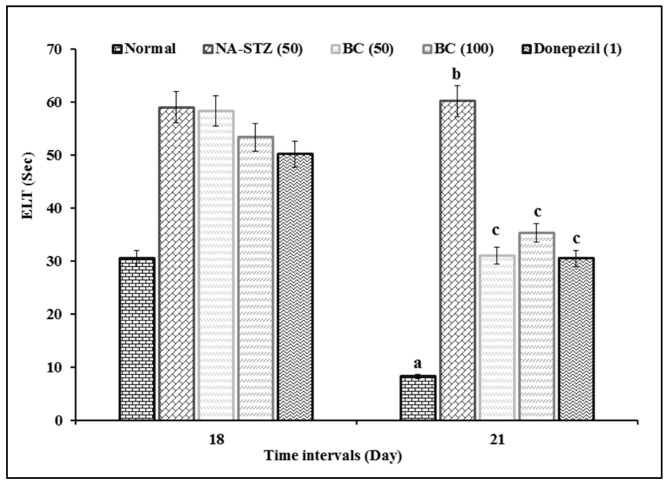
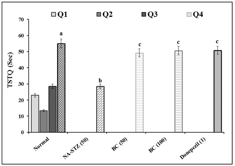
The administration of BC showed a significant (p < 0.05) ameliorative effect against the diabetic VaD condition in the MWM test. There is a significant difference in ELT between groups [F (4,25) = 38.777; p < 0.001] and between days [F (3,75) = 125.04; p < 0.001]. Further, the interaction between groups and days is significant [F (12,75) = 7.354; p < 0.001]. On day 18, there is no difference in ELT across groups, except normal group. Rats in normal group showed significant reduction in ELT from day 18 to day 21. This showed that normal rats have normal learning ability. However, VaD control group had longer ELT compared to all other groups from day 18 to day 21. The TSTQ (Q4) of normal rats is longer on day 22 when compared to other quadrants (Q1, Q2, and Q3) [F (3,15) = 29.608; p < 0.001]. Hence, normal rats have significant memory retention ability (increased in TSTQ) on day 22. The TSTQ between groups showed significant difference [F (4,25) = 11.167; p < 0.001], where the TSTQ of VaD control rats on day 22 was longer than that of other groups. This indicates that the VaD control group had potential impairment of memory retention. The treatment with BC (50 and 100 mg/kg) and DP (1 mg/kg) showed a significant (p < 0.001) ameliorative effect against the NA-STZ-induced increased ELT and decreased TSTQ. Therefore, both BC and DP attenuated the NA-STZ-induced impairment of learning and memory retention (Figure 1 and Figure 2). There is no statistically significant difference in ELT and TSTQ between groups treated with BC and DP.
Figure 1.
Effect of BC on ELT in MWM. Digits in parenthesis indicated dose in mg/kg. Data were expressed as mean ± SD; n = 12 rats. a p < 0.05 vs. ELT of normal group on day 18; b p < 0.05 vs. ELT of normal group on day 21; and c p < 0.05 vs. ELT of VaD group on day 21. Abbreviations: BC, beta-carotene; ELT, escape latency time; NA, nicotinamide; and STZ, streptozotocin.
Figure 2.
Effect of BC on TSTQ in MWM. Digits in parenthesis indicated dose in mg/kg. Data were expressed as mean ± SD; n = 12 rats. a p < 0.05 vs. the time spent in Q1 of normal group; b p < 0.05 vs. TSTQ (Q4) of normal group; c p < 0.05 vs. TSTQ (Q4) of VaD group. Abbreviations: BC, beta-carotene; NA, nicotinamide; Q, quadrant; STZ, streptozotocin; TSTQ, time spent in target quadrant.
2.2. Effect of Beta Carotene on Tissue Biomarkers
In VaD control group, the AChE activity and the level of thiobarbituric acid reactive substance (TBARS) were increased, while the level of reduced glutathione (GSH) was decreased in the hippocampus when compared to the normal control group, as shown in Table 1. The one-way ANOVA of AChE activity showed statistically significant difference between groups: F (4,25) = 30.277; p < 0.001. Similarly, the levels of GSH [F (4,25) = 24.73; p < 0.001] and TBARS [F (4,25) = 15.882; p < 0.001] were significantly different between groups. Treatment with BC (50 and 100 mg/kg) and DP (1 mg/kg) hampered the alteration of above tissue biomarkers in NA-STZ-induced diabetic VaD. Hence, BC and DP potentially attenuate the VaD-associated changes in tissue biomarkers via regulation of cellular oxidative stress and enzymatic activity.
Table 1.
Effect of BC on hippocampal biomarkers.
| Groups | ACHE (μmol of Acetylthiocholine Iodide/min/mg of Protein) | GSH (μmol/L/mg of Protein) | TBARS (μM/mg of Protein) |
|---|---|---|---|
| Normal | 159.73 ± 29.05 | 35.68 ± 3.47 | 1.05 ± 0.16 |
| NA-STZ | 311.21 ± 26.58 a | 23.53 ± 2.54 a | 1.67 ± 0.12 a |
| BC (50) | 227.11 ± 18.82 a,b | 31.41 ± 2.45 b | 1.18 ± 0.16 b |
| BC (100) | 234.56 ± 20.97 a,b | 33.51 ± 2.36 b | 1.05 ± 0.22 b |
| DP (1) | 231.29 ± 22.60 a,b | 35.88 ± 1.10 b | 1.01 ± 0.17 b |
Digits in parenthesis indicated dose in mg/kg. Data were expressed as mean ± SD; n = 12 rats. a p < 0.05 vs. normal group; b p < 0.05 vs. VaD group. Abbreviations: AChE, acetylcholinesterase; BC, beta-carotene; DP, donepezil; GSH, reduced glutathione; NA, nicotinamide; STZ, streptozotocin; TBARS, thiobarbituric acid reactive substances.
3. Discussion
Our study showed that the VaD control group had longer ELT and shorter TSTQ in the MWM test, increased AChE activity and TBARS level, as well as reduced GSH level compared to the normal group. This indicates that the administration of STZ resulted in impairment of spatial memory and an increase in oxidative stress. The administration of BC and DP improved the performance in behavioral test and, hence, the spatial memory of animals in MWM tests. This finding was associated with attenuation of VaD-induced changes in AChE activity and oxidative stress marker levels. VaD is heterogenous in nature and involves complex mechanisms that are involved in the pathophysiology. Generally, VaD is a result of decreased blood flow to certain parts of the brain [12], which causes ischemic lesions to occur in neuronal networks supplying the brain. Multiple facets (mitochondrial dysfunction [13], endothelial dysfunction [14], blood brain barrier disruption [15], white matter damage [16], and neuroinflammation [17]) are involved throughout the development and progression of VaD. The cholinergic system is crucial for cognition, including execution, memory and emotion control [18]. Deficiency of this system is associated with significant decline in executive function [19]. Cholinergic dysfunction most commonly occurs in the basal forebrain cholinergic nuclei and reduces cholinergic connections to the cortex [12]. Cholinergic dysfunction may also be due to a reduction in affinity of receptor towards ligand. It was reported that cerebral blood flow (CBF) and cholinergic signaling affect each other reciprocally [20]. The widespread WM and vascular lesions result in disruption of cholinergic connections, ultimately leading to dysfunction of the cholinergic system. This contributes to the decrease in CBF and, subsequently, brain hypoperfusion [21]. Impairment of the cholinergic system was evident in studies of VaD [18,22,23], where there was deafferentation of frontal and limbic cortical structures, as well as disturbance of basal ganglia, thalamus, white matter and sub frontal areas [24]. In VaD, there are loss of cholinergic neurons, reduced choline acetyltransferase (ChAT) activity, decrement in m3 and m5 muscarinic acetylcholine (ACh) receptor expression, lowered ACh level and worsened memory and learning [25].
Apart from the cholinergic system, the regulation of oxidative stress is also crucial to the development and progression of VaD [26]. In our study, we used STZ for the induction of diabetic VaD. STZ was known to alter the cerebral oxidative stress as well as energy metabolism through impairment of adenosine triphosphate (ATP) and acetyl CoA synthesis [27]. STZ enters pancreatic beta-cells via GLUT 2 glucose transporter [28]. The pancreas is very susceptible to oxidative damage by STZ because of the low level of antioxidant enzymes [29]. Further, STZ also reduces the expression of nuclear factor-erythroid factor 2-related factor 2 (Nrf2), which is responsible for regulating antioxidative response. This happens owing to the increased reactive oxygen species (ROS) and the ratio of oxidized glutathione (GSSG) to GSH [30]. In a study investigating the molecular mechanisms of STZ damage on pancreatic beta-cells, it was found that STZ inhibits the mitochondrial respiratory enzymes. Parallelly, there is an increase in the generation of ROS and reactive nitrogen species (RNS), causing lipid peroxidation. On top of that, caspase-3 and caspase-9 are activated, which leads to DNA fragmentation and apoptosis [31]. Diabetes is one of the risk factors of VaD. It hinders the hippocampal long-term potentiation, and hence impairs learning ability as well as memory. Diabetes also decreases the level of nitric oxide (NO), which leads to impaired vasodilation and reduced CBF [32]. In diabetic condition, there is increased production of ROS, which attack the membranous polyunsaturated fatty acids. Consequently, there is an increased level of malondialdehyde, which is reflected by increased TBARS level [33]. GSH is one of the most potent endogenous antioxidants. Its production requires nicotinamide adenine dinucleotide phosphate (NADPH). However, diabetes leads to an increased level of reduced nicotinamide adenine dinucleotide (NADH). This, in turn, depletes the NADPH and, therefore, decreases GSH level [34].
Although the frontal cortex was not investigated in this study, it plays an important role in spatial working memory. In a study using a photothrombosis-induced frontal cortex stroke mice model, the authors reported that neuronal cell degeneration, reactive astrogliosis and infiltration of immune cells occurred in the frontal cortex, which ultimately leads to behavioral impairment [35]. In another study using frozen post-mortem human brains of VaD, AD and mixed dementia, it was found that the extent of hypoperfusion in frontal cortex was most significant in VaD brains. The myelin-associated glycoprotein:proteolipid protein-1 (MAG:PLP-1) ratio was inversely associated with level of endothelin-1 and proportional to the level of vascular endothelial growth factor (VEGF) in frontal cortex [36]. Our finding is in line with a study conducted in 2019, where BC improved the spatial memory of rats with obstructive sleep apnea syndrome, as evident in shorter ELT in the MWM test when compared to the disease group. This is due to a decrease in the expression of caspase-3 and phosphorylated tau (pτ) protein [37]. The administration of BC at a dose of 2.05 mg/kg inhibited the increase in AChE activity in STZ-induced Alzheimer’s disease in mice. The authors also performed in silico docking studies and proved that BC has high binding affinity towards AChE [38]. Another enzyme that is related to AChE is butyrylcholinesterase (BuChE). BuChE is claimed to be involved in the cell proliferation and growth of nervous system. In cases of AD, the level of BuChE correlates with the expression of neuritic plaques and neurofibrillary tangles [39]. BuChE is also involved in neuroinflammation, as the cholinergic anti-inflammatory pathway regulates the expression of proinflammatory cytokines, such as tumor necrosis factor alpha, interleukin-1, interleukin-6 and Prostaglandin E2 [40]. Hence, it is worth incorporating BuChE in future studies.
In a study using an animal model of two-kidney one-clip (2K1C) renovascular hypertension-induced vascular dementia, there is an increase in TBARS level and decrease in the GSH level in the disease control group [41]. In another study using a mice model of STZ-induced AD, the administration of all-trans retinoic acid resulted in improved memory, reduction in STZ-induced increase in AChE activity, TBARS and reduction in GSH [42]. BC is a reddish orange color pigment and it belongs to the carotenoid family. It consists of eight isoprene units and is rich in conjugated double bond [43]. BC has potent antioxidative activity due to the presence of double bonds. BC can chelate ROS and RNS through energy transfer during the formation and cleavage of bonds [44]. It was also reported that BC can reduce membrane lipid peroxidation by suppressing the expression of inflammatory cytokines and nuclear factor-kappa β (NF-κβ) [45]. Interestingly, BC possesses neuroprotective activity by modulating the Nrf2/Kelch-like ECH-associated protein 1 (KEAP1) pathway [46], where the expression of Nrf2 increases, while the expression of KEAP1, repressor of Nrf2, decreases [47]. Therefore, BC could ameliorate VaD through regulation of oxidative stress.
4. Materials and Methods
4.1. Animals
Disease-free healthy male Sprague Dawley rats (220 ± 20 g) were obtained from Lab-rat Breeders Farm PVT Ltd., Selangor, Malaysia, and housed in Central Animal House of AIMST University, Malaysia. The conditions of Central Animal house were maintained as follows: temperature (22 ± 1 °C), relative humidity (60%) and 12-h light/dark cycle. The animals had access to food and water ad libitum and were kept for 7 days for adaptation before starting the experiment. All the animal handling, dosing, behavioral assessments as well as sacrifice were conducted during daytime.
4.2. Induction of Diabetic Vascular Dementia
The animals were first administered with nicotinamide (NA; 50 mg/kg) through the intraperitoneal (i.p.) route, followed by streptozotocin (STZ; 50 mg/kg; i.p.), with a 15 min interval in between for the induction of diabetic VaD [48,49]. Tail vein blood samples were taken on the 3rd and 7th day for measurement of non-fasting blood glucose level. Animals with a blood glucose level of more than 200 mg/dL were considered diabetic and were used in this study [50].
4.3. Experimental Protocol
The diabetic animals were distributed into 5 groups of 12 animals each, with different interventions.
Group 1: Normal, healthy rats (normal control).
Group 2: NA (50 mg/kg; i.p.), followed by (15 min later) STZ (50 mg/kg; i.p.) were administered for the induction of VaD (disease control group).
Group 3 and 4: BC (Nacalai Tesque Inc.) (50 and 100 mg/kg; for 15 consecutive days) was administered orally (p.o.) for the treatment of VaD (induced animal) for the respective group (treatment groups).
Group 5: DP (1 mg/kg; p.o.; for 15 consecutive days) was administered per oral (p.o.) for the treatment of VaD (induced animal) (reference group).
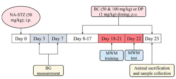
The details of the experimental protocol are illustrated in Figure 3. The dose of BC was chosen based on the fact that rodents have a good capacity to convert BC into vitamin A. Hence, they can be fed a large amount of BC. In a study that developed a novel BC formulation, BC at a dose of 50 mg/kg was administered to rats for 7 days [51]. To the best of our knowledge, there is no research article published on the investigation of BC in the rat model of VaD.
Figure 3.
The experimental protocol. VaD was induced by NA and STZ injection on day 0. The blood glucose levels were measured on day 3 and day 7. BC and DP were administered from day 8 to day 23. The MWM training (learning phase) was conducted from day 18 to day 21. The MWM test (retrieval phase) was conducted on day 22. All the animals were sacrificed on day 23. Abbreviations: NA, nicotinamide; STZ, streptozotocin; BC, beta-carotene. DP, donepezil; BG, blood glucose; EPM, elevated plus maze; i.p., intraperitoneal, p.o., per oral; mg/kg, milligram per kilogram.
4.4. Collection of Biological Samples
Briefly, the animals were anesthetized with diethyl ether and sacrificed by cervical dislocation. The head was isolated and the skull was cut open to collect the brain samples. Then, the hippocampus was isolated from the whole brain [52]. Each hippocampus was homogenized (10% w/v) with phosphate buffer saline (pH 7.4) and subjected to centrifugation at 3500 rpm (1720 g) for 15 min [53]. The supernatant obtained was used for biochemical assessment.
4.5. Assessment of Spatial Learning and Memory by Morris Water Maze
The MWM test was conducted as described in the method of Morris [54]. Briefly, a circular water pool (150 cm in diameter; 45 cm in height) was used. The water pool was divided into 4 quadrants with distinct visual cues on the inner wall of each quadrant. A platform (10 × 10 cm square and 28 cm in height) was placed in one of the quadrants and water was filled until the water level was 2 cm above the platform. During the training session, each animal was placed in each quadrant to assess the escape latency time (ELT): the time needed to find the hidden platform. Each animal was allowed to explore the pool for 120 s. Once the animals found the platform, they were allowed to stay on the platform for 15 s. Otherwise, they were guided to the platform after 120 s. During the MWM test day, each animal was placed in the middle of the pool and allowed to swim for 120 s. The time spent in the target quadrant (TSTQ) by each animal was noted and it served as the index of retrieval (memory retention) [55].
4.6. Estimation of Acetylcholinesterase (AChE) Activity
The hippocampal AChE activity was estimated as described in the method of Ellman et al. [56]. Concisely, 0.5 mL of brain supernatant was added with 2.5 mL of phosphate buffer saline (pH 7.4). Thereafter, 0.1 mL of 5,5-dithio-bis-(2-nitrobenzoic acid) (DTNB) solution was added. The changes in absorbance (optical density; OD) were measured spectrophotometrically (Shimadzu UV-1800 UV/Visible Scanning Spectrophotometer, Kyoto, Japan) at 412 nm wavelength. Next, 20 μL of acetylthiocholine iodide was added. The changes in absorbance were measured instantly and at an interval of every 2 min until the absorbance value became constant. The acetylcholinesterase activity was calculated using the following formula:
where ∆A, changes in absorbance/min; P, protein content (mg/mL); VBs, volume of brain supernatant (VBs = 500 µL); TVts, total volume of test samples (TVts = 3120 µL); and 13,600 refers to the molar extinction coefficient of DTNB (M−1 cm−1). The AChE activity results were expressed as μM of acetylthiocholine hydrolyzed/mg of protein/minute.
4.7. Estimation of Reduced Glutathione (GSH)
The GSH content was estimated using the method of Beutler et al. [57]. Briefly, the aliquots (0.5 mL) were mixed with 2 mL of 0.3 M disodium hydrogen phosphate. Thereafter, 0.25 mL of 0.001 M freshly prepared DTNB (5, 5′-dithiobis (2-nitrobenzoic acid) dissolved in 1% w/v sodium citrate) was added. The changes in absorbance were noted spectrophotometrically (Shimadzu UV-1800 UV/Visible Scanning Spectrophotometer, Kyoto, Japan) at 412 nm. A standard curve was plotted using 10–100 µM of reduced form of glutathione. The results of GSH were expressed as micromoles of GSH per mg of protein.
4.8. Estimation of Thiobarbituric Acid Reactive Substances (TBARS)
The TBARS content was estimated using the method of Buege and Aust [58]. Briefly, 0.2 mL of brain supernatant was mixed with 2 mL of thiobarbituric acid (TBA), trichloroacetic acid (TCA), and hydrochloric acid (HCL) reagent mixture. The mixture was heated for 10 min in a boiling water bath to develop pink colour chromogen. Thereafter, tubes were cooled with tap water and centrifuged at 5500 rpm (3252 g) for 15 min. The changes in absorbance were noted spectrophotometrically (Shimadzu UV-1800 UV/Visible Scanning Spectrophotometer, Kyoto, Japan) at 532 nm wavelength. A standard curve was plotted using 0–100 µM of tetramethoxypropane. The results of TBARS were expressed as micromoles of TBARS per mg of protein.
4.9. Statistical Analysis
The data collected for both EPM test and AChE activity estimation were expressed as mean ± standard deviation (SD) and analyzed by two-way and one-way analysis of variance (ANOVA) test correspondingly. The post hoc analysis was performed using Tukey’s honestly significant difference (HSD) test. The statistical analysis was conducted using Statistical Package for the Social Sciences (SPSS) software (version 25). The alpha value was set at 0.05.
5. Conclusions
Our study showed that BC improves the performance of rats in MWM and attenuates the elevated AChE activity, TBARS level and reduced GSH level. These signify that BC, as a natural compound that is widely available, is helpful in ameliorating VaD. As ACh is a crucial neurotransmitter that regulates bodily functions, it is worth exploring and investigating the potential of BC in other neurodegenerative diseases in which the cholinergic system is affected. This study can also be extrapolated to a clinical stage once more fruitful results are reported.
Acknowledgments
We thank Sohrab Akhtar Shaikh, Aswinprakash Subramanian and Jagadeesh Dhamodharan for their help in the execution of this study.
Author Contributions
Conceptualization, A.M.; methodology, A.M.; software, K.G.L.; validation, A.M. and R.V.; formal analysis, K.G.L.; investigation, K.G.L.; resources, A.M.; data curation, K.G.L. and R.V.; writing—original draft preparation, K.G.L.; writing—review and editing, A.M. and R.V.; visualization, K.G.L.; supervision, A.M. and R.V.; project administration, A.M.; funding acquisition, A.M. All authors have read and agreed to the published version of the manuscript.
Institutional Review Board Statement
The animal study protocol was approved by the AIMST University Animal Ethics Committee of AIMST University (AUAEC/FOP/2020/13, 9 November 2020).
Informed Consent Statement
Not applicable.
Data Availability Statement
The data presented in this study are available on request from the corresponding author.
Conflicts of Interest
The authors declare no conflict of interest. The funders had no role in the design of the study; in the collection, analyses, or interpretation of data; in the writing of the manuscript, or in the decision to publish the results.
Sample Availability
Samples of the compounds are available from the authors.
Funding Statement
This research was funded by Malaysian Ministry of Education through Fundamental Research Grant Scheme, FRGS/1/2019/SKK08/AIMST/02/3.
Footnotes
Publisher’s Note: MDPI stays neutral with regard to jurisdictional claims in published maps and institutional affiliations.
References
- 1.Calabrese V., Giordano J., Signorile A., Laura Ontario M., Castorina S., De Pasquale C., Eckert G., Calabrese E.J. Major Pathogenic Mechanisms in Vascular Dementia: Roles of Cellular Stress Response and Hormesis in Neuroprotection. J. Neurosci. Res. 2016;94:1588–1603. doi: 10.1002/jnr.23925. [DOI] [PubMed] [Google Scholar]
- 2.Vandepitte S., Van Wilder L., Putman K., Van Den Noortgate N., Verhaeghe S., Trybou J., Annemans L. Factors Associated with Costs of Care in Community-Dwelling Persons with Dementia from a Third Party Payer and Societal Perspective: A Cross-Sectional Study. BMC Geriatr. 2020;20:18. doi: 10.1186/s12877-020-1414-6. [DOI] [PMC free article] [PubMed] [Google Scholar]
- 3.Kalaria R.N. The Pathology and Pathophysiology of Vascular Dementia. Neuropharmacology. 2018;134:226–239. doi: 10.1016/j.neuropharm.2017.12.030. [DOI] [PubMed] [Google Scholar]
- 4.Iadecola C. The Pathobiology of Vascular Dementia. Neuron. 2013;80:844–866. doi: 10.1016/j.neuron.2013.10.008. [DOI] [PMC free article] [PubMed] [Google Scholar]
- 5.O’Brien J.T., Thomas A. Vascular Dementia. Lancet. 2015;386:1698–1706. doi: 10.1016/S0140-6736(15)00463-8. [DOI] [PubMed] [Google Scholar]
- 6.Venkat P., Chopp M., Chen J. Models and Mechanisms of Vascular Dementia. Exp. Neurol. 2015;272:97–108. doi: 10.1016/j.expneurol.2015.05.006. [DOI] [PMC free article] [PubMed] [Google Scholar]
- 7.Murphy M.P., Corriveau R.A., Wilcock D.M. Vascular Contributions to Cognitive Impairment and Dementia (VCID) Biochim. Biophys. Acta (BBA)-Mol. Basis Dis. 2016;1862:857–859. doi: 10.1016/j.bbadis.2016.02.010. [DOI] [PubMed] [Google Scholar]
- 8.Strobel M., Tinz J., Biesalski H.-K. The Importance of β-Carotene as a Source of Vitamin A with Special Regard to Pregnant and Breastfeeding Women. Eur. J. Nutr. 2007;46:1–20. doi: 10.1007/s00394-007-1001-z. [DOI] [PubMed] [Google Scholar]
- 9.Gong X., Marisiddaiah R., Rubin L.P. Inhibition of Pulmonary β-Carotene 15, 15’-Oxygenase Expression by Glucocorticoid Involves PPARα. PLoS ONE. 2017;12:e0181466. doi: 10.1371/journal.pone.0181466. [DOI] [PMC free article] [PubMed] [Google Scholar]
- 10.Kawata A., Murakami Y., Suzuki S., Fujisawa S. Anti-Inflammatory Activity of β-Carotene, Lycopene and Tri-n-Butylborane, a Scavenger of Reactive Oxygen Species. In Vivo. 2018;32:255–264. doi: 10.21873/invivo.11232. [DOI] [PMC free article] [PubMed] [Google Scholar]
- 11.Cho S.O., Kim M.-H., Kim H. β-Carotene Inhibits Activation of NF-ΚB, Activator Protein-1, and STAT3 and Regulates Abnormal Expression of Some Adipokines in 3T3-L1 Adipocytes. J. Cancer Prev. 2018;23:37–43. doi: 10.15430/JCP.2018.23.1.37. [DOI] [PMC free article] [PubMed] [Google Scholar]
- 12.Vijayan M., Reddy P.H. Stroke, Vascular Dementia, and Alzheimer’s Disease: Molecular Links. J. Alzheimer’s Dis. 2016;54:427–443. doi: 10.3233/JAD-160527. [DOI] [PMC free article] [PubMed] [Google Scholar]
- 13.Sun C., Liu M., Liu J., Zhang T., Zhang L., Li H., Luo Z. ShenmaYizhi Decoction Improves the Mitochondrial Structure in the Brain and Ameliorates Cognitive Impairment in VCI Rats via the AMPK/UCP2 Signaling Pathway. Neuropsychiatr. Dis. Treat. 2021;17:1937–1951. doi: 10.2147/NDT.S302355. [DOI] [PMC free article] [PubMed] [Google Scholar]
- 14.Wang F., Cao Y., Ma L., Pei H., Rausch W.D., Li H. Dysfunction of Cerebrovascular Endothelial Cells: Prelude to Vascular Dementia. Front. Aging Neurosci. 2018;10:376. doi: 10.3389/fnagi.2018.00376. [DOI] [PMC free article] [PubMed] [Google Scholar]
- 15.Hussain B., Fang C., Chang J. Blood–Brain Barrier Breakdown: An Emerging Biomarker of Cognitive Impairment in Normal Aging and Dementia. Front. Neurosci. 2021;15:688090. doi: 10.3389/fnins.2021.688090. [DOI] [PMC free article] [PubMed] [Google Scholar]
- 16.Venkat P., Chopp M., Zacharek A., Cui C., Zhang L., Li Q., Lu M., Zhang T., Liu A., Chen J. White Matter Damage and Glymphatic Dysfunction in a Model of Vascular Dementia in Rats with No Prior Vascular Pathologies. Neurobiol. Aging. 2017;50:96–106. doi: 10.1016/j.neurobiolaging.2016.11.002. [DOI] [PMC free article] [PubMed] [Google Scholar]
- 17.Grande G., Qiu C., Fratiglioni L. Prevention of Dementia in an Ageing World: Evidence and Biological Rationale. Ageing Res. Rev. 2020;64:101045. doi: 10.1016/j.arr.2020.101045. [DOI] [PubMed] [Google Scholar]
- 18.Ballinger E.C., Ananth M., Talmage D.A., Role L.W. Basal Forebrain Cholinergic Circuits and Signaling in Cognition and Cognitive Decline. Neuron. 2016;91:1199–1218. doi: 10.1016/j.neuron.2016.09.006. [DOI] [PMC free article] [PubMed] [Google Scholar]
- 19.Bohnen N.I., Grothe M.J., Ray N.J., Müller M.L.T.M., Teipel S.J. Recent Advances in Cholinergic Imaging and Cognitive Decline—Revisiting the Cholinergic Hypothesis of Dementia. Curr. Geri. Rep. 2018;7:1–11. doi: 10.1007/s13670-018-0234-4. [DOI] [PMC free article] [PubMed] [Google Scholar]
- 20.Yu D., Swardfager W., Black S.E. Pathophysiology of Vascular Cognitive Impairment (I): Theoretical Background. In: Lee S.-H., Lim J.-S., editors. Stroke Revisited: Vascular Cognitive Impairment. Springer; Singapore: 2020. pp. 71–86. Stroke Revisited. [Google Scholar]
- 21.Jellinger K.A. Pathology and Pathogenesis of Vascular Cognitive Impairment—A Critical Update. Front. Aging Neurosci. 2013;5:17. doi: 10.3389/fnagi.2013.00017. [DOI] [PMC free article] [PubMed] [Google Scholar]
- 22.Kim S.H., Kang H.S., Kim H.J., Moon Y., Ryu H.J., Kim M.Y., Han S.-H. The Effect of Ischemic Cholinergic Damage on Cognition in Patients With Subcortical Vascular Cognitive Impairment. J. Geriatr. Psychiatry Neurol. 2012;25:122–127. doi: 10.1177/0891988712445089. [DOI] [PubMed] [Google Scholar]
- 23.Liu Q., Zhu Z., Teipel S.J., Yang J., Xing Y., Tang Y., Jia J. White Matter Damage in the Cholinergic System Contributes to Cognitive Impairment in Subcortical Vascular Cognitive Impairment, No Dementia. Front. Aging Neurosci. 2017;9:47. doi: 10.3389/fnagi.2017.00047. [DOI] [PMC free article] [PubMed] [Google Scholar]
- 24.Bohnen N.I., Bogan C.W., Müller M.L.T.M. Frontal and Periventricular Brain White Matter Lesions and Cortical Deafferentation of Cholinergic and Other Neuromodulatory Axonal Projections. Eur. Neurol. J. 2009;1:33–50. [PMC free article] [PubMed] [Google Scholar]
- 25.Marzoughi S., Banerjee A., Jutzeler C.R., Prado M.A.M., Rosner J., Cragg J.J., Cashman N. Tardive Neurotoxicity of Anticholinergic Drugs: A Review. J. Neurochem. 2021;158:1334–1344. doi: 10.1111/jnc.15244. [DOI] [PubMed] [Google Scholar]
- 26.Benisty S. Current Concepts in Vascular Dementia. Gériatrie Psychol. Neuropsychiatr. Viellissement. 2013;11:171–180. doi: 10.1684/pnv.2013.0410. [DOI] [PubMed] [Google Scholar]
- 27.Ishrat T., Khan M.B., Hoda M.N., Yousuf S., Ahmad M., Ansari M.A., Ahmad A.S., Islam F. Coenzyme Q10 Modulates Cognitive Impairment against Intracerebroventricular Injection of Streptozotocin in Rats. Behav. Brain Res. 2006;171:9–16. doi: 10.1016/j.bbr.2006.03.009. [DOI] [PubMed] [Google Scholar]
- 28.Karunanayake E.H., Baker J.R.J., Christian R.A., Hearse D.J., Mellows G. Autoradiographic Study of the Distribution and Cellular Uptake of (14C)-Streptozotocin in the Rat. Diabetologia. 1976;12:123–128. doi: 10.1007/BF00428976. [DOI] [PubMed] [Google Scholar]
- 29.Jang Y.Y., Song J.H., Shin Y.K., Han E.S., Lee C.S. Protective Effect of Boldine on Oxidative Mitochondrial Damage in Streptozotocin-Induced Diabetic Rats. Pharmacol. Res. 2000;42:361–371. doi: 10.1006/phrs.2000.0705. [DOI] [PubMed] [Google Scholar]
- 30.Elango B., Dornadula S., Paulmurugan R., Ramkumar K.M. Pterostilbene Ameliorates Streptozotocin-Induced Diabetes through Enhancing Antioxidant Signaling Pathways Mediated by Nrf2. Chem. Res. Toxicol. 2016;29:47–57. doi: 10.1021/acs.chemrestox.5b00378. [DOI] [PubMed] [Google Scholar]
- 31.Nahdi A.M.T.A., John A., Raza H. Elucidation of Molecular Mechanisms of Streptozotocin-Induced Oxidative Stress, Apoptosis, and Mitochondrial Dysfunction in Rin-5F Pancreatic β-Cells. Oxidative Med. Cell. Longev. 2017;2017:7054272. doi: 10.1155/2017/7054272. [DOI] [PMC free article] [PubMed] [Google Scholar]
- 32.Kennedy D. B Vitamins and the Brain: Mechanisms, Dose and Efficacy—A Review. Nutrients. 2016;8:68. doi: 10.3390/nu8020068. [DOI] [PMC free article] [PubMed] [Google Scholar]
- 33.Mirza R., Sharma B. Beneficial Effects of Pioglitazone, a Selective Peroxisome Proliferator-activated Receptor-γ Agonist in Prenatal Valproic Acid-induced Behavioral and Biochemical Autistic like Features in Wistar Rats. Int. J. Dev. Neurosci. 2019;76:6–16. doi: 10.1016/j.ijdevneu.2019.05.006. [DOI] [PubMed] [Google Scholar]
- 34.Muriach M., Flores-Bellver M., Romero F.J., Barcia J.M. Diabetes and the Brain: Oxidative Stress, Inflammation, and Autophagy. Oxidative Med. Cell. Longev. 2014;2014:102158. doi: 10.1155/2014/102158. [DOI] [PMC free article] [PubMed] [Google Scholar]
- 35.Houlton J., Barwick D., Clarkson A.N. Frontal Cortex Stroke-Induced Impairment in Spatial Working Memory on the Trial-Unique Nonmatching-to-Location Task in Mice. Neurobiol. Learn. Mem. 2021;177:107355. doi: 10.1016/j.nlm.2020.107355. [DOI] [PubMed] [Google Scholar]
- 36.Tayler H., Miners J.S., Güzel Ö., MacLachlan R., Love S. Mediators of Cerebral Hypoperfusion and Blood-brain Barrier Leakiness in Alzheimer’s Disease, Vascular Dementia and Mixed Dementia. Brain Pathol. 2021;31:e12935. doi: 10.1111/bpa.12935. [DOI] [PMC free article] [PubMed] [Google Scholar]
- 37.Zhou T., Liu H.-J., Xu P. Effect of beta-carotene on learning, memory and expression of caspase-3 and phosphorylated tau in hippocampus of rats with obstructive sleep apnea syndrome. J. Clin. Neurol. 2019;6:50–53. [Google Scholar]
- 38.Hira S., Saleem U., Anwar F., Sohail M.F., Raza Z., Ahmad B. β-Carotene: A Natural Compound Improves Cognitive Impairment and Oxidative Stress in a Mouse Model of Streptozotocin-Induced Alzheimer’s Disease. Biomolecules. 2019;9:441. doi: 10.3390/biom9090441. [DOI] [PMC free article] [PubMed] [Google Scholar]
- 39.Darvesh S., Hopkins D.A., Geula C. Neurobiology of Butyrylcholinesterase. Nat. Rev. Neurosci. 2003;4:131–138. doi: 10.1038/nrn1035. [DOI] [PubMed] [Google Scholar]
- 40.Ha Z.Y., Mathew S., Yeong K.Y. Butyrylcholinesterase: A Multifaceted Pharmacological Target and Tool. CPPS. 2020;21:99–109. doi: 10.2174/1389203720666191107094949. [DOI] [PubMed] [Google Scholar]
- 41.Singh P., Gupta S., Sharma B. Antagonism of Endothelin (ETA and ETB) Receptors During Renovascular Hypertension-Induced Vascular Dementia Improves Cognition. CNR. 2016;13:219–229. doi: 10.2174/1567202613666160518122534. [DOI] [PubMed] [Google Scholar]
- 42.Sodhi R.K., Singh N. All-Trans Retinoic Acid Rescues Memory Deficits and Neuropathological Changes in Mouse Model of Streptozotocin-Induced Dementia of Alzheimer’s Type. Prog. Neuro-Psychopharmacol. Biol. Psychiatry. 2013;40:38–46. doi: 10.1016/j.pnpbp.2012.09.012. [DOI] [PubMed] [Google Scholar]
- 43.Institute of Medicine (US) β-Carotene and Other Carotenoids. National Academies Press (US); Washington, DC, USA: 2000. Panel on Dietary Antioxidants and Related Compound. [Google Scholar]
- 44.Heymann T., Heinz P., Glomb M.A. Lycopene Inhibits the Isomerization of β-Carotene during Quenching of Singlet Oxygen and Free Radicals. J. Agric. Food Chem. 2015;63:3279–3287. doi: 10.1021/acs.jafc.5b00377. [DOI] [PubMed] [Google Scholar]
- 45.Kim Y., Kim Y.J., Lim Y., Oh B., Kim J.Y., Bouwman J., Kwon O. Combination of Diet Quality Score, Plasma Carotenoids, and Lipid Peroxidation to Monitor Oxidative Stress. Oxidative Med. Cell. Longev. 2018;2018:8601028. doi: 10.1155/2018/8601028. [DOI] [PMC free article] [PubMed] [Google Scholar]
- 46.Chen P., Li L., Gao Y., Xie Z., Zhang Y., Pan Z., Tu Y., Wang H., Han Q., Hu X., et al. β-Carotene Provides Neuro Protection after Experimental Traumatic Brain Injury via the Nrf2-ARE Pathway. J. Integr. Neurosci. 2019;18:153–161. doi: 10.31083/j.jin.2019.02.120. [DOI] [PubMed] [Google Scholar]
- 47.Deshmukh P., Unni S., Krishnappa G., Padmanabhan B. The Keap1-Nrf2 Pathway: Promising Therapeutic Target to Counteract ROS-Mediated Damage in Cancers and Neurodegenerative Diseases. Biophys. Rev. 2017;9:41–56. doi: 10.1007/s12551-016-0244-4. [DOI] [PMC free article] [PubMed] [Google Scholar]
- 48.Ghasemi A., Khalifi S., Jedi S. Streptozotocin-Nicotinamide-Induced Rat Model of Type 2 Diabetes (Review) Acta Physiol. Hung. 2014;101:408–420. doi: 10.1556/APhysiol.101.2014.4.2. [DOI] [PubMed] [Google Scholar]
- 49.Masiello P., Broca C., Gross R., Roye M., Manteghetti M., Hillaire-Buys D., Novelli M., Ribes G. Experimental NIDDM: Development of a New Model in Adult Rats Administered Streptozotocin and Nicotinamide. Diabetes. 1998;47:224–229. doi: 10.2337/diab.47.2.224. [DOI] [PubMed] [Google Scholar]
- 50.Birgani G.A., Ahangarpour A., Khorsandi L., Moghaddam H.F. Anti-Diabetic Effect of Betulinic Acid on Streptozotocin-Nicotinamide Induced Diabetic Male Mouse Model. Braz. J. Pharm. Sci. 2018;54:1–7. doi: 10.1590/s2175-97902018000217171. [DOI] [Google Scholar]
- 51.Bagle S., Muke S., Saha S., Jayakodi S. Evaluation of Novel and Superior Formulation CaroTexTM Developed by Biofusion Technology. IJRPS. 2019;10:1868–1873. doi: 10.26452/ijrps.v10i3.1385. [DOI] [Google Scholar]
- 52.Spijker S. Dissection of Rodent Brain Regions. In: Li K.W., editor. Neuroproteomics. Volume 57. Humana Press; Totowa, NJ, USA: 2011. pp. 13–26. (Neuromethods). [Google Scholar]
- 53.Bhatia P., Singh N. Tadalafil Ameliorates Memory Deficits, Oxidative Stress, Endothelial Dysfunction and Neuropathological Changes in Rat Model of Hyperhomocysteinemia Induced Vascular Dementia. Int. J. Neurosci. 2020:1–13. doi: 10.1080/00207454.2020.1817009. [DOI] [PubMed] [Google Scholar]
- 54.Morris R. Developments of a Water-Maze Procedure for Studying Spatial Learning in the Rat. J. Neurosci. Methods. 1984;11:47–60. doi: 10.1016/0165-0270(84)90007-4. [DOI] [PubMed] [Google Scholar]
- 55.Singh M., Prakash A. Possible Role of Endothelin Receptor against Hyperhomocysteinemia and β-Amyloid Induced AD Type of Vascular Dementia in Rats. Brain Res. Bull. 2017;133:31–41. doi: 10.1016/j.brainresbull.2017.02.012. [DOI] [PubMed] [Google Scholar]
- 56.Ellman G.L., Courtney K.D., Andres V., Featherstone R.M. A New and Rapid Colorimetric Determination of Acetylcholinesterase Activity. Biochem. Pharmacol. 1961;7:88–95. doi: 10.1016/0006-2952(61)90145-9. [DOI] [PubMed] [Google Scholar]
- 57.Beutler E., Duron O., Kelly B.M. Improved Method for the Determination of Blood Glutathione. J. Lab. Clin. Med. 1963;61:882–888. [PubMed] [Google Scholar]
- 58.Buege J.A., Aust S.D. Methods in Enzymology. Volume 52. Elsevier; Amsterdam, The Netherlands: 1978. Microsomal Lipid Peroxidation; pp. 302–310. [DOI] [PubMed] [Google Scholar]
Associated Data
This section collects any data citations, data availability statements, or supplementary materials included in this article.
Data Availability Statement
The data presented in this study are available on request from the corresponding author.


