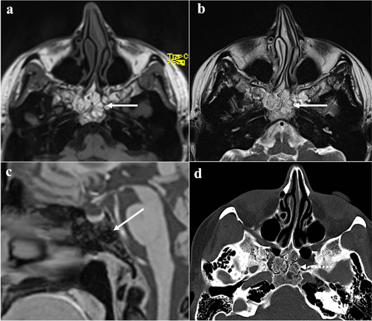
Fig. 3.
Arrested pneumatisation of the sphenoid sinus as an incidental finding in a 51-year-old female patient with headache. The sphenoid sinus is replaced by a non-expansile solid lesion (white arrows) showing high SI on MRI axial T1W (a) and T2W images (b), and homogeneous low SI on sagittal T1W fat-saturated sequence (c). Axial bone algorithm reconstruction CT image (d) shows a lesion with sclerotic margins, internal curvilinear calcifications, foci of fat, and loss of bone trabeculae (white dotted arrow). Note the absence of a cortical bone breach

