Abstract
Myelodysplastic syndromes (MDS) and myelodysplastic/myeloproliferative neoplasms (MDS/MPN) are bone marrow disorders characterized by cytopenias and progression to acute myeloid leukemia. Hypomethylating agents (HMAs) are Food and Drug Administration-approved therapies for MDS and MDS/MPN patients. HMAs have improved patients’ survival and quality of life when compared with other therapies. Although HMAs are effective in MDS and MDS/MPN patients, they are associated with significant toxicities that place a large burden on patients. Our goal is to develop a safer and more effective HMA from natural products. We previously reported that black raspberries (BRBs) have hypomethylating effects in the colon, blood, spleen, and bone marrow of mice. In addition, BRBs exert hypomethylating effects in patients with colorectal cancer and familial adenomatous polyposis. In the current study, we conducted a pilot clinical trial to evaluate the hypomethylating effects of BRBs in patients with low-risk MDS or MDS/MPN. Peripheral blood mononuclear cells (PBMCs) were isolated before and after three months of BRB intervention. CD45+ cells were isolated from PBMCs for methylation analysis using a reduced-representation bisulfite sequencing assay. Each patient served as their own matched control, with their measurements assessed before intervention providing a baseline for post-intervention results. Clinically, our data showed that BRBs were well-tolerated with no side effects. When methylation data was combined, BRBs significantly affected methylation levels of 477 promoter regions. Pathway analysis suggests that BRB-induced intragenic hypomethylation drives leukocyte differentiation. A randomized, placebo-controlled clinical trial of BRB use in low-risk MDS or MDS/MPN patients is warranted.
Keywords: Rubus, Myelodysplastic syndromes, Leukocytes, mononuclear, Peripheral blood stem cells, Clinical trial
INTRODUCTION
Myelodysplastic syndromes (MDS) and myelodysplastic syndromes/myeloproliferative neoplasm (MDS/MPN) are a group of bone marrow disorders characterized by progressive cytopenias and progression to acute myeloid leukemia (AML) [1]. Clinicians use the French-American-British (FAB) and the World Health Organization systems, in addition to the International Prognostic Scoring System (IPSS) and revised IPSS (R-IPSS), to classify MDS and MDS/MPN [2]. The R-IPSS is the most commonly used prognostic scoring system and includes the percentage of blasts/morphology, the number of cytopenias, and cytogenetic abnormalities. The R-IPSS system divides patients into five prognostic groups—very low, low, intermediate-high, and very high-risk disease. The median survival is 8.8, 5.3, 3, 1.6, and 0.8 years for patients with very low, low, intermediate-high, and very high-risk diseases, respectively. More than 80% of these patients are above the age of 65 [3]. As most patients with MDS and MDS/MPN are older, therapy with low toxicity is desirable.
Genetic and epigenetic alterations are the molecular hallmarks of cancer. Genetic alterations include mutations or deletions that alter the primary sequence of DNA. In contrast, epigenetic alterations result from biochemical modification of the composition of chromatin. DNA methylation and alteration of the histone code are two epigenetic changes that can result in transcriptional deregulation and gene silencing. Reversal of this process with hypomethylating agents (HMAs) results in reactivation of transcription and killing of leukemic cells, a phenomenon widely exploited in human clinical trials [4-6].
Hematopoietic stem cell transplantation remains the only cure for MDS and MDS/MPN. However, clinicians also have other therapy options corresponding to disease risk in treating MDS and MDS/MPN. For example, supportive care, which may consist of transfusions and growth factor therapy, remains the backbone of low-risk MDS and MDS/MPN treatment [7]. Azacitidine (AZA) and decitabine (DAC) [8,9] are first-line, Food and Drug Administration (FDA)-approved HMAs for high-risk MDS and MDS/MPN patients who can tolerate them. These agents were developed as cytarabine derivatives with disappointing results, especially at higher concentrations. However, both drugs have shown efficacy as therapies in follow-up studies and with lower-dose schedules. HMAs have yielded a response rate ranging from 30-50% for an average of 12 months.
Although AZA and DAC are effective therapies for patients with MDS and MDS/MPN, they are associated with significant cytopenias and gastrointestinal toxicity. In the AZA-001 trial [8], 91%, 85%, and 57% of patients developed grade 3 or 4 neutropenia, anemia, or thrombocytopenia, respectively. In the alternative dosing for outpatient treatment trial [9], 31%, 18%, and 12% of patients receiving decitabine developed grade 3 or 4 neutropenia, thrombocytopenia, and anemia, respectively. In addition, other non-hematologic toxicities occurring in > 10% of patients manifested as fatigue, nausea, fever, diarrhea, constipation, pneumonia, vomiting, and chills.
One of the most desirable features of a chemopreventive agent is minimal toxicity at concentrations producing chemopreventive efficacy. Our group reported that black raspberries have hypomethylating effects in patients with colon cancer [10] and familial adenomatous polyposis [11]. Twenty patients with colorectal cancer were treated with a daily dose of 60 g black raspberries (BRBs) for 1 to 9 weeks before surgical resection of the tumor. In this study, BRBs were well-tolerated while exerting hypomethylating effects on the adjacent normal colorectal mucosa of all patients and carcinomas in patients with > 4 weeks of BRB treatment [10]. These observations regarding the hypomethylating effects of BRBs with prolonged therapy are consistent with what is seen in patients with MDS treated with conventional hypomethylating agents. In almost all patients, 2-3 cycles of azacitidine or decitabine are needed (up to 6 cycles) before a response is seen. In 14 patients with familial adenomatous polyposis (FAP), daily doses of 60 g BRBs were tolerated in a 9-month intervention study, and BRBs led to fewer and smaller polyps via hypomethylation [11]. This hypomethylating effect in colon cancer and FAP patients is most likely due to the local effects of BRBs in colon cells and rectal cells. However, BRBs are systemically absorbed and have been shown to have a systemic hypomethylating effect. In the colon cancer study previously mentioned, none of the patients had detectable urinary levels of BRB-derived anthocyanins before therapy. In contrast, all had detectable levels in urine after administration of BRBs, confirming the systemic absorption of BRBs [10]. In addition, the systemic hypomethylating effects of BRBs have been demonstrated in interleukin-10 knockout mice [12] and dextran sodium sulfate-treated mice [13]. In both mouse studies, four weeks of a 5% BRB diet reversed aberrant promoter methylation of genes in the Wnt pathway in the colon, bone marrow, and spleen [12,13]. This study aims to evaluate if daily consumption of 50 g (25 g × 2) BRBs in patients with MDS or MDS/MPN is tolerated and if BRBs exert hypomethylating effects in these patients.
MATERIALS AND METHODS
Clinical trial
This study was approved by the Medical College of Wisconsin Institutional Review Board (IRB No. 28985), FDA Investigational New Drug application (IND) (No. 134752), and registered on ClinicalTrials.org (NCT03140280). Written informed consent was obtained from patients before enrollment.
1) Key inclusion criteria included
(1) Patients must have a confirmed diagnosis of MDS or MDS/MPN proven by bone marrow biopsy/aspirate; (2) Patients with cytopenias (blood cell counts lower than the institutional lower limit of normal within the eight weeks prior to the study) who are receiving or received: red blood cell transfusions, observation, platelet transfusions, erythropoietin, granulocyte colony-stimulating factors, granulocyte-macrophage colony-stimulating factors, hydroxyurea (Hydrea); (3) Age >18 years; (4) Predicted life expectancy of at least 12 weeks; (5) Patients should be expected to stay on the same therapy for the period of the study; (6) Patients who do not have an indication for and/or cannot tolerate a hypomethylating agent are eligible for the study; (7) Ability to understand a written informed consent document and willingness to sign it.
2) Key exclusion criteria included
(1) Previously received hypomethylating agents; (2) Taking nonsteroidal anti-inflammatory drugs within 48 hours of day one of BRB treatment; (3) Allergy to black raspberries. (4) Inability to swallow oral medication; (5) Inability or unwillingness to comply with BRB administration requirements; (6) Uncontrolled intercurrent illness, including, but not limited to, ongoing or active infection, symptomatic congestive heart failure, or psychiatric illness/social situations, that, at the treating investigator’s discretion, would limit compliance with study requirements. Concurrent active malignancy is not an exclusion criterion; (7) Active infection not well-controlled by antibacterial or antiviral therapy.
3) Dietary restrictions
There were no dietary restrictions. Patients took BRB powder orally in 8 ounces of water. If the patient could not tolerate the taste of the BRB powder mixed in water, they were permitted to mix it with 8 ounces of milk, ice cream, or smoothie.
4) Prohibited medications
It includes nonsteroidal anti-inflammatory drugs, other chemotherapy/radiation, or participating in clinical trials with other investigational agents not included in this trial, within 14 days of the start of this trial and throughout the duration of this trial.
5) Monitoring subject compliance
Twenty five grams of packed fresh frozen BRBs (Jewel variety, harvested in 2010) were purchased from BerriProducts, Inc. (Corvallis, OR, USA) and stored at –20. BRB powder was consumed twice a day, once in the morning and once at night. Fifty grams of BRB powder approximate 1.2 lbs of fresh BRBs per day and are equivalent to a rodent diet of ~6% berry powder. BRB powder was found to be chemopreventive in multiple animal cancer models, such as esophageal, colon, skin, lung, etc. when provided in the diet at concentrations of 5%-10% [14,15].
The patients were seen every four weeks by the treating physician for compliance. Patients were also required to complete a food diary at home while taking BRBs and to return the diary to the clinic for assessment every four weeks. Treatment was administered on an outpatient basis.
Measurement of BRB anthocyanins in plasma and urine
At baseline and after three months of BRB intervention, plasma and urine samples were collected and stored at –80until analysis. Plasma or urine samples (100 μL) were mixed with 50 μL of 20% v/v formic acid in water and 10 μL of kaempferol-3-O-rutinoside aqueous solution (160 pg/μL), the internal standard. The mixture was centrifuged at 12,000 g for 15 min. 5 μL of the supernatant was directly injected onto an 1290 Infinity II High Speed Pump (Agilent, Santa Clara, CA, USA) attached to an Agilent 6460 Triple Quadrupole Mass Spectrometer (Aglient). Separation was accomplished using gradient elution on a Jupiter RP column (300 Å C18, 250 × 2.0 mm, 5 μm particle diameter; Phenomenex, Torrance, CA, USA) at 0.3 mL/min. The first 3 min of the elution were diverted to waste. Mobile phase A was 1% aqueous formic acid, and B was acetonitrile. The gradient started at 5% B, increased linearly to 15% B at 10 min, then increased to 40% at 14 minutes, held at 40% for 1 minute, returned to 5% at 16 minutes, and then held at 5% until 20 minutes. Selective reaction monitoring (SRM) in positive ion mode through a Jet SprayTM (Agilent, Santa Clara, CA, USA) source. Nitrogen gas was used throughout. The capillary voltage was 3,500 V, and the nozzle voltage 1,500 V. Gas temperature and sheath gas temperature were 350 with gas flows of 10 L/min. Common SRM parameters at unit mass resolution were dwell 200 ms and fragmentor voltage 140. The individual compound SRM parameters were as follows: cyanidin-3-O-rutinoside, 595.2 → 287.1, collision energy (CE) 26; cyanidin-3-O-glucoside, 449.1 → 287.1, CE 18; and kaempferol-3-O-rutinoside, 595.2 → 287.1, CE 14. MassHunterTM software (https://www.agilent.com/en/product/software-informatics/mass-spectrometry-software) was used to control the LC-MS/MS system and process data.
Reduced representation bisulfite sequencing (RRBS)
DNA was isolated from CD45+ cells from peripheral blood using the QIAamp DNA Mini and Blood Mini Kit (51304; Qiagen, Venol, Netherlands). RRBS was performed by the Genomic Science and Precision Medicine Center (GSPMC) at Medical College of Wisconsin. Briefly, 150 ng of extracted DNA was digested at CpG-motifs with MspI (10 Units, R0106L; New England Biolabs, Ipswich, MC, USA). Digested fragments underwent end repair and A-tailing with Klenow fragment polymerase (5 Units, M0212S; New England Biolabs) and overnight ligation with TruSeq adapters (Illumina, San Diego, CA, USA). Samples were size selected and processed twice for bisulfite conversion using the EpiTect Bisulfite conversion kit (Qiagen). Final libraries were amplified (10-15 cycles with Pfu Turbo Cx hotstart DNA polymerase; Agilent) and size selected to enrich for 150-500 bp products with quantity and quality checked by fragment analysis (Agilent) and quantitative PCR (Kapa Library Quantification Kit; Kapa Biosystems, Wilmingtom, MA, USA), respectively. Next-Generation Sequencing was completed at GSPMC on the HiSeq2500 (Illumina) with paired-end 150 base pair reads at 10-20 million reads per sample. Raw data were further analyzed using Streamlined Analysis and Annotation Pipeline for reduced representation bisulfite sequencing data [16], a streamlined analysis and annotation pipeline for RRBS. Briefly, FASTQ files were trimmed to remove adaptor sequences, and reads less than 15 bp were discarded. Trimmed FASTQ files were then aligned against the reference genome hg38 using BSMAP [17].
Statistical analysis
1) DNA methylation data analysis
For each CpG site, the methylation rate was calculated as the percentage of unconverted cytosines in each library. Sixty libraries were merged based on CpG coordinates (hg38). 3,274,038 CpG sites with an average coverage of 17 were detected. Among them, 2,229,834 CpG sites with at least 5X among 80% libraries were taken into downstream analysis. A modified software based on Metilene [18] and Wilcoxon Signed Rank Test was applied to identify the differentially methylated region (DMR) de novo between samples before and after 3-month BRB intervention. Specifically, the Wilcoxon rank-sum test in the original version of Metilene for comparing the median difference between two independent groups was replaced by the Wilcoxon signed-rank test for paired samples in our study. A DMR is required to contain at least five CpG sites and exhibit a 5mC difference of greater than 10% between samples before and after a 3-month BRB intervention. The methylation rate of each region was calculated as the average methylation rate of each site across the region. Benjamin-Hochberg procedure was used to control false discovery rates (FDR). DMR with FDR < 0.05 from Wilcoxon signed-rank test was considered significant. We divided genomic regions of interest into three genomic features: Transcription start site (TSS), intragenic regions, and intergenic regions. TSS regions were defined as 1,000 bp upstream and downstream of a transcription start site. Intragenic regions were defined as regions between the start and end of a transcript. The remaining regions not covered by TSS and intragenic regions were defined as intergenic regions. Then, the identified significant DMRs were assigned to one of the above three regions.
2) Pathway analysis
Enriched Gene Ontology terms and pathways for genes with significant DMR that are located in TSS or intragenic regions were identified using the web portal Metascape [19] with default parameter settings (https://metascape.org/gp/index.html).
Plasma and urine anthocyanins, blood chemistry, and blood cells were compared using Student’s t-test. All analyses were two-sided and paired, and a P-value of less than 0.05 was significant. Linear regression was used to determine the correlation between average hypomethylation level and changes in neutrophils and white blood cells.
RESULTS
Patient demographics, anthocyanins, and blood chemistry
Among 15 patients, 12 were MDS, and 3 were MPN (Table 1). The age range of patients was 43-83 years old. Five were females, and 10 were males. Only one patient had prior chemotherapy. Cyanidin-3-O-glucoside and cyanidin-3-O-rutinoside, the two major anthocyanins in BRBs, in plasma and urine were significantly increased after three months of BRB intervention, suggesting the systemic absorption of BRBs (Fig. S1). Regarding blood chemistry, there were no significant differences after three months of BRB intervention in plasma levels of alanine aminotransferase, aspartate aminotransferase, bilirubin total, blood urea nitrogen, and creatinine (Fig. S2). In all patients, BRBs were well-tolerated with no adverse events.
Table 1.
Patient demography
| Patient code | MDS or MPN | Age (yr) | Gender | Race | Prior chemotherapy | Use of aspirin or other NSAIDs |
|---|---|---|---|---|---|---|
| BRB01 | MDS | 76 | Female | White | None | No |
| BRB03 | MDS | 68 | Female | White | None | No |
| BRB04 | MDS | 83 | Male | White | None | No |
| BRB05 | MDS | 78 | Male | White | None | No |
| BRB06 | MDS | 77 | Male | African American | None | No |
| BRB08 | MDS | 76 | Female | White/Hispanic | None | No |
| BRB11 | MDS | 43 | Female | White | Yes | No |
| BRB12 | MDS | 76 | Male | White | None | No |
| BRB13 | MDS | 70 | Male | White | None | No |
| BRB15 | MDS | 62 | Male | White | None | Yes |
| BRB16 | MPN | 73 | Male | White | None | No |
| BRB17 | MDS | 64 | Male | White | None | Yes |
| BRB18 | MDS | 76 | Female | White/Hispanic | None | Yes |
| BRB19 | MPN | 82 | Male | White | None | No |
| BRB20 | MPN | 71 | Male | White | None | No |
BRB, black raspberry; MDS, myelodysplastic syndromes; MPN, myeloproliferative neoplasm; NSAID, nonsteroidal anti-inflammatory drugs.
Genome-wide methylation rates before and after BRB intervention
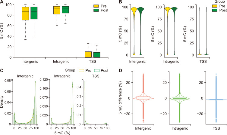
We first examined the global methylation rates in different genomic regions before and after BRB intervention. Distribution of methylation showed that methylations occurred mainly in intragenic and intergenic regions, but very few occurred in transcription start site (TSS) regions (Fig. 1A-1C). In all three regions, BRB intervention resulted in both hypo- and hypermethylation (Fig. 1D).
Figure 1. Genome-wide methylation rates before and after BRB intervention.
Global methylation rates in different genomic regions before and after BRB intervention. Distribution of methylation showed that methylations occurred mainly in intragenic and intergenic regions but very few in TSS regions (A-C). In all three regions, BRB intervention resulted in both hypo- and hypermethylation (D). TSS, transcription start site; BRB, black raspberry.
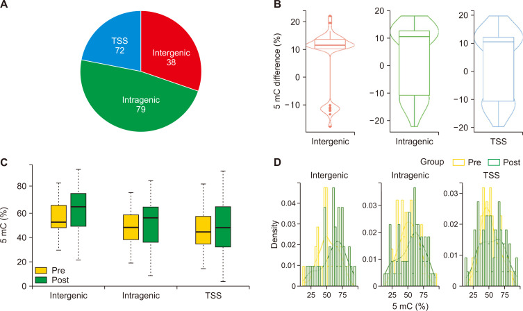
We then applied modified software based on Metilene to identify de novo DMRs before and after BRB intervention. Among 189 identified DMRs reaching false discovery rates (FDRs) < 0.05, 72, 79, and 38 were located at TSS, intragenic, and intergenic regions, respectively (Fig. 2A). The methylation rate at each CpG site within a DMR was averaged to represent the methylation rate of a DMR. On average, the methylation rate of DMRs after BRB intervention was higher than the methylation rate before BRB intervention in the intergenic, intragenic, and TSS regions (Fig. 2B-2D).
Figure 2. Differentially methylation region (DMR) de novo before and after BRB intervention.
Metilene was used to identify de novo differentially methylated regions before and after BRB intervention. Among 189 identified DMRs reaching FDR < 0.05, 72, 79, and 38 were located at TSS, Intragenic and Intergenic regions, respectively (A). The methylation rate of DMRs after BRB intervention was higher than before BRB intervention (B-D), especially in the region of Intergenic (B). DMR, differentially methylated region; FDR, false discovery rates; TSS, transcription start site; BRB, black raspberry.
BRB-intervention induced methylation changes in leukocyte differentiation pathways
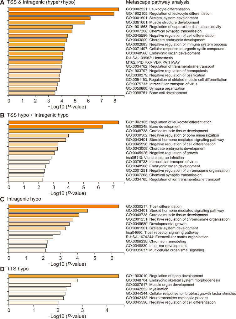
We performed metascape pathway analysis using DMRs that were hypo- and hypermethylated in their TSS or intragenic regions. The most significantly changed pathways were associated with leukocyte differentiation and regulation of leukocyte differentiation (Fig. 3A). Further analysis was performed to determine if hypo- or hypermethylated genes were the key players. This analysis suggested that hypomethylated TSS or intragenic regions were most associated with leukocyte differentiation (Fig. 3B). Interestingly, the intragenic hypomethylated regions (Fig. 3C), but not TSS hypomethylated regions (Fig. 3D), were associated with T cell differentiation.
Figure 3. Metascape pathway analysis of DMRs.
Metascape pathway analysis was performed using DMRs that were hypo- and hypermethylated in their TSS or intragenic regions. The most significantly changed pathways were associated with leukocyte differentiation and regulation of leukocyte differentiation in DMRs from combining TSS and intragenic regions (A). Hypomethylated TSS or intragenic regions were most associated with leukocyte differentiation (B). Interestingly, the intragenic hypomethylated regions (C), but not TSS hypomethylated regions (D), were associated with T cell differentiation. DMR, differentially methylated region; TSS, transcription start site.
BRBs-induced differential methylation changes in each MDS/MPN patient
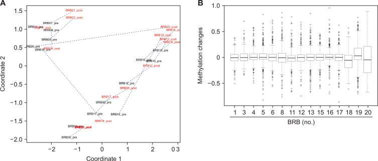
Each patient’s pre- and post-methylation levels were displayed in a principal component analysis plot. Each line connected global pre- and post-methylation data from each patient (Fig. 4A); the longer the line, the more the differences between global pre- and post-methylation levels. Lines connecting pre- and post-samples for BRB18, BRB19, and BRB20 were much longer than those in the other 12 patients (Fig. 4A). We then identified methylation changes 500 bp up- and downstream of the TSS (FDR < 0.01, P < 0.05). Among all 15 patients, 477 common genes were significantly altered. The results shown in Figure 4B suggest that BRB18, BRB19, and BRB20 had more significant changes in methylation; this agreed with the visualization demonstrated in Figure 4A.
Figure 4. BRBs-induced differential changes in methylation in each MDS/MPN patient.
(A) The PCA plot, with each line connecting global pre- and post-methylation data from each patient; the longer the line, the larger the differences between global pre- and post-methylation levels. In particular, lines connecting pre- and post-samples in BRB18, BRB19, and BRB20 were much longer than those in the other 12 patients. (B) Methylation changes 500 bp up- and downstream of the TSS (FDR < 0.01, P < 0.05) were identified. In particular, BRB18, BRB19, and BRB20 had more significant changes in methylation; this corresponded with the visualization demonstrated in (A). BRB, black raspberry; MDS/MPN, myelodysplastic syndromes/myeloproliferative neoplasms; PCA, principal component analysis; FDR, false discovery rates; TSS, transcription start site.
BRBs-induced intragenic hypomethylation is associated with increased neutrophils and white blood cells
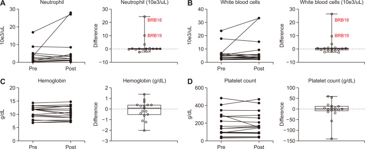
The circulating blood from all 15 patients before and after BRB intervention was collected to measure the numbers of neutrophils, white blood cells, and platelets, as well as the level of hemoglobin. There were no significant differences in these blood cell counts after BRB intervention (Fig. 5). BRB18 and BRB 19 had the most dramatic increases in white blood cell and neutrophil counts (Fig. 5). Strikingly, BRB-induced intragenic hypomethylation was associated with increased neutrophils and white blood cells (Fig. 6).
Figure 5. Effects of BRB intervention on blood cells.
The circulating blood from all 15 patients before and after BRB intervention was collected to measure the numbers of neutrophils (A), white blood cells (B), hemoglobin (C), and platelets (D). BRB18 and BRB 19 had the most dramatic increases in white blood cell and neutrophil counts. BRB, black raspberry.
Figure 6. Association of BRB-induced intragenic hypomethylation with changes in neutrophils and white blood cells.
The greater the degree of hypomethylation, the greater the increase in neutrophils (A) and white blood cells (B). BRB, black raspberry.
DISCUSSION
In this phase II clinical trial, three months of BRB dietary intervention (daily dose of 50 g) elicited no adverse events in MDS and MDS/MPN patients. Pathway analysis of BRB-induced hypomethylation of intragenic regions demonstrated regulation of leukocyte and T cell differentiation. Further, BRB-induced intragenic hypomethylation was associated with increased neutrophil and white blood cell counts. Given the small sample size of this pilot trial, the results from this study will need to be confirmed in larger clinical settings.
MDS is characterized by abnormal and failed differentiation leading to a lack of proper cell maturation; mutated hematopoietic stem cells can promote the development of clonal mutated leukocytes before the development of MDS [18]. Therefore, hypomethylation of regions associated with the regulation of leukocyte differentiation may modulate this abnormal development pathway and encourage the development of diverse mature leukocytes. Additionally, higher-risk MDS patients show increased natural killer T cells and reduced CD4+ T cells as opposed to lower-risk patients, suggesting that altered T cell differentiation may be implicated in the risk of MDS [19]. Specific T cell-related therapies have demonstrated success in AML, a condition that may result from the transformation of MDS [20]. Regulation of T cell differentiation thus may have the potential to benefit MDS patients by reducing dysfunctional proliferation and potential for transformation into AML. Still, more research is warranted before drawing definitive conclusions to this end.
Normal cells are characterized by an open chromatin structure at promoter regions leading to a transcriptionally active conformation [21]. It is appreciated that hypermethylation and histone deacetylation at repetitive regions maintain chromosome structure and genomic integrity in these normal cells. However, hypermethylation is also common in cancer and aging [21]. Tumor cells are frequently hypermethylated at promoter regions, leading to downregulated expression of tumor suppressor genes, and, at the same time, hypomethylated at repetitive DNA regions, leading to chromosomal instability and eventually malignant transformation [21]. Global methylation depicts the sum of hyper- and hypomethylation in different regions.
Interestingly, BRB intervention induced both hypo- and hypermethylation in all patients. On the other hand, pathway analysis of the hypermethylated regions did not result in pathways that regulate leukocytes or other blood cells (data not shown). Therefore, our results suggest that BRB-induced hypomethylation could directly impact the disease state of MDS and MDS/MPN patients by regulating the cell types in which these diseases arise. There were no differences in global methylation in MDS patients achieving complete and partial remission versus those resistant to treatment of HMAs [22,23]. Likewise, the reduction in marrow blasts did not correlate with the decrease in global methylation levels in patients with AML, suggesting that hypomethylation was related to the activity of DAC but not cytoreduction, leading to a decrease in leukemia burden [20]. One explanation could be that the presence of concurrent genomic hypermethylation and hypomethylation may impair the predictive power of current detection techniques. Our results suggest that it could be possible that the combined effects from both global hypermethylation and hypomethylation, but not at any specific sites, regulate immune cells and, ultimately, hematologic responses. Although only 10% of differentially methylated regions were located at promoter regions, the current evidence suggests that long non-coding DNA regions and corresponding RNAs may have a critical role in gene expression regulation at the histone level [24]. Therefore, analysis of the methylation status of these long non-coding regions may help predict response to HMAs. AZA and DAC have been shown to actively demethylate DNA, but the use of the methylation markers as a predictor of response needs to be validated in extensive prospective studies [24].
Overall, this trial demonstrated that a daily dose of 50 g BRBs taken over three months by MDS and MDS/MPN patients holds the potential to regulate cell differentiation via hypomethylation of intragenic regions. These effects manifested without significant side effects or adverse events in our study patients. Hypermethylation of intragenic and intergenic regions was also seen in the study cohort, but further research is necessary to precisely determine the impact of hypermethylation. Given the desired safety profile of BRB intervention and the potential beneficial effects, placebo-controlled randomized trials are warranted to validate and confirm the findings from this pilot study.
SUPPLEMENTARY MATERIALS
Supplementary materials can be found via https://doi.org/10.15430/JCP.2022.27.2.129.
Footnotes
FUNDING
This work was supported by Kurtis Froedtert Clinical Trials Seed Grant, Medical College of Wisconsin Cancer Center, NIH grants (No. CA148818) and USDA/NIFA (No. 2020-67017-30843) (to L.-S. Wang), (No. CA185301), (No. AI129582), (No. NS106170), (to J. Yu).
CONFLICTS OF INTEREST
No potential conflicts of interest were disclosed.
REFERENCES
- 1.Atallah E, Garcia-Manero G. Treatment strategies in myelodysplastic syndromes. Cancer Invest. 2008;26:208–16. doi: 10.1080/07357900701788122. [DOI] [PubMed] [Google Scholar]
- 2.Greenberg P, Cox C, LeBeau MM, Fenaux P, Morel P, Sanz G, et al. International scoring system for evaluating prognosis in myelodysplastic syndromes. Blood. 1997;89:2079–88. doi: 10.1182/blood.V89.6.2079. Erratum in: Blood 1998;91:1100. [DOI] [PubMed] [Google Scholar]
- 3.Ma X, Does M, Raza A, Mayne ST. Myelodysplastic syndromes: incidence and survival in the United States. Cancer. 2007;109:1536–42. doi: 10.1002/cncr.22570. [DOI] [PubMed] [Google Scholar]
- 4.Jones PA, Taylor SM. Cellular differentiation, cytidine analogs and DNA methylation. Cell. 1980;20:85–93. doi: 10.1016/0092-8674(80)90237-8. [DOI] [PubMed] [Google Scholar]
- 5.Uchida T, Kinoshita T, Nagai H, Nakahara Y, Saito H, Hotta T, et al. Hypermethylation of the p15INK4B gene in myelodysplastic syndromes. Blood. 1997;90:1403–9. doi: 10.1182/blood.V90.4.1403. [DOI] [PubMed] [Google Scholar]
- 6.Yang AS, Doshi KD, Choi SW, Mason JB, Mannari RK, Gharybian V, et al. DNA methylation changes after 5-aza-2'-deoxycytidine therapy in patients with leukemia. Cancer Res. 2006;66:5495–503. doi: 10.1158/0008-5472.CAN-05-2385. [DOI] [PubMed] [Google Scholar]
- 7.Hellstrom-Lindberg E, Negrin R, Stein R, Krantz S, Lindberg G, Vardiman J, et al. Erythroid response to treatment with G-CSF plus erythropoietin for the anaemia of patients with myelodysplastic syndromes: proposal for a predictive model. Br J Haematol. 1997;99:344–51. doi: 10.1046/j.1365-2141.1997.4013211.x. [DOI] [PubMed] [Google Scholar]
- 8.Fenaux P, Mufti GJ, Hellstrom-Lindberg E, Santini V, Finelli C, Giagounidis A, et al. Efficacy of azacitidine compared with that of conventional care regimens in the treatment of higher-risk myelodysplastic syndromes: a randomised, open-label, phase III study. Lancet Oncol. 2009;10:223–32. doi: 10.1016/S1470-2045(09)70003-8. [DOI] [PMC free article] [PubMed] [Google Scholar]
- 9.Steensma DP, Baer MR, Slack JL, Buckstein R, Godley LA, Garcia-Manero G, et al. Multicenter study of decitabine administered daily for 5 days every 4 weeks to adults with myelodysplastic syndromes: the alternative dosing for outpatient treatment (ADOPT) trial. J Clin Oncol. 2009;27:3842–8. doi: 10.1200/JCO.2008.19.6550. [DOI] [PMC free article] [PubMed] [Google Scholar]
- 10.Wang LS, Arnold M, Huang YW, Sardo C, Seguin C, Martin E, et al. Modulation of genetic and epigenetic biomarkers of colorectal cancer in humans by black raspberries: a phase I pilot study. Clin Cancer Res. 2011;17:598–610. doi: 10.1158/1078-0432.CCR-10-1260. [DOI] [PMC free article] [PubMed] [Google Scholar]
- 11.Wang LS, Burke CA, Hasson H, Kuo CT, Molmenti CL, Seguin C, et al. A phase Ib study of the effects of black raspberries on rectal polyps in patients with familial adenomatous polyposis. Cancer Prev Res (Phila) 2014;7:666–74. doi: 10.1158/1940-6207.CAPR-14-0052. [DOI] [PubMed] [Google Scholar]
- 12.Wang LS, Kuo CT, Huang TH, Yearsley M, Oshima K, Stoner GD, et al. Black raspberries protectively regulate methylation of Wnt pathway genes in precancerous colon tissue. Cancer Prev Res (Phila) 2013;6:1317–27. doi: 10.1158/1940-6207.CAPR-13-0077. [DOI] [PMC free article] [PubMed] [Google Scholar]
- 13.Wang LS, Kuo CT, Stoner K, Yearsley M, Oshima K, Yu J, et al. Dietary black raspberries modulate DNA methylation in dextran sodium sulfate (DSS)-induced ulcerative colitis. Carcinogenesis. 2013;34:2842–50. doi: 10.1093/carcin/bgt310. [DOI] [PMC free article] [PubMed] [Google Scholar]
- 14.Wang LS, Stoner GD. Anthocyanins and their role in cancer prevention. Cancer Lett. 2008;269:281–90. doi: 10.1016/j.canlet.2008.05.020. [DOI] [PMC free article] [PubMed] [Google Scholar]
- 15.Golovinskaia O, Wang CK. Review of functional and pharmacological activities of berries. Molecules. 2021;26:3904. doi: 10.3390/molecules26133904.3d83c7688ae24bc9900d343cd43a1c96 [DOI] [PMC free article] [PubMed] [Google Scholar]
- 16.Sun Z, Baheti S, Middha S, Kanwar R, Zhang Y, Li X, et al. SAAP-RRBS: streamlined analysis and annotation pipeline for reduced representation bisulfite sequencing. Bioinformatics. 2012;28:2180–1. doi: 10.1093/bioinformatics/bts337. [DOI] [PMC free article] [PubMed] [Google Scholar]
- 17.Xi Y, Li W. BSMAP: whole genome bisulfite sequence MAPping program. BMC Bioinformatics. 2009;10:232. doi: 10.1186/1471-2105-10-232. [DOI] [PMC free article] [PubMed] [Google Scholar]
- 18.Jühling F, Kretzmer H, Bernhart SH, Otto C, Stadler PF, Hoffmann S. metilene: fast and sensitive calling of differentially methylated regions from bisulfite sequencing data. Genome Res. 2016;26:256–62. doi: 10.1101/gr.196394.115. [DOI] [PMC free article] [PubMed] [Google Scholar]
- 19.Zhou Y, Zhou B, Pache L, Chang M, Khodabakhshi AH, Tanaseichuk O, et al. Metascape provides a biologist-oriented resource for the analysis of systems-level datasets. Nat Commun. 2019;10:1523. doi: 10.1038/s41467-019-09234-6.09b6ff0a5f1c4d52a1f792ba98c8531e [DOI] [PMC free article] [PubMed] [Google Scholar]
- 20.Yan P, Frankhouser D, Murphy M, Tam HH, Rodriguez B, Curfman J, et al. Genome-wide methylation profiling in decitabine-treated patients with acute myeloid leukemia. Blood. 2012;120:2466–74. doi: 10.1182/blood-2012-05-429175. [DOI] [PMC free article] [PubMed] [Google Scholar]
- 21.Teschendorff AE, Menon U, Gentry-Maharaj A, Ramus SJ, Weisenberger DJ, Shen H, et al. Age-dependent DNA methylation of genes that are suppressed in stem cells is a hallmark of cancer. Genome Res. 2010;20:440–6. doi: 10.1101/gr.103606.109. [DOI] [PMC free article] [PubMed] [Google Scholar]
- 22.Blum W, Klisovic RB, Hackanson B, Liu Z, Liu S, Devine H, et al. Phase I study of decitabine alone or in combination with valproic acid in acute myeloid leukemia. J Clin Oncol. 2007;25:3884–91. doi: 10.1200/JCO.2006.09.4169. [DOI] [PubMed] [Google Scholar]
- 23.Soriano AO, Yang H, Faderl S, Estrov Z, Giles F, Ravandi F, et al. Safety and clinical activity of the combination of 5-azacytidine, valproic acid, and all-trans retinoic acid in acute myeloid leukemia and myelodysplastic syndrome. Blood. 2007;110:2302–8. doi: 10.1182/blood-2007-03-078576. [DOI] [PubMed] [Google Scholar]
- 24.Voso MT, Santini V, Fabiani E, Fianchi L, Criscuolo M, Falconi G, et al. Why methylation is not a marker predictive of response to hypomethylating agents. Haematologica. 2014;99:613–9. doi: 10.3324/haematol.2013.099549. [DOI] [PMC free article] [PubMed] [Google Scholar]
Associated Data
This section collects any data citations, data availability statements, or supplementary materials included in this article.


