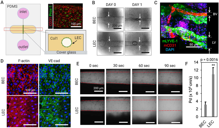
Figure 1. Engineered 3D lymphatic and blood vessels show distinct vessel structure and barrier function.
(A) A schematic of an organotypic 3D lymphatic vessel model (LV-on-chip). Prox-1 (green) and CD31 (red) expression confirms lymphatic endothelial identity and cell morphology in the channel. (B) Morphologic changes in human dermal microvascular blood endothelial cells (BECs) with lymphatic endothelial cells (LECs) after one day of cell seeding. BECs become more contractile than LECs, forming a smaller vessel diameter compared to LECs. (C) BVs and LVs observed in mouse ear tissues. mLYVE-1, anti-mouse LYVE-1 antibody; mCD31, anti-mouse CD31 antibody. (D) Phalloidin (red) and anti-VE-cad (VE-cadherin) antibody (green) staining to visualize F-actin and adherens junctions. (E) Lymphatic and blood vessel barrier function. 70 kDa dextran was introduced into the vessel lumens and dextran diffusion was observed in real time under microscopy. Superimposed red dashed lines represent the edges of the vessel lumens. (F) Quantification of the permeability of BEC-generated engineered BVs and LEC-generated LVs. ** p = 0.0016, two tailed unpaired Student t-test, n = 5 per group. Data are expressed as mean ± S.E.M.

