Key Points
Question
What is the efficacy and safety of berdazimer gel, 10.3%, in the treatment of molluscum contagiosum (MC)?
Findings
This phase 3 randomized clinical trial of 891 patients with MC found greater complete lesion clearance in patients treated with berdazimer gel vs vehicle (32.4% vs 19.7%). Mild transient application-site pain was the most frequently reported adverse event; mild to moderate erythema was the most commonly observed local skin reaction.
Meaning
Berdazimer gel, a novel topical nitric oxide–releasing medication, appears to demonstrate favorable efficacy and safety in patients with MC who are 6 months or older.
Abstract
Importance
Molluscum contagiosum (MC) is a highly contagious skin condition. Lesions may persist for months to years, and no US Food and Drug Administration–approved medications are currently available in the US.
Objective
To assess the efficacy and safety of berdazimer gel, 10.3%, a novel topical nitric oxide–releasing medication, in the treatment of MC.
Design, Setting, and Participants
This was a multicenter, vehicle-controlled, double-blind, phase 3 randomized clinical trial (B-SIMPLE4) conducted in 55 clinics (mostly dermatology and pediatric) in the US from September 1, 2020, to July 21, 2021. Eligible participants were 6 months or older and had from 3 to 70 raised MC lesions. Patients with sexually transmitted MC or with MC only in the periocular area were excluded.
Interventions
Patients were randomized to treatment with berdazimer gel, 10.3%, or vehicle gel, applied as a thin layer to all lesions once daily for 12 weeks.
Main Outcomes and Measures
The primary efficacy end point was complete clearance of all MC lesions at week 12. Safety and tolerability measures included adverse event frequency and severity, and assessment of local skin reactions and scarring. Data analyses were performed from August 31, 2021, to September 14, 2021.
Results
A total of 891 participants were randomized, 444 to berdazimer, 10.3% (mean [range] age, 6.6 [0.9-47.5] years; 228 [51.4%] male; 387 [87.2%] White individuals), and 447 to vehicle (mean [range] age, 6.5 [1.3-49.0] years; 234 [52.3%] female; 382 [85.5%] White individuals). In the intention-to-treat population, 88.5% (393 patients) in the berdazimer group and 88.8% (397 patients) in the vehicle group had a lesion count performed at week 12. At week 12, 32.4% (144 patients) in the berdazimer group achieved complete clearance of MC lesions compared with 19.7% (88 patients) in the vehicle group (absolute difference, 12.7%; odds ratio, 2.0; 95% CI, 1.5-2.8; P < .001) with 14.4% (64 patients) of the berdazimer group discontinuing treatment because of MC clearance compared with 8.9% (40 patients) of the vehicle group. Adverse event rates were low. The most common adverse events were application-site pain and erythema, mostly mild in severity. Adverse events leading to discontinuation affected 4.1% (18 patients) of the berdazimer group and 0.7% (3 patients) of the vehicle group. The most common local skin reaction was mild to moderate erythema.
Conclusions and Relevance
Use of berdazimer gel, 10.3%, for MC appears to demonstrate favorable efficacy and safety with low adverse event rates.
Trial Registration
ClinicalTrials.gov Identifier: NCT04535531
This multicenter, vehicle-controlled, double-blind, phase 3 randomized clinical trial examined the clearance rates of molluscum contagiosum and adverse events of berdazimer gel, 10.3%, for patients 6 months and older during 12 weeks.
Introduction
Molluscum contagiosum (MC) is a common, persistent, and highly contagious skin infection caused by the molluscipoxvirus.1 It affects approximately 6 million people in the US annually, with the greatest incidence among children 1 to 14 years of age.2 Molluscipoxvirus exclusively infects humans, and it replicates in the cytoplasm of keratinocytes.1 The virus infects only the epidermis after contact with infected people or virus-contaminated objects.1,3 Once infected, keratinocyte proliferation manifests as the round, skin-colored to red papules with a central, umbilicated viral core characterized by hyalinized, aggregated molluscum bodies (Henderson-Paterson bodies) in the keratinocyte cytoplasm.1 Molluscum contagiosum proteins evade host immunity, which may contribute to virus’ persistence.1,4
Molluscum contagiosum infection is usually self-limited, yet may persist for months to years, generating a substantial health care burden and quality-of-life concerns necessitating therapeutic intervention.1,5,6 Treatment may also be warranted because of its highly contagious nature and concern for infecting peers or household members.7 Additionally, outwardly visible lesions may be associated with discomfort and psychosocial stigma, and may scar after resolution.6,8 In patients with underlying atopic dermatitis, MC lesions may be more widespread, persistent, and prone to infection.9 There are currently no therapeutic treatments approved by the US Food and Drug Administration (FDA) for MC. Treatment options include in-office, clinician-administered physical procedures (eg, physical ablation of lesions by curettage or cryotherapy) or chemical destruction using topically applied cantharidin.1,8,10 These therapies may require multiple clinic visits and may be painful.8,11 Off-label prescriptions (eg, tretinoin, imiquimod, and over-the-counter products) also may be used but have not been proven to be efficacious against MC.7,12,13,14,15
Nitric oxide functions as both a short-lived immune modulator and a direct broad-spectrum antimicrobial agent to provide localized immunity against foreign organisms.16 Nitric oxide has regulatory functions that affect NF-κB, immunomodulation, inflammation, cytokine production, and apoptosis likely through S-nitrosylation of proteins.17 Nitric oxide also has cytotoxic functions that affect viral replication through reactive oxygen and/or nitrogen molecules.16 Topical nitric oxide, therefore, has therapeutic potential, but the inability to store and safely deliver a stable form of nitric oxide to the site of infection or inflammation has limited development of topical nitric oxide treatments.18
Berdazimer gel, 10.3% (SB206; Novan Inc) is a novel, topical nitric oxide–releasing agent under investigation as a first-in-class therapy for the treatment of MC. Coadministration of 2 components, the new chemical entity berdazimer sodium (gel) and a hydrogel functioning as a proton donor, promotes nitric oxide release from the berdazimer sodium macromolecule (comprised of a polysiloxane backbone with covalently bound N-diazeniumdiolate nitric oxide donors) at the time and site of application.16 The nitric oxide is stably released and targeted to the skin, thus minimizing systemic exposure. Berdazimer likely exerts its antiviral effects on MC through protein nitrosylation and NF-κB modulation.16,17,18,19
A dose-finding phase 2 study in children and adults with MC identified berdazimer sodium 12%, equivalent to berdazimer free base 10.3%, applied once daily as a suitable candidate for phase 3 development.20 A post hoc integrated efficacy analysis of 2 phase 3 randomized clinical trials (B-SIMPLE [berdazimer sodium in molluscum patients with lesions] 1 and 2) revealed greater MC complete clearance rates at week 12 with once-daily berdazimer gel, 10.3%, vs vehicle (27.9% [132 of 473 patients] vs 20.9% [49 of 234 patients]; P < .04). Herein, we report the efficacy and safety findings of the B-SIMPLE4 trial, a third phase 3 study designed after discussions with the FDA to confirm the efficacy and safety of topical berdazimer gel, 10.3%, compared with vehicle applied once daily for up to 12 weeks in patients 6 months or older with MC.
Methods
Trial Design
The B-SIMPLE4 was a multicenter, double-blind, vehicle-controlled, parallel-group (1:1) phase 3 randomized clinical trial of the efficacy and safety of berdazimer gel, 10.3%, completed at 55 sites in the US. The relevant ethics committees and institutional review boards provided study approval for all study sites, and the FDA reviewed and approved the trial protocol (Supplement 1). The study was conducted in accordance with the Declaration of Helsinki. Patients or their caregiver voluntarily provided written informed consent or assent before screening procedures were initiated. We followed the Consolidated Standards of Reporting Trials (CONSORT) reporting guidelines for reporting the results of randomized clinical trials. The investigators classified each participant’s race and ethnicity.
Participants
Eligible participants were patients with MC who were 6 months or older, in generally good health, immunocompetent, and had 3 to 70 raised and palpable MC lesions at baseline. Patients with atopic dermatitis were included. Exclusion criteria included (1) sexually transmitted MC; (2) MC only in the periocular area; and (3) inability to treat and accurately count active lesions (eg, obscuration of lesions by an exuberant surrounding dermatitis response). In addition, patients who had received any of the following treatments within 14 days before baseline were excluded: topical treatment for MC or within 2 cm of an MC lesion (eg, calcineurin inhibitors, glucocorticoids, retinoids, or zinc); podophyllotoxin, imiquimod, cantharidin, sinecatechins, oral zinc, or other homeopathic or over-the-counter product, including but not limited to ZymaDerm, tea tree oil, and H2 receptor antagonists; and surgical procedures (eg, cryotherapy, curettage). Full inclusion and exclusion criteria are listed in the trial protocol (Supplement 1).
The intention-to-treat (ITT) set included all randomized patients. The safety set included all patients who received at least 1 application of study medication. The efficacy analysis was conducted on the ITT set. The safety analysis was conducted on the safety set.
Interventions
Study visits occurred at screening or baseline and weeks 2, 4, 8, 12, and 24. Participants from a 2-patient household were required to have baseline visits on the same day. Patients or caregivers applied a thin layer of study medication once daily to the top of all MC lesions identified at baseline and any new lesions that arose, for a maximum of 12 weeks. Patients and caregivers were instructed to continue treating an area until the next scheduled visit, even if the lesion(s) cleared. Study medication was applied during clinic visits at baseline and weeks 2, 4, 8, and 12, and at home on other days. If all lesions were cleared at a clinic visit, the patient’s treatment period ended, and patients were followed for recurrence or appearance of new lesions until week 24. If lesions recurred or new lesions occurred between visits after treatment was stopped because of clearance, the patient or caregiver was instructed to reinitiate treatment until week 12.
Patients were randomized 1:1 to berdazimer or vehicle and stratified by the number of randomized patients per household (1 vs 2); patients from 1-patient households were further stratified by investigator type (dermatologist vs other) and the absence or presence of the beginning-of-the-end (BOTE) sign at baseline (ie, a BOTE score of 0 vs ≥1). A clinical sign of inflammation, BOTE precedes resolution of MC lesions.21 Patients from the same household were randomized to receive the same treatment, and 2 or fewer patients from the same household were enrolled. A computer-generated randomization schedule used a permuted block algorithm to randomly allocate patients to randomization numbers assigned through a central Interactive Web Response System. Clinicians, patients and caregivers, and study site employees were blinded to treatment allocation. The vehicle was identical to berdazimer in color, consistency, and smell. The berdazimer formulation has been described previously.20
Assessments
For a given patient, active (palpable) MC lesions were counted at baseline and each study visit through week 12 by the same assessor, if possible, using a body map for documentation. Only treatable lesions, defined as active (palpable) MC lesions at least 2 cm away from the ocular region, were counted (eMethods 1 in Supplement 2). Lesion clearance was defined as resolution of the active MC lesion. The investigator’s overall impression of MC severity was assessed on a 5-point scale from 0 (none) to 4 (very severe) using the Investigator Global Severity Assessment (IGSA) at baseline and at weeks 12 and 24 (exploratory assessment).
Adverse events (AEs) were assessed throughout the study. Investigator−evaluators assessed local skin reactions (LSRs) at baseline (≥30 minutes post dose) and at weeks 2, 4, 8, and 12. The 6 individual components of LSRs (eg, erythema, flaking/scaling, crusting, swelling, vesiculation/pustulation, and erosion/ulceration) were scored separately from 0 (not present) to 4 (severe) (eTable in Supplement 2). The LSR composite score was the sum of individual component scores (range, 0-24). Investigators reported clinically important LSRs (ie, interfered with patient’s daily activities) as AEs (eg, application-site erythema). Investigators also assessed treated areas for scars (including small pitted scars as a residual sign of MC resolution) and keloidal or hypertrophic scar formation at weeks 4, 8, 12, and 24, using the lesion body map. All scars, including pitted scars (indentations), were considered AEs.
Main Outcomes and Measures
The primary efficacy end point was the difference in the percentage of berdazimer and vehicle patients who achieved complete clearance of all treatable MC lesions at week 12 (ie, lesion count of 0). The secondary efficacy end points were the percentage of patients who achieved: a MC lesion count of 0 or 1 at week 12; a 90% or greater reduction from baseline in the number of lesions at week 12; complete clearance of lesions at week 8; and/or percent change from baseline in the number of lesions at week 4. Safety end points were AEs and LSRs through week 12 and scarring through week 24.
Statistical Analysis
A sample size of 850 patients (425 patients per group) provided 90% power using a 2-sided test with α = .05 to detect an absolute difference of 9.5% between berdazimer and a 20% vehicle response rate. The primary efficacy analysis of the ITT set compared the percentage of patients with complete clearance of all MC lesions at week 12 for berdazimer with vehicle using logistic regression with adjustments for the following factors: investigator type (dermatologist vs other), number of patients per household (1 vs 2), baseline BOTE score (0 vs ≥1), age in years (6 months to <3, 3 to <4 , 4 to <5, 5 to <6, 6 to <7, 7 to <8, 8 to <9, 9 to <12, and ≥12), and baseline lesion count. The analysis used nonresponder imputation, in which patients with missing lesion count data at week 12 were counted as nonresponders.
Secondary efficacy analyses of the ITT set were completed using a hierarchical fixed-sequence testing strategy. More details are available in eMethods 2 in Supplement 2.
Unless otherwise noted, all statistical testing was 2-sided and performed at α = .05. Odds ratios (ORs) and 95% CIs for berdazimer compared with vehicle are reported for logistic regression analyses. All analyses and tabulations were performed using SAS, version 9.4 or higher (SAS Institute) from August 31, 2021, to September 14, 2021.
Results
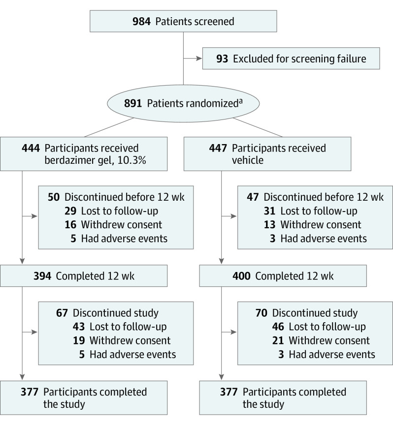
Of 984 patients screened, 891 participants were randomized, 444 to berdazimer (mean [range] age, 6.6 [0.9-47.5] years; 228 [51.4%] male; 216 [48.6%] female; 6 [1.4%] Asian, 21 [4.7%] Black, 387 [87.2%] White, and 30 [6.8%] individuals of other race) and 447 to vehicle (mean [range] age, 6.5 [1.3-49] years; 234 [52.3%] female; 213 [47.7%] male; 6 [1.3%] Asian, 28 [6.3%] Black, 382 [85.5%] White, and 31 [6.9%] individuals of other race). Figure 1 shows participant enrollment. Baseline demographic information and clinical characteristics for the ITT population were similar in berdazimer and vehicle groups (Table 1). Ethnicity information was collected separately: among the berdazimer group, 94 patients (21.2%) were Hispanic or Latino, 345 (77.7%) were not, 4 (0.9%) did not respond, and 1 (0.2%) did not know; and among the vehicle group, 87 patients (19.5%) were Hispanic or Latino, 357 (79.9%) were not, 1 (0.2%) did not respond, and 2 (0.4%) did not know. The first patient was enrolled in the trial on September 1, 2020, and the last patient completed the study on July 20, 2021.
Figure 1. CONSORT Diagram.
aAfter 200 patients were randomized, a data safety monitoring board reviewed all available unblinded safety data, including completed patch testing results for allergic dermatitis, and recommended that the study proceed without modification.
Table 1. Baseline Demographic Information and Clinical Characteristics of Participants.
| Characteristic | No. (%) | |
|---|---|---|
| Berdazimer gel, 10.3% (n = 444) | Vehicle (n = 447) | |
| Age, mean (range), y | 6.6 (0.9-47.5) | 6.5 (1.3-49) |
| Sex | ||
| Male | 228 (51.4) | 213 (47.7) |
| Female | 216 (48.6) | 234 (52.3) |
| Race and ethnicity | ||
| Asian | 6 (1.4) | 6 (1.3) |
| Black or African American | 21 (4.7) | 28 (6.3) |
| Hispanica | 94 (21.2) | 87 (19.5) |
| Otherb | 30 (6.8) | 31 (6.9) |
| White | 387 (87.2) | 382 (85.5) |
| Baseline lesion count, mean (range) | 23.1 (3-70) | 20.5 (3-69) |
| Baseline BOTE scorec | ||
| 0 (no inflammation) | 225 (50.7) | 223 (49.9) |
| ≥1 (mild to very severe) | 219 (49.3) | 224 (50.1) |
| Age at awareness of lesions, median (range), y | 4.8 (0.2-46.9) | 4.9 (0-37.3) |
| Months since awareness of lesions, mean (range) | 12.0 (0.2-153.3) | 13.1 (0-192.8) |
| Patients randomized per householdd | ||
| 1 Patient | 403 (90.8) | 406 (90.8) |
| 2 Patients | 41 (9.2) | 41 (9.2) |
Abbreviation: BOTE, beginning-of-the-end.
Individuals of Hispanic and Latino ethnicity are also included in the numbers by race because race and ethnicity were treated separately for data collection.
Includes American Indian, Alaska Native, Native Hawaiian, other Pacific Islander, more than 1 race, and not reported.
Average for all lesions.
Percentages based on total number of households.
Primary Efficacy
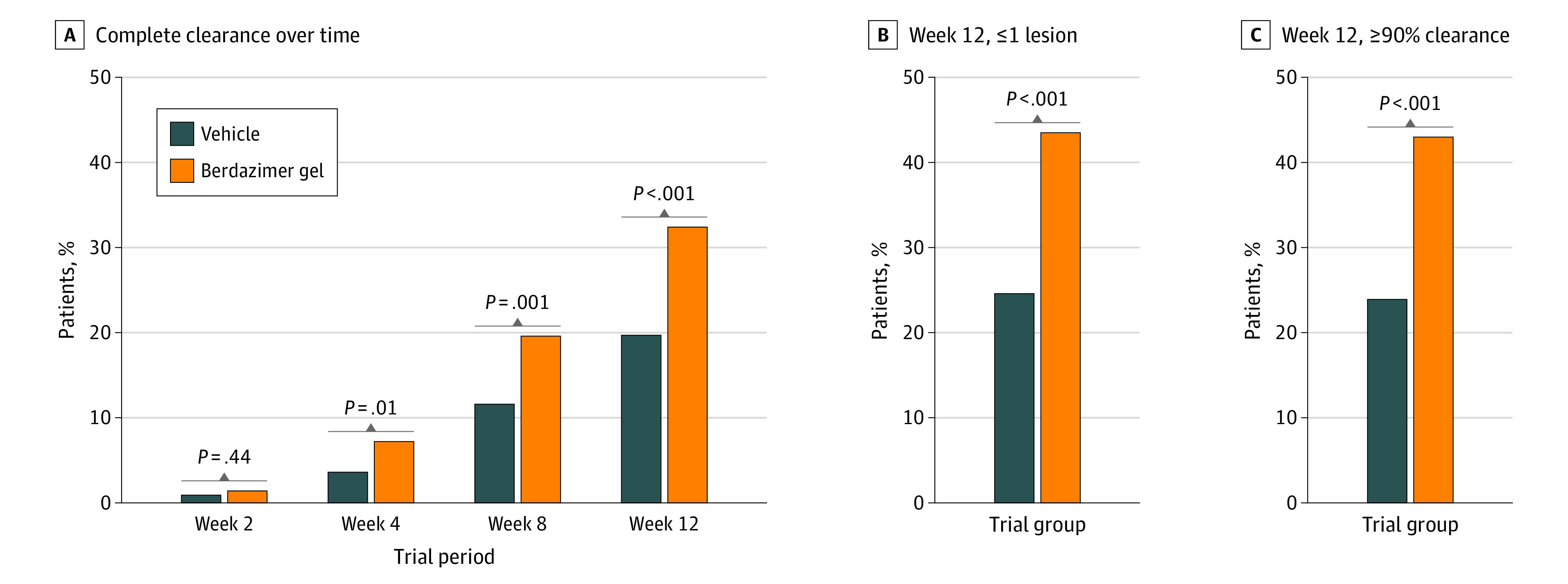
In the ITT population, 88.5% (393 patients) of the berdazimer group and 88.8% (397 patients) of the vehicle group had a lesion count performed at week 12. For the primary efficacy analysis of the ITT population at week 12 using nonresponder imputation, berdazimer demonstrated statistically significant efficacy, with 32.4% (144 patients) achieving complete clearance of all MC lesions compared with 19.7% (88 patients) of the vehicle group (absolute difference, 12.7%; OR, 2.0; 95% CI, 1.5-2.8; P < .001; Figure 2A).
Figure 2. Molluscum Contagiosum Lesion Clearance Through Week 12.

A, Percentage of patients in the ITT population who achieved complete clearance of molluscum contagiosum lesions (defined as having a lesion count of 0) through week 12. B, Percentage of patients in the ITT population with 0 or 1 lesion(s) at week 12. C, Percentage of patients in the ITT population with ≥90% clearance at week 12. P values are based on logistic regression analysis of the treatment differences (berdazimer gel to vehicle) adjusted for the following factors: investigator type (dermatologist vs other), number of patients per household (1 vs 2), baseline BOTE inflammation score (0 vs ≥1), age in years (6 months to <3, 3 to <4, 4 to <5, 5 to <6, 6 to <7, 7 to <8, 8 to <9, 9 to <12, and ≥12), and baseline lesion count. BOTE denotes beginning-of-the-end and ITT, intention-to-treat.
Secondary Efficacy
In the ITT population, 43.5% (193 patients) of the berdazimer group compared with 24.6% (110 patients) of the vehicle group achieved a lesion count of 0 or 1 at week 12 (absolute difference, 18.9%; OR, 2.5; 95% CI, 1.9-3.4; P < .001; Figure 2B). In addition, 43.0% (191 patients) of the berdazimer group compared with 23.9% (107 patients) of the vehicle group achieved a reduction of 90% or greater from baseline MC lesion number at week 12 (OR, 2.4; 95% CI, 1.8-3.3; P < .001; Figure 2C).
At week 8, 88.5% (393 patients) of the berdazimer group and 89.5% (400 patients) of the vehicle group in the ITT population had a lesion count performed. Complete clearance at week 8 was achieved by 19.6% (87 patients) of berdazimer and 11.6% (52 patients) of the vehicle group (OR, 1.88; 95% CI, 1.3-2.8; P = .001; Figure 2A).
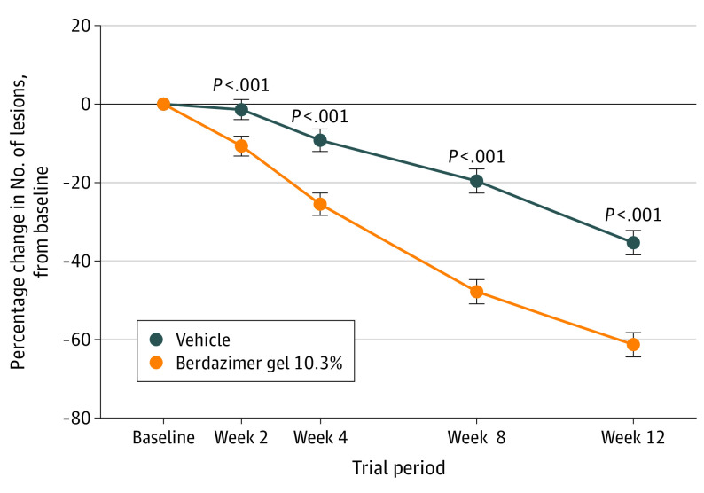
Throughout the study, in the ITT population, mixed-model repeated measures analysis of the least-squares (LS) mean percent change from baseline in MC lesion count showed a greater reduction for berdazimer vs vehicle, beginning as early as week 2 and continuing to week 12 (Figure 3). At week 4, the mean percent reduction was 27.1% for the berdazimer vs 5.6% for vehicle (LS mean adjusted treatment difference, –16.3; 95% CI, –22.6 to –10.0; P < .001). The percentage reduction from baseline was greater for berdazimer than for vehicle at week 8 (mean reduction, 49.3% vs 14.5%; 95% CI, –35.4 to –21.2; P < .001) and at week 12 (mean reduction, 57.5% vs 31.3%; 95% CI, –33.1 to –19.0; P < .001). The IGSA was 0 (no MC lesions) for 33.1% (147 patients) of the berdazimer group vs 19.9% (89 patients) of the vehicle group at week 12, and 51.8% (230 patients) vs 39.6% (177 patients), respectively, at week 24.
Figure 3. Molluscum Contagiosum Lesion Count, Mean Percent Change From Baseline to Week 12.
Data points are LS mean percent change in lesion count from baseline for the ITT population. Error bars indicate the standard error; P values for LS mean differences from vehicle (berdazimer gel to vehicle) are from mixed-model repeated-measures analyses with treatment, visit, treatment-by-visit interaction, investigator type (dermatologist vs other), household number of randomized patients (1 vs 2), baseline BOTE score (0 vs ≥1), age, and baseline lesion count as factors with an unstructured covariance matrix. BOTE denotes beginning-of-the-end; ITT, intention-to-treat, and LS, least-squares.
Safety
Overall, berdazimer treatment was well tolerated, as demonstrated by low AE-related discontinuation rates, and mostly mild or moderate treatment-emergent AEs (TEAE). At least 1 TEAE was reported by 43.0% (191 patients) of the berdazimer group and 23.0% (103 patients) of the vehicle group (Table 2). Most TEAEs were rated as mild or moderate in intensity. Four of 5 berdazimer patients who reported a TEAE of severe intensity had application-site–related events, including pain (n = 1), erythema (n = 1), and dermatitis (n = 2). Application-site reactions were the most common TEAEs overall and the most common reason for treatment discontinuation (Table 2).
Table 2. Summary of Treatment-Emergent Adverse Events, Local Skin Reactions, and Scarring for the Safety Set.
| Event or reaction | Patients, No. (%) | |
|---|---|---|
| Berdazimer gel, 10.3% (n = 444) | Vehicle (n = 447) | |
| Patients with ≥1 TEAEa | 191 (43.0) | 103 (23.0) |
| Mild | 108 (24.3) | 75 (16.8) |
| Moderate | 78 (17.6) | 26 (5.8) |
| Severe | 5 (1.1) | 2 (0.4) |
| TEAEs at application site affecting ≥5% of patients in group, by severity | ||
| Pain | ||
| Mild | 64 (14.4) | 21 (4.7) |
| Moderate | 18 (4.1) | 2 (0.4) |
| Severe | 1 (0.2) | 0 |
| Erythema | ||
| Mild | 25 (5.6) | 5 (1.1) |
| Moderate | 26 (5.9) | 1 (0.2) |
| Severe | 1 (0.2) | 0 |
| Pruritus | ||
| Mild | 25 (5.6) | 4 (0.9) |
| Moderate | 8 (1.8) | 1 (0.2) |
| Severe | 0 | 0 |
| Exfoliation | ||
| Mild | 11 (2.5) | 0 |
| Moderate | 16 (3.6) | 0 |
| Severe | 0 | 0 |
| Dermatitis | ||
| Mild | 8 (1.8) | 1 (0.2) |
| Moderate | 16 (3.6) | 2 (0.4) |
| Severe | 2 (0.5) | 0 |
| Scarb | ||
| Mild | 20 (4.5) | 28 (6.3) |
| Moderate | 1 (0.2) | 0 |
| Severe | 0 | 0 |
| Patients with ≥1 serious TEAE | 0 | 1 (0.2)c |
| Patients with ≥1 TEAE and discontinuation | 18 (4.1) | 3 (0.7) |
| Application site | ||
| Pain | 10 (2.3) | 3 (0.7) |
| Dermatitis | 4 (0.9) | 0 |
| Eczema | 1 (0.2) | 0 |
| Erythema | 1 (0.2) | 1 (0.2) |
| Pruritus | 1 (0.2) | 0 |
| Vesicles | 1 (0.2) | 0 |
| Molluscum contagiosum | 1 (0.2) | 0 |
| Dermatitis contact | 1 (0.2) | 0 |
| LSRs at weeks 2, 4, 8, and 12 | ||
| Mean LSR composite score (0-24)d | ||
| 2 | 2.3 | 0.6 |
| 4 | 2.4 | 0.5 |
| 8 | 1.8 | 0.6 |
| 12 | 1.0 | 0.5 |
| Individual LSR component score e , f | ||
| Erythema, No.g | ||
| 2 | 206 (50) | 100 (24) |
| 4 | 195 (47) | 89 (21) |
| 8 | 166 (42) | 81 (20) |
| 12 | 110 (28) | 82 (21) |
| Severity at week 12, %h | ||
| 0 | 71.8 | 79.3 |
| 1 | 16.9 | 14.1 |
| 2 | 7.9 | 5.5 |
| 3 | 2.6 | 0.5 |
| 4 | 0.8 | 0.5 |
| Flaking/scaling, No.g | ||
| 2 | 141 (34) | 48 (12) |
| 4 | 142 (35) | 37 (9) |
| 8 | 125 (32) | 37 (9) |
| 12 | 79 (20) | 37 (9) |
| Severity at week 12, %h | ||
| 0 | 79.7 | 90.7 |
| 1 | 15.6 | 8.3 |
| 2 | 3.3 | 1.0 |
| 3 | 1.0 | 0 |
| 4 | 0.3 | 0 |
| Crusting, No.g | ||
| 2 | 92 (22) | 25 (6) |
| 4 | 89 (22) | 19 (5) |
| 8 | 82 (21) | 23 (6) |
| 12 | 36 (9) | 20 (5) |
| Severity at week 12, %h | ||
| 0 | 90.8 | 95.0 |
| 1 | 6.9 | 4.3 |
| 2 | 1.3 | 0.8 |
| 3 | 1.0 | 0 |
| 4 | 0 | 0 |
| Swelling, No.g | ||
| 2 | 90 (22) | 17 (4) |
| 4 | 84 (20) | 16 (4) |
| 8 | 64 (16) | 25 (6) |
| 12 | 30 (8) | 17 (4) |
| Severity at week 12, %h | ||
| 0 | 92.3 | 95.7 |
| 1 | 4.6 | 2.8 |
| 2 | 1.8 | 1.0 |
| 3 | 1.3 | 0.5 |
| 4 | 0 | 0 |
| Scarsi | ||
| Week 12 | 13 (2.9) | 10 (2.2) |
| Week 24 | 12 (2.7) | 18 (4.0) |
| Hypo- and hyperpigmentation | ||
| Through week 12 | 6 (1.4) | 0 |
| Through week 24 | 3 (0.7) | 0 |
Abbreviations: AE, adverse event; LSR, local skin reaction; TEAE, treatment-emergent adverse event.
Events that occurred or worsened on or after first application through last application of medication.
The US Food and Drug Administration required that temporary epidermal atrophy from the resolution of a space occupying lesion be captured as a scar.
Humerus fracture deemed not treatment related.
Sum of 6 LSR component scores (range, 0-4 for each component).
Most patients (≥92%) in both groups had an absence of vesiculation/pustulation and erosion/ulceration at week 12; therefore, these data are not shown.
In the berdazimer group, 7 patients had local skin reactions suggestive of allergic contact dermatitis; however, only 1 patient underwent patch testing that confirmed a sensitization reaction.
Percentages for LSRs over time are based on observed data at each time point and represent the sum of all patients with a score of 1, 2, 3, or 4.
Based on a 5-point scale, from 0 (not present) to 4 (severe) described in the eTable in Supplement 2.
Scar assessment was performed at each visit independent of AE assessment. Clinically important scars were reported as AEs. No patients in either group had keloidal or hypertrophic scars during the 24-week study period.
Local Tolerability and Scarring
Mean LSR composite scores (LSR component scores summed, range: 0-24) were highest in the berdazimer group at weeks 2 and 4 (mean, 2.3 and 2.4, respectively), and lowest at week 12 (mean, 1.0; Table 2). If present, LSRs were mostly scored as 1 or 2 on the 5-point scale (Table 2). Throughout the study period, erythema rated as 1 or 2 was the most frequently observed LSR in patients in the berdazimer group, affecting up to 50% of patients at week 2 compared with 25% of patients in the vehicle group. Crusting, swelling, vesiculation or pustulation, and erosion or ulceration were absent in most patients (>90%) at week 12. No patients in either group had keloidal or hypertrophic scars during the 24-week study period (Table 2).
Discussion
Highly contagious, MC is a viral infection that may persist for months to years, with lesions appearing and/or spreading to different areas of the body over time. Thus, control of lesion persistence and spread is a cornerstone of therapeutic success and is indicated by complete lesion clearance.
The B-SIMPLE4 trial demonstrated that berdazimer, applied topically once daily for 12 weeks by patients or caregivers, was significantly more effective than vehicle in achieving complete lesion clearance and reduced lesion counts. The B-SIMPLE4 differs from previous phase 3 study designs (B-SIMPLE1 and B-SIMPLE2) in 4 key aspects: (1) a larger sample size (larger than B-SIMPLE1 + B-SIMPLE2 combined); (2) 1:1 vs 2:1 randomization (berdazimer:vehicle); (3) stratification by baseline BOTE sign; and (4) patient-retention efforts at week 12 to ensure that lesion clearance and AEs were captured at this time point, even in patients who had experienced complete clearance or had an AE before week 12. In the absence of thresholds to interpret meaningful within-patient changes in lesion count, the totality of the B-SIMPLE4 data, including statistically significant improvements in the primary and all secondary end points, suggest clinical meaningfulness. Complete clearance at week 12 is a rigorous end point, and the 27% reduction in mean lesion count by week 4 would likely be perceived by patients and caregivers as meaningful and would encourage them to maintain compliance with the prescribed 12-week therapy. Indeed, an exit interview revealed that 23 of 30 patients or caregivers in B-SIMPLE4 who did not achieve complete clearance considered the lesion count reduction at week 12 to be meaningful. No safety concerns were observed in this study and berdazimer was well tolerated.
The safety profile of berdazimer was consistently favorable, with most TEAEs, including application-site pain and erythema, being mild to moderate in severity. Transient and reversible erythema, possibly accompanied by a stinging or burning sensation, is an expected pharmacologic effect of topical nitric oxide application. No systemic treatment-related AEs, except crying and insomnia, were reported with topical application of berdazimer. The mean LSR composite score was highest at weeks 2 to 4 and then declined, indicating minimal potential for cumulative irritant contact dermatitis. Together, to our knowledge, these 3 phase 3 randomized clinical studies of berdazimer represent the largest interventional study cohort of patients with MC (N = 1598), with 917 exposed to berdazimer.
Currently, there is no FDA-approved medication for MC treatment and available treatments have notable limitations, such as requiring repeated in-office visits for administration.8,11 Cantharidin application, often the therapy of choice for pediatric patients, may result in pain. For example, application-site pain of varying severity affected approximately two-thirds of patients in 2 clinical trials of an in-office cantharidin drug-device combination.11 In addition, cantharidin has the potential to produce severe, painful blistering with inappropriate administration or aftercare. Nitric oxide is a ubiquitous gas with antiviral and immunomodulatory properties.18 Berdazimer gel overcomes the challenge of targeted topical nitric oxide delivery and holds promise as an effective and safe treatment for MC. If approved, berdazimer would provide the only self- or caregiver-administered prescription medication option for MC treatment.
Limitations
While to our knowledge, BSIMPLE4 included the largest cohort of patients with MC randomized in a controlled trial (n = 891), sample sizes for robust subgroup analyses by race, ethnicity, or age were insufficient. Berdazimer efficacy in patients with sexually transmitted MC is unknown, and concomitant use with other topical therapies for MC was not evaluated. Primary efficacy and safety assessments were limited to 12 weeks; after discontinuation of treatment at week 12, week 24 response based on an IGSA score of 0 suggests continuous improvement and durability of effect in the berdazimer group. Long-term safety of continuous topical berdazimer sodium use for 52 weeks has been evaluated in acne.22 Finally, although the antiviral effects of berdazimer have been demonstrated,19,20,21,23 the exact mechanism of action is unknown.
Conclusions
This phase 3 randomized clinical trial of berdazimer, 10.3%, found that this topical, once-daily, nitric oxide−releasing medication may be a novel approach to MC treatment. To date, the B-SIMPLE4 is the largest randomized clinical trial of a medication for MC treatment to our knowledge, and it demonstrated the efficacy and safety of berdazimer in patients 6 months and older. Berdazimer is under consideration as a first-in-class therapeutic agent for MC and may provide a topical prescription alternative to other therapies used for this highly contagious and psychosocially challenging skin condition.
Trial Protocol
eMethods 1: Investigator Training for Diagnosis and Molluscum Contagiosum (MC) Lesion Counts
eMethods 2: Analyses of Secondary End Points
eTable 1. Local Skin Reaction Score
Data Sharing Statement
References
- 1.Chen X, Anstey AV, Bugert JJ. Molluscum contagiosum virus infection. Lancet Infect Dis. 2013;13(10):877-888. doi: 10.1016/S1473-3099(13)70109-9 [DOI] [PubMed] [Google Scholar]
- 2.Schofield JK, Fleming D, Grindlay D, Williams H. Skin conditions are the commonest new reason people present to general practitioners in England and Wales. Br J Dermatol. 2011;165(5):1044-1050. doi: 10.1111/j.1365-2133.2011.10464.x [DOI] [PubMed] [Google Scholar]
- 3.Dohil MA, Lin P, Lee J, Lucky AW, Paller AS, Eichenfield LF. The epidemiology of molluscum contagiosum in children. J Am Acad Dermatol. 2006;54(1):47-54. doi: 10.1016/j.jaad.2005.08.035 [DOI] [PubMed] [Google Scholar]
- 4.Shisler JL. Immune evasion strategies of molluscum contagiosum virus. Adv Virus Res. 2015;92:201-252. doi: 10.1016/bs.aivir.2014.11.004 [DOI] [PubMed] [Google Scholar]
- 5.Butala N, Siegfried E, Weissler A. Molluscum BOTE sign: a predictor of imminent resolution. Pediatrics. 2013;131(5):e1650-e1653. doi: 10.1542/peds.2012-2933 [DOI] [PubMed] [Google Scholar]
- 6.Olsen JR, Gallacher J, Finlay AY, Piguet V, Francis NA. Time to resolution and effect on quality of life of molluscum contagiosum in children in the UK: a prospective community cohort study. Lancet Infect Dis. 2015;15(2):190-195. doi: 10.1016/S1473-3099(14)71053-9 [DOI] [PubMed] [Google Scholar]
- 7.van der Wouden JC, van der Sande R, Kruithof EJ, Sollie A, van Suijlekom-Smit LW, Koning S. Interventions for cutaneous molluscum contagiosum. Cochrane Database Syst Rev. 2017;5(5):CD004767. [DOI] [PMC free article] [PubMed] [Google Scholar]
- 8.Gerlero P, Hernández-Martín Á. Update on the treatment of molluscum contagiosum in children. Actas Dermosifiliogr (Engl Ed). 2018;109(5):408-415. Engl Ed. doi: 10.1016/j.ad.2018.01.007 [DOI] [PubMed] [Google Scholar]
- 9.Olsen JR, Piguet V, Gallacher J, Francis NA. Molluscum contagiosum and associations with atopic eczema in children: a retrospective longitudinal study in primary care. Br J Gen Pract. 2016;66(642):e53-e58. doi: 10.3399/bjgp15X688093 [DOI] [PMC free article] [PubMed] [Google Scholar]
- 10.Leung AKC, Barankin B, Hon KLE. Molluscum contagiosum: an update. Recent Pat Inflamm Allergy Drug Discov. 2017;11(1):22-31. doi: 10.2174/1872213X11666170518114456 [DOI] [PubMed] [Google Scholar]
- 11.Eichenfield LF, McFalda W, Brabec B, et al. Safety and efficacy of VP-102, a proprietary, drug-device combination product containing cantharidin, 0.7% (w/v), in children and adults with molluscum contagiosum: two phase 3 randomized clinical trials. JAMA Dermatol. 2020;156(12):1315-1323. doi: 10.1001/jamadermatol.2020.3238 [DOI] [PMC free article] [PubMed] [Google Scholar]
- 12.Katz KA. Imiquimod is not an effective drug for molluscum contagiosum. Lancet Infect Dis. 2014;14(5):372-373. doi: 10.1016/S1473-3099(14)70728-5 [DOI] [PubMed] [Google Scholar]
- 13.Katz KA. Dermatologists, imiquimod, and treatment of molluscum contagiosum in children: righting wrongs. JAMA Dermatol. 2015;151(2):125-126. doi: 10.1001/jamadermatol.2014.3335 [DOI] [PubMed] [Google Scholar]
- 14.Katz KA, Swetman GL. Imiquimod, molluscum, and the need for a better “Best Pharmaceuticals for Children” Act. Pediatrics. 2013;132(1):1-3. doi: 10.1542/peds.2013-0116 [DOI] [PubMed] [Google Scholar]
- 15.Go U, Nishimura-Yagi M, Miyata K, Mitsuishi T. Efficacy of combination therapies of topical 5% imiquimod and liquid nitrogen for penile molluscum contagiosum. J Dermatol. 2018;45(10):e268-e269. doi: 10.1111/1346-8138.14319 [DOI] [PubMed] [Google Scholar]
- 16.Stasko N, McHale K, Hollenbach SJ, Martin M, Doxey R. Nitric oxide-releasing macromolecule exhibits broad-spectrum antifungal activity and utility as a topical treatment for superficial fungal infections. Antimicrob Agents Chemother. 2018;62(7):e01026-17. doi: 10.1128/AAC.01026-17 [DOI] [PMC free article] [PubMed] [Google Scholar]
- 17.Matthews JR, Botting CH, Panico M, Morris HR, Hay RT. Inhibition of NF-kappaB DNA binding by nitric oxide. Nucleic Acids Res. 1996;24(12):2236-2242. doi: 10.1093/nar/24.12.2236 [DOI] [PMC free article] [PubMed] [Google Scholar]
- 18.Banerjee NS, Moore DW, Wang HK, Broker TR, Chow LT. NVN1000, a novel nitric oxide-releasing compound, inhibits HPV-18 virus production by interfering with E6 and E7 oncoprotein functions. Antiviral Res. 2019;170:104559. doi: 10.1016/j.antiviral.2019.104559 [DOI] [PubMed] [Google Scholar]
- 19.Tyring SK, Rosen T, Berman B, Stasko N, Durham T, Maeda-Chubachi T. A phase 2 controlled study of SB206, a topical nitric oxide-releasing drug for extragenital wart treatment. J Drugs Dermatol. 2018;17(10):1100-1105. [PubMed] [Google Scholar]
- 20.Hebert AA, Siegfried EC, Durham T, et al. Efficacy and tolerability of an investigational nitric oxide-releasing topical gel in patients with molluscum contagiosum: a randomized clinical trial. J Am Acad Dermatol. 2020;82(4):887-894. doi: 10.1016/j.jaad.2019.09.064 [DOI] [PubMed] [Google Scholar]
- 21.Maeda-Chubachi T, Hebert D, Messersmith E, Siegfried EC. SB206, a nitric oxide-releasing topical medication, induces the beginning of the end sign and molluscum clearance. JID Innov. 2021;1(3):100019. doi: 10.1016/j.xjidi.2021.100019 [DOI] [PMC free article] [PubMed] [Google Scholar]
- 22.Hebert AA, Del Rossi J, Rico M, et al. Evaluation of the efficacy, safety and tolerability of SB204 4% once-daily in subjects with moderate to severe acne vulgaris treated topically for up to 52 weeks. 2018 American Academy of Dermatology Annual Meeting; February 16-20, 2018. San Diego, CA. [Google Scholar]
- 23.McHale KBK, Wang HK, Hollenbach S, et al. In vitro and in vivo efficacy of nitric oxide-releasing antiviral therapeutic agents. Society for Investigative Dermatology Annual Meeting; May 14, 2016; Scottsdale, AZ. [Google Scholar]
Associated Data
This section collects any data citations, data availability statements, or supplementary materials included in this article.
Supplementary Materials
Trial Protocol
eMethods 1: Investigator Training for Diagnosis and Molluscum Contagiosum (MC) Lesion Counts
eMethods 2: Analyses of Secondary End Points
eTable 1. Local Skin Reaction Score
Data Sharing Statement


