Abstract
Background
Malaria remains endemic in Bangladesh, with the majority of cases occurring in forested, mountainous region in the Chittagong Hill Tracts (CHT). This area is home to Bengali and diverse groups of indigenous people (Pahari) residing largely in mono-ethnic villages.
Methods
1002 individuals of the 9 most prominent Pahari and the Bengali population were randomly selected and screened by RDT and qPCR. Parasites were genotyped by msp2 and deep sequencing of 5 amplicons (ama1-D3, cpmp, cpp, csp, and msp7) for Plasmodium falciparum (n = 20), and by microsatellite (MS) typing of ten loci and amplicon sequencing of msp1 for Plasmodium vivax (n = 21). Population structure was analysed using STRUCTURE software. Identity-by-state (IBS) was calculated as a measure of parasite relatedness and used to generate relatedness networks.
Results
The prevalence of P. falciparum and P. vivax infection was 0.7% by RDT (P. falciparum 6/1002; P. vivax 0/1002, mixed: 1/1002) and 4% by qPCR (P. falciparum 21/1002; P. vivax 16/1002, mixed: 5/1002). Infections were highly clustered, with 64% (27/42) of infections occurring in only two Pahari groups, the Khumi and Mro. Diversity was high; expected heterozygosity was 0.93 for P. falciparum and 0.81 for P. vivax. 85.7% (18/21) of P. vivax and 25% (5/20) of P. falciparum infections were polyclonal. No population structure was evident for either species, suggesting high transmission and gene flow among Pahari groups.
Conclusions
High subclinical infection prevalence and genetic diversity mirror ongoing transmission. Control activities should be specifically directed to Pahari groups at greatest risk.
Supplementary Information
The online version contains supplementary material available at 10.1186/s12936-022-04236-0.
Keywords: Malaria, Plasmodium falciparum, Plasmodium vivax, Bangladesh, Chittagong Hill Tracts, Pahari, Ethnic groups, Genotyping, Population structure, Clustering
Background
The Bangladesh National Malaria Control Programme has made significant progress towards controlling malaria within the country, reducing the incidence of malaria and mortality by 40% between 2015 and 2020 [1]. However, malaria remains hypoendemic in several eastern districts with an estimated 17 million people at risk [1]. The epidemiology of malaria in Bangladesh is complex, with four parasite species present. Infections with Plasmodium falciparum account for 86% of all clinical cases, Plasmodium vivax for 12%, and Plasmodium malariae and Plasmodium ovale account for remaining cases [1]. The spatial distribution of clinical cases is highly heterogeneous, with the greatest incidence recorded in the mountainous, forested Chittagong Hill Tracts (CHT) encompassing Bandarban, Rangamati, and Khagrachhari districts in the southeastern part of the country neighbouring Myanmar and India. Despite this region contributing only 7% of the national population, approximately 90% of malaria cases in Bangladesh are reported from the CHT [2]. Distribution of malaria is heterogeneous within the CHT as well, with 85% of malaria cases attributable to 11% of the population at risk for malaria [2].
The CHTs are home to at least 12 diverse indigenous groups, collectively known as Pahari, as well as increasing numbers of non-indigenous Bengali. Specific risk factors for malaria include jhum cultivation (the practice of forest-based shifting cultivation) and residing close to dense forest and at higher elevations [3–5]. The risk of malaria and subclinical P. falciparum infection vary among Pahari groups, likely due to differences in lifestyle and geographic location [3, 5, 6].
To ensure ongoing progress towards malaria elimination, it is critical that malaria control efforts are targeted to those at the greatest risk of infection, including parasite reservoirs that sustain ongoing transmission [7, 8]. Subclinical infections represent a key challenge for malaria elimination, as they are not identified by passive detection strategies. For instance, in a mass survey of tribal communities in the Indian Balaghat district, slide positivity rates of 32.4% and 29.0% were reported for febrile and afebrile individuals, respectively [9]. Furthermore, in nearly all transmission settings, many subclinical infections are below the limit of detection of microscopy or rapid diagnostic test [10, 11]. A survey in the CHT conducted between 2009 and 2012 reported a 1% prevalence of subclinical P. falciparum infection by light microscopy, which persisted year-round [5]. The true infection rate was likely higher; more sensitive molecular methods, such as qPCR, are required to detect these infections. The prevalence of subclinical infections and associated risk factors have been well described for regions where the transmission intensity of P. falciparum and P. vivax is moderate or high; however, few studies have been conducted in low endemic, pre-elimination settings, and in regions where P. malariae and P. ovale are also present.
Parasite genotyping is being applied increasingly to provide a better understanding of residual transmission [12–14]. If infections among villages or at-risk groups are closely related and separated from other groups, most transmission is predicted to occur within these groups. In contrast, high parasite genetic diversity and absence of population structure indicate transmission among groups [13, 15–17].
To understand transmission patterns and differences in infection prevalence, residents of the CHT were enrolled into a cross sectional survey and blood samples were screened for any Plasmodium spp. infection using qPCR [18]. Genotyping by size-polymorphic markers and amplicon deep-sequencing was applied to assess relationships among infections within and between ethnic groups.
Methods
Ethics statement
The study obtained ethical approval from the Ethics Review Committee of the icddr,b, Bangladesh (PR-15021), the Human Research Ethics Committee of the Northern Territory Department of Health and Menzies School of Health Research, Australia (HREC 2015–2336), and the University of Notre Dame Institutional Review Board (18-09-4875). Written informed consent was collected from all participants or their legal guardians prior to enrollment and in addition, written assent was collected from all minors above the age of 11 years [19].
Sample collection
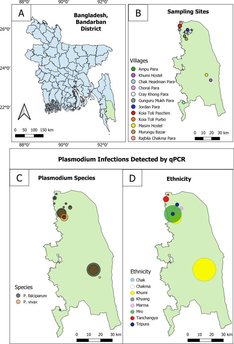
As the aim of this study was to understand malaria epidemiology among different ethnic groups in the CHT, villages were purposively selected to represent different ethnicities [19]. Most villages in the CHT are mono-ethnic. Multiple villages were visited when necessary to sample roughly 100 individuals among the Bengali and each of the 9 targeted Pahari groups. The collection area spanned a range of approximately 150 km from the north to the south of the CHT (Fig. 1). Between August 2015 and January 2016 (overlapping with the June to October monsoon season), venous blood samples (5 mL) were collected in EDTA Vacutainers (BD, USA) from individuals aged more than five years old irrespective of the presence of symptoms of malaria. Blood for rapid diagnostic tests (RDTs) was collected by finger prick from the same individuals.
Fig. 1.
A Location of Bandarban District in Bangladesh. B Villages sampled for this study (24 of 28). Villages in which Plasmodium spp. samples were detected are labelled by colour. Villages in which no infections were detected are marked with a (+). Not pictured are Faruk Para, Hebron Para, Hati Bhanga Para, Paschim Antaha Para, and Purbo Antaha Para for which GPS location was not recorded. C Distribution of Plasmodium spp. infections detected by qPCR. Plasmodium falciparum and P. vivax were both found throughout the study area. D The majority of infections were clustered among the Khumi and Mro
Laboratory methods
DNA was extracted from 200 µL blood using the Qiagen DNeasy kit according to manufacturer’s instructions. 4 µL of DNA, corresponding to 4 µL of blood, were screened by qPCR for P. falciparum using the varATS assay [20], and for P. vivax using the cox1 assay [21]. The varATS assay targets multicopy genes and amplifies approximately 20 copies per genome. Cox1 is a mitochondrial gene and present in approximately 10 copies per genome. Samples were screened for P. ovale and P. malariae using single gene assays [22].
Parasite genotyping
Plasmodium falciparum-positive samples were genotyped by size-polymorphic msp2 and 5 highly diverse amplicons. msp2 typing was done by nested PCR followed by capillary electrophoresis [23]. Amplicon deep sequencing was done for markers apical membrane antigen 1 (ama1-D3, PF3D7_1133400), circumsporozoite surface protein (csp, PF3D7_0304600), merozoite surface protein 7 (msp7, PF3D7_1335100), a conserved Plasmodium membrane protein (cpmp, PF3D7_0104100), and a conserved Plasmodium protein (cpp, PF3D7_1475800) [24].
Plasmodium vivax-positive samples were typed by 10 microsatellite markers (MS1, MS2, MS4, MS5, MS6, MS7, MS9, MS10, MS15, MS20) [25] and one amplicon (Pvmsp1). Peaks higher than 300 units of relative fluorescent intensity and above background noise were considered true clones. Peaks less than 1/3 the height of the highest peak were not recorded. Alleles were grouped using TANDEM software [26]. Amplicon deep sequencing was done for merozoite surface protein 1 (Pvmsp1, PVP01_0728900) as described [27] with the following modifications: the 5′ ends of the forward and reverse primers for the nested PCR included the overhang sequences 5′-GTGACCTATGAACTCAGGA-3′ and 5′-CTGAGACTTGCACATCGCAGC-3′, respectively.
PCR products were run on a 2% agarose gel, then 4uL of PCR product from each sample was combined to create pools of similar marker concentration as estimated from band intensities on the gel. These pools were purified using AMPure XP beads (Beckman Coulter) and DNA concentrations were measured using a Qubit fluorometer (Thermo Fisher Scientific). Pools were diluted to 0.1ug/uL and combined to form the sequencing library. The library was sequenced in paired-end mode in one run using the Illumina MiSeq reagent kit v3 600 cycle (2 × 300 bp) with 15% Enterobacteria phage phiX control v3 (Illumina).
Plasmodium falciparum positive samples were typed for pfhrp2 deletion using a protocol based on droplet digital PCR (ddPCR) [28].
Data analysis
Analyses were conducted using R software version 4.0.0 (Additional file 2) [29]. Haplotypes were determined using the bioinformatic pipeline from HaplotypR package version 0.3.3 [30]. Amplicon sequencing reads were demultiplexed by sample and marker. Overlapping paired end reads were merged via the vsearch package and clustered via the swarm package. Samples with ≤ 10 reads were excluded from further analysis. Single nucleotide polymorphisms were required to have a mismatch rate of at least 0.5 occurring in two or more samples for further consideration. Chimeric reads, singletons, and reads containing an insertion or deletion were excluded. Haplotype calling from the remaining reads required within-host haplotype frequency ≥ 1% and minimum coverage of 3 reads per haplotype and 25 reads per sample.
Statistical analyses were done in the R base package. Prevalence and parasitaemia were compared across age groups (5–10, 11–20, 21–30, 31–40, 41–50, 51–60, and > 60 years) by analysis of variance (ANOVA) and Tukey Post Hoc tests. Pearson’s Chi-squared tests were performed to test for differences in prevalence by sex, ethnicity, and village. Simple logistic regression was performed to separately compare participants’ village and ethnicity as predictors of infection. Multivariate regression was not performed due to high correlation between villages and ethnicities. Multiplicity of infection (MOI) for P. falciparum was defined as the maximum number of alleles detected for any one amplicon in a sample. MOI for P. vivax was defined as the number of alleles detected for the MS with the second most number of alleles to prevent overestimating MOI due to presence of PCR artifacts [31]. Expected heterozygosity (He) and a modified identity-by-state (IBS) metric were calculated using R software version 3.6.3 (Company, Country) [32]. A modified IBS metric from Tessema, 2019 [32] was used to measure pairwise relatedness between infections based on all alleles detected, giving consideration to minor clones in polyclonal infections. For P. falciparum, IBS was calculated using data from the 5 amplicons. For P. vivax, IBS was calculated using data from the 10 MSs and the one amplicon. Structure plots were created with the rmaverick package in R, using no admixture and considering only the dominant clone in each sample. Relatedness networks were generated using R package igraph, with an IBS threshold of 0.5 for each link. This value is the theoretical relatedness of meiotic siblings; however, it is a less restrictive threshold than the more rigorous IBS ≥ 0.6 used for identification of highly related infections in some studies [33, 34]. Principal components analysis (PCA) was conducted using R package ade4 considering the dominant alleles at 5 amplicon markers for P. falciparum and at 1 amplicon and 10 MS markers for P. vivax.
Results
Prevalence of infection
In total, 1002 samples were collected from 28 villages, with 9–100 individuals sampled per village (Fig. 1, Additional file 1). 900 individuals were surveyed from 9 Pahari groups (99–101 individuals per group) as well as 100 individuals who identified as Bengali. Two individuals from a tenth Pahari group, the Lushai, were sampled in a majority Bawm village. Median age of participants was 31 years (range 5–80). Overall, 59.9% (600/1002) of individuals were female. Two individuals reported travel in the prior 4 weeks, one individual reported fever within the previous 48 h, and two individuals reported having had malaria in the last 90 days. These numbers were too low to assess them as risk factors for infection.
In total 42/1002 (4.2%) of individuals tested positive by qPCR for P. falciparum and/or P. vivax (21 P. falciparum mono-infection, 16 P. vivax mono-infection, 5 mixed) (Table 1). No P. ovale or P. malariae infections were detected. Only 7/1002 (0.70%) individuals tested positive by RDT or microscopy, one of which was not confirmed by qPCR. RDT and microscopy detected 6/26 P. falciparum infections and 1/5 mixed infections (Table 1). All individuals who tested positive by RDT or microscopy were aged 7 to 20 years.
Table 1.
Plasmodium spp. infections detected by each diagnostic method
| Diagnostic | P. falciparum | P. vivax | Mixed | Total |
|---|---|---|---|---|
| qPCR | 21 | 16 | 5 | 42 |
| RDT | 6 | 0 | 1 | 7 |
| Microscopy | 3 | 0 | 1 | 4 |
Many more infections were detected by qPCR than by RDT or microscopy
One P. falciparum infection detected by RDT was not confirmed by qPCR
Prevalence by qPCR was highest in individuals aged between 11 and 20 years (7.69%, 16/208), and lowest in individuals older than 60 years (1.25%, 1/80). The only significant difference was between the 11–20 years and 21–30 years (1.62%, 3/185) age groups (ANOVA: p = 0.045; Tukey Post Hoc: p = 0.043) (Table 2). The prevalence in men was 5.97%, (24/402) compared to 3.00% (18/600) in females (p = 0.021). Parasitaemia did not differ among age groups for either P. falciparum (p = 0.545) or P. vivax (p = 0.749).
Table 2.
Age distribution of participants with Plasmodium spp. Infections
| Age (yrs) | n | Infections | Prevalence (%) |
|---|---|---|---|
| 5–10 | 98 | 5 | 5.10 |
| 11–20 | 208 | 16 | 7.69 |
| 21–30 | 185 | 3 | 1.62 |
| 31–40 | 207 | 8 | 3.86 |
| 41–50 | 143 | 4 | 2.80 |
| 51–60 | 81 | 5 | 6.17 |
| > 60 | 80 | 1 | 1.25 |
| Total | 1002 | 42 |
The highest prevalence was in the 11–20 age group (7.69%) and the lowest prevalence was in the > 60 age group (1.25%)
There were significant differences in the prevalence of parasitaemia between ethnic groups (P < 0.0001), with 0% (0/100) of Bengali participants infected and 64% (27/42) infections concentrated among two of the ten Pahari groups (Table 3). In total, 18% (18/100) of Khumi and 9% (9/100) of Mro participants were infected. Among the other 8 ethnic groups, less than 4 individuals had parasitaemia detected by qPCR in each group. The differences in ethnicity were not reflected in RDT results. Among 10 Pahari groups, one or two positive RDTs were recorded in five groups.
Table 3.
Distribution of Plasmodium spp. infections among ethnicities as detected by qPCR and RDT
| qPCR | RDT | ||||
|---|---|---|---|---|---|
| Ethnicity | P. vivax | P. falciparum | Mixed | Positive (%) | Positive (%) |
| Khumi | 9 | 7 | 2 | 18 | 1 |
| Mro | 4 | 4 | 1 | 9 | 1 |
| Tripura | 1 | 2 | 1 | 4 | 0 |
| Tanchangya | 0 | 3 | 1 | 4 | 2 |
| Marma | 0 | 3 | 0 | 3 | 2 |
| Chak | 1 | 1 | 0 | 2 | 0 |
| Chakma | 1 | 0 | 0 | 1 | 0 |
| Khyang | 0 | 1 | 0 | 1 | 1 |
| Bawm | 0 | 0 | 0 | 0 | 0 |
| Lushai | 0 | 0 | 0 | 0 | 0 |
| Bengali | 0 | 0 | 0 | 0 | 0 |
| Total | 16 | 21 | 5 | 4.2 | 0.70 |
A much higher infection prevalence was detected by qPCR than by RDT
Plasmodium vivax and P. falciparum were detected in similar numbers (Pv: n = 16, Pf: n = 21, Mixed: n = 5)
Infections were highly clustered, with 64% occurring in the Khumi and Mro groups (27/42)
No infections were detected among Bengalis (0/100)
There were significant differences in the prevalence of parasitaemia between villages (p < 0.0001), with no infections detected in 50% (14/28) of villages. All Mro were sampled in one village (Ampu Para), where prevalence was 9% (9/100). Khumi were sampled in four villages, and prevalence was high in two of them: 12% (6/51) in Murungu Bazar, and 36% (12/33) Masim Hostel (Fig. 1).
Simple logistic regression identified two ethnicities as significant predictors of infection, Khumi (p < 0.0001) and Mro (P = 0.001), while village was not a significant predictor (p = 0.006).
Parasite genotyping
All 21 individuals with P. vivax parasitaemia were successfully genotyped. Among 12 samples successfully typed by amplicon deep sequencing, the median coverage for amplicon marker msp1 was 11,342 reads. Genetic diversity was high, with 5–11 alleles per MS marker (mean He = 0.81) and 7 alleles for msp1. MOI ranged from 1 to 3 (median = 2.0) and 85% (18/21) were polyclonal.
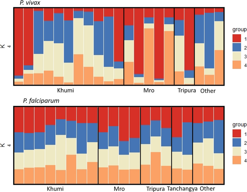
STRUCTURE plots were generated to infer population structure based on alleles of the major clones in each sample. Plots were generated using k = 2–5 as the majority of infection samples came from 2 Pahari groups, with less representation from a few other groups. There was no obvious population structure for P. vivax (Fig. 2).
Fig. 2.
Structure plots showed no structure among P. falciparum or P. vivax populations. Plots were generated using k = 2–5, and all plots looked very similar
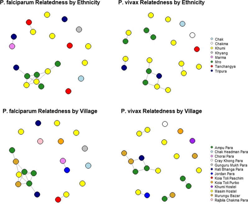
The mean P. vivax IBS within ethnicities was 0.14 for the Khumi (n = 11), 0.31 for the Mro (n = 5), and 0.29 for Tripura (n = 2) (Table 4). No two samples had all 11 alleles in common. Relatedness networks were generated using a minimum threshold of IBS ≥ 0.5 for each link to generate the theoretical relatedness of meiotic siblings. Only one pair and one triplet of infections showed IBS > 0.5. These small networks included four of the five samples collected from Mro, and one Tripura sample. The Tripura sample was taken from the Hati Bhanga village adjacent to the Mro Ampu village from which all five Mro samples were collected.
Table 4.
Identity-by-state among Plasmodium spp. samples
| Ethnicity | n | Mean IBS |
|---|---|---|
| P. vivax | ||
| Khumi | 11 | 0.14 |
| Mro | 5 | 0.31 |
| Tripura | 2 | 0.29 |
| All P. vivax | 21 | 0.15 |
| P. falciparum | ||
| Khumi | 8 | 0.12 |
| Mro | 4 | 0.50 |
| Tripura | 3 | 0.00 |
| Tanchangya | 2 | 0.05 |
| All P. falciparum | 20 | 0.11 |
Parasite relatedness within Pahari groups as measured by IBS. For both P. vivax and P. falciparum, mean IBS was greatest among parasites detected among the Mro
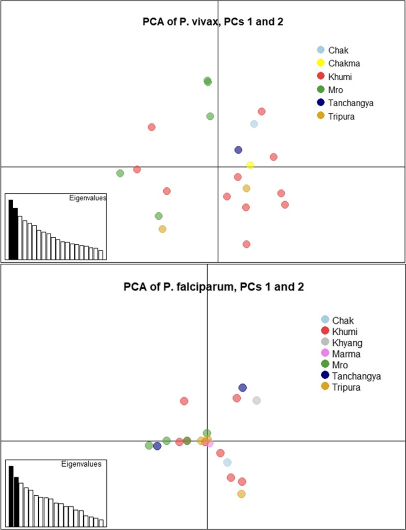
By PCA, P. vivax showed some separation of samples by ethnicity with the Khumi and Mro forming loose clusters, although the Khumi samples appeared evenly throughout most of the plot.
In total 76.9% (20/26) of individuals with P. falciparum parasitaemia were successfully genotyped by msp2 size-polymorphic marker and 5 amplicons. The median coverage for amplicon markers was 14,729 reads (range 364–68,910, 2.5 and 97.5 percentiles) per amplicon marker and sample. Genetic diversity was high with 14 msp2 alleles (3 FC27 and 11 3D7) and 8–15 alleles per amplicon marker (mean He = 0.93 across all amplicon markers). The MOI ranged from 1 to 5 (median = 1) and 20% (4/20) of infections were polyclonal. None of the isolates had a pfhrp2 deletion.
As for P. vivax, STRUCTURE analysis revealed no population structure for P. falciparum (Fig. 2), and none of P. falciparum isolates (with haplotypes called at more than 1 marker) had identical haplotypes. The mean IBS within ethnicities was 0.12 for the Khumi (n = 8), 0.50 for Mro (n = 4), 0.0 for Tripura (n = 3), and 0.05 Tanchangya (n = 2) (Table 4). Only the P. falciparum from the Mro had a mean IBS of at least 0.5, indicative of close relatedness.
Plasmodium falciparum infections showed a low level of clustering, with 40% (8/20) of isolates linked in a small network including samples from the Khumi (n = 3), Mro (n = 4), and Tripura (n = 1) (Fig. 3). Parasite isolates from Khumi individuals in this small network included 2/2 from the northern Murungu Bazar village and 1/6 from the southern village near Masim Hostel 75 km away. The two samples from Murungu Bazar were not linked to each other, but were connected in the network by the sample from Masim Hostel.
Fig. 3.
Relatedness networks based on pairwise IBS ≥ 0.5. In the top row, samples are colored according to ethnicity; in the bottom row, samples are colored by village. Among P. falciparum there is a small network of related infections from the Khumi and Mro, plus one infection from the Tripura. Among P. vivax there are two smaller networks of related infections from the Mro plus one infection from the Tripura. There are very few linkages for either parasite species
By PCA, the P. falciparum haplotypes showed very little separation of samples, with samples appearing closely around the origin (Fig. 4). All 5 Mro samples clustered fairly close together, although they overlapped with samples from the Khumi, Tripura, and Tanchangya as well.
Fig. 4.
Principal components analysis based on principal components 1 and 2. Plasmodium vivax haplotypes showed some separation by ethnicity, with the Khumi and Mro forming loose clusters. P. falciparum samples showed very little separation, with samples appearing closely around the origin
Discussion
In the CHT in Bangladesh, infections were highly clustered among Pahari groups, with nearly two thirds of all infections occurring in just two out of ten Pahari groups surveyed, the Khumi (42%, 18/42) and Mro (21%, 9/42). The 1% overall prevalence measured by RDT in this study was the same as that measured by RDT and microscopy on samples collected from the CHT in 2009 to 2012 [5]. As observed in other low-transmission sites [35], the prevalence by qPCR was substantially higher than by RDT or microscopy.
There is significant spatial heterogeneity in the prevalence of infectious diseases, including that for malaria [36–38]. Where transmission intensity is low, control interventions are most cost-effective if targeted towards those at greatest risk [53]; however, few studies have investigated the contribution of ethnic diversity to spatial heterogeneity [39, 40]. These findings suggest that clustering of infections is driven by ethnicity but not by village. Plasmodium spp. infections were far more prevalent among the Mro in Ampu Para and the Khumi in Murungu Bazar than in any of the nearby villages in northern Bandarban. Additionally, prevalence was elevated in a second Khumi village Masim Hostel, despite its location roughly 50 km to the south. In the dense forests of Indian subdistrict or Keshkal, a cross-sectional survey of three closely situated tribal groups demonstrated similar small-scale heterogeneity, with a significantly higher incidence of malaria in one group compared to the other two groups [41].
Despite the clustering of infections within ethnic groups, there was high genetic diversity and limited population structure for both Plasmodium species. Locally increased prevalence of infection in low transmission settings can arise from localized, small outbreaks, in which case, expansion of clonal or nearly related lineages would be expected [42–44]; however, this was not the case in this study in the CHT. The high diversity of parasite populations within villages and absence of population structure reflects ongoing transmission at a sufficiently high level to maintain genetic diversity, and that increased prevalence among certain ethnicities is not the result of within-village transmission of possibly imported cases. A similar pattern of high diversity and lack of population structure despite low transmission has also been observed in other sites, such as in Senegal, South Africa, and Eswanti [34, 45, 46], and suggests that local transmission is likely to be higher than previously predicted [46].
The causes for the pronounced differences in paras ite prevalence yet absence of any indication of small-scale transmission are unknown. Differences in behaviour, housing structure, and treatment-seeking are likely to play a role. A survey of health-seeking behaviour among five ethnic groups in the CHT in 2001 reported that the Mro had the lowest incidence of malaria (based on self-reported symptoms) and were the least likely to seek external healthcare when ill; this could reflect the greater distance to health facilities for the Mro [6]. Although this previous study did not survey any individuals from the Khumi group, both the Khumi and Mro may miss medical interventions and malaria control efforts due to their particular geographic isolation compared to other Pahari groups [6, 47]. Anecdotally, some Khumi believe that they have been excluded from government and NGO programmes aimed at Pahari due to lack of education or ability to speak Bangla [48]. National programmes are typically delivered in Bangla and sometimes with major Pahari languages e.g. Chakma and Marma [48]. An inability to communicate in these languages could present a major obstacle for seeking treatment and participating in vector control programmes. Future studies should consider social factors including participants’ bed net usage, distance to a health facility, and ability to communicate in Bangla. Alternatively, parasite heterogeneity may be attributable to human genetic factors resulting in a decreased risk of infection [49]. Lastly, stochastic effects owing to the inherent heterogeneity of infectious diseases should not be ruled out. A survey across multiple nearby villages in Papua New Guinea found prevalence of infection by PCR to range from 0 to 22% for P. falciparum and from 11 to 34% for P. vivax [50]. These villages were inhabited by the same ethnicities and similar in terms of housing and climate factors.
Although this study did not find any pfhrp2 deletions in P. falciparum samples from the CHT, a deletion has been detected in Sylet in northeastern Bangladesh near the Indian border [51]. Additionally, deletions of pfhrp2 and pfhrp3 genes have been detected in northern India [52]. Continued surveillance for such gene deletions is warranted in this area.
Conclusions
This study detected a substantial burden of subclinical Plasmodium infections clustered among the Khumi and Mro Pahari groups in the CHT. Infections were highly diverse and showed no population structure, indicating sustained transmission of parasites rather than a single, clonal outbreak. To ensure sustained progress towards malaria elimination in Bangladesh, the potential reservoir for ongoing transmission should be identified and targeted for public health control interventions.
Supplementary Information
Additional file 1: Ethnicity, sex, age, and infection data for each participant; Haplotype data for P. vivax and P. falciparum.
Additional file 2: R code used for haplotyping and statistical analysis.
Acknowledgements
We would like to thank all participants for their time and support as well as all staff that has made this survey possible. We thank Anita Lerch for support with amplicon sequencing data analysis.
Abbreviations
- ama1-D3
Apical membrane antigen 1
- CHT
Chittagong Hill Tracts
- cox1
Cytochrome C oxidase subunit 1
- cpmp
Conserved Plasmodium membrane protein
- cpp
Conserved Plasmodium protein
- csp
Circumsporozoite surface protein
- DNA
Deoxyribonucleic acid
- He
Expected heterozygosity
- icddr,b
International Centre for Diarrhoeal Disease Research, Bangladesh
- IBS
Identity-by-state
- MOI
Multiplicity of infection
- MS
Microsatellite
- msp1
Merozoite surface protein 1
- msp2
Merozoite surface protein 2
- msp7
Merozoite surface protein 7
- PCA
Principal component analysis
- pfhrp2
Plasmodium falciparum Histidine rich protein 2
- pfhrp3
Plasmodium falciparum Histidine rich protein 3
- qPCR
Quantitative polymerase chain reaction
- RDT
Rapid diagnostic test
- varATS
var Gene acidic terminal sequence
Author contributions
Conceptualization: WAK, RNP, BL, CK. Investigation: TH, MGK, FTJ, NJ, MSH. Data analysis: TH, MSA. Supervision: CSP, MSA, CK. Writing–original draft: TH and CK. All authors read and approved the final manuscript.
Funding
This project was supported, in part, by the Indiana Clinical and Translational Sciences Institute, funded, in part by Grant Number UL1TR002529 from the National Institutes of Health, National Center for Advancing Translational Sciences, Clinical and Translational Sciences Award; by NIH Grant R21AI137891, the Bill and Melinda Gates Foundation (OPP1054404 and OPP1164105), and the Eck Institute for Global Health at the University of Notre Dame. BL is funded by the Australian Department of Foreign Affairs and Trade. RNP is funded by the Wellcome Trust (Senior Fellowship in Clinical Science, 200909). icddr,b is grateful to the Governments of Bangladesh, Canada, Sweden and the UK for providing core/unrestricted support. No funding bodies had any role in study design, data collection and analysis, decision to publish, or preparation of the manuscript.
Availability of data and materials
The datasets supporting the conclusions of this article are included in Additional File 1. Raw sequence data are available in the Sequence Read Archive repository, BioProject ID PRJNA826830 at http://www.ncbi.nlm.nih.gov/bioproject/826830.
Declarations
Ethics approval and consent to participate
The study obtained ethical approval from the Ethics Review Committee of the icddr,b, Bangladesh (PR-15021), the Human Research Ethics Committee of the Northern Territory Department of Health and Menzies School of Health Research, Australia (HREC 2015–2336), and the University of Notre Dame Institutional Review Board (18–09-4875).
Consent for publication
Not applicable.
Competing interests
The authors declare that they have no competing interests.
Footnotes
Publisher's Note
Springer Nature remains neutral with regard to jurisdictional claims in published maps and institutional affiliations.
Tiffany Huwe and Mohammad Golam Kibria contributed equally to this work
Contributor Information
Benedikt Ley, Email: Benedikt.Ley@menzies.edu.au.
Mohammad Shafiul Alam, Email: shafiul@icddrb.org.
Cristian Koepfli, Email: ckoepfli@nd.edu.
References
- 1.WHO. World malaria report . 20 years of global progress and challenges. Geneva: World Health Organization; 2020. p. 2020. [Google Scholar]
- 2.Noé A, Zaman SI, Rahman M, Saha AK, Aktaruzzaman MM, Maude RJ. Mapping the stability of malaria hotspots in Bangladesh from 2013 to 2016. Malar J. 2018;17:259. doi: 10.1186/s12936-018-2405-3. [DOI] [PMC free article] [PubMed] [Google Scholar]
- 3.Haque U, Soares Magalhães RJ, Mitra D, Kolivras KN, Schmidt WP, Haque R, et al. The role of age, ethnicity and environmental factors in modulating malaria risk in Rajasthali, Bangladesh. Malar J. 2011;10:367. doi: 10.1186/1475-2875-10-367. [DOI] [PMC free article] [PubMed] [Google Scholar]
- 4.Galagan SR, Prue CS, Khyang J, Khan WA, Ahmed S, Ram M, et al. The practice of jhum cultivation and its relationship to Plasmodium falciparum infection in the Chittagong Hill Districts of Bangladesh. Am J Trop Med Hyg. 2014;91:374–383. doi: 10.4269/ajtmh.13-0592. [DOI] [PMC free article] [PubMed] [Google Scholar]
- 5.Shannon KL, Khan WA, Sack DA, Alam MS, Ahmed S, Prue CS, et al. Subclinical Plasmodium falciparum infections act as year-round reservoir for malaria in the hypoendemic Chittagong Hill districts of Bangladesh. Int J Infect Dis. 2016;49:161–169. doi: 10.1016/j.ijid.2016.06.019. [DOI] [PubMed] [Google Scholar]
- 6.Ahmed SM. Differing health and health-seeking behaviour: ethnic minorities of the Chittagong Hill Tracts, Bangladesh. Asia Pac J Public Health. 2001;13:100–108. doi: 10.1177/101053950101300208. [DOI] [PubMed] [Google Scholar]
- 7.Chaumeau V, Kajeechiwa L, Fustec B, Landier J, Naw Nyo S, Nay Hsel S, et al. Contribution of asymptomatic Plasmodium Infections to the transmission of malaria in Kayin State, Myanmar. J Infect Dis. 2019;219:1499–1509. doi: 10.1093/infdis/jiy686. [DOI] [PMC free article] [PubMed] [Google Scholar]
- 8.Sumner KM, Freedman E, Abel L, Obala A, Pence BW, Wesolowski A, et al. Genotyping cognate Plasmodium falciparum in humans and mosquitoes to estimate onward transmission of asymptomatic infections. Nat Commun. 2021;12:909. doi: 10.1038/s41467-021-21269-2. [DOI] [PMC free article] [PubMed] [Google Scholar]
- 9.Chaturvedi N, Krishna S, Bharti PK, Gaur D, Chauhan VS, Singh N. Prevalence of afebrile parasitaemia due to Plasmodium falciparum and P. vivax in district Balaghat (Madhya Pradesh): implication for malaria control. Indian J Med Res. 2017;146:260–6. doi: 10.4103/ijmr.IJMR_1538_16. [DOI] [PMC free article] [PubMed] [Google Scholar]
- 10.Okell LC, Bousema T, Griffin JT, Ouedraogo AL, Ghani AC, Drakeley CJ. Factors determining the occurrence of submicroscopic malaria infections and their relevance for control. Nat Commun. 2012;3:1237. doi: 10.1038/ncomms2241. [DOI] [PMC free article] [PubMed] [Google Scholar]
- 11.Cheng Q, Cunningham J, Gatton ML. Systematic review of sub-microscopic P. vivax infections: prevalence and determining factors. PLoS Negl Trop Dis. 2015;9:e3413. doi: 10.1371/journal.pntd.0003413. [DOI] [PMC free article] [PubMed] [Google Scholar]
- 12.Auburn S, Barry AE. Dissecting malaria biology and epidemiology using population genetics and genomics. Int J Parasitol. 2017;47:77–85. doi: 10.1016/j.ijpara.2016.08.006. [DOI] [PubMed] [Google Scholar]
- 13.Noviyanti R, Miotto O, Barry A, Marfurt J, Siegel S, Thuy-Nhien N, Quang HH, Anggraeni ND, Laihad F, Liu Y, et al. Implementing parasite genotyping into national surveillance frameworks: feedback from control programmes and researchers in the Asia-Pacific region. Malar J. 2020;19:271. doi: 10.1186/s12936-020-03330-5. [DOI] [PMC free article] [PubMed] [Google Scholar]
- 14.Taylor AR, Schaffner SF, Cerqueira GC, Nkhoma SC, Anderson TJC, Sriprawat K, et al. Quantifying connectivity between local Plasmodium falciparum malaria parasite populations using identity by descent. PLoS Genet. 2017;13:e1007065. doi: 10.1371/journal.pgen.1007065. [DOI] [PMC free article] [PubMed] [Google Scholar]
- 15.Koepfli C, Mueller I. Malaria epidemiology at the clone level. Trends Parasitol. 2017;33:974–985. doi: 10.1016/j.pt.2017.08.013. [DOI] [PMC free article] [PubMed] [Google Scholar]
- 16.Wesolowski A, Taylor AR, Chang HH, Verity R, Tessema S, Bailey JA, et al. Mapping malaria by combining parasite genomic and epidemiologic data. BMC Med. 2018;16:190. doi: 10.1186/s12916-018-1181-9. [DOI] [PMC free article] [PubMed] [Google Scholar]
- 17.Barry AE, Waltmann A, Koepfli C, Barnadas C, Mueller I. Uncovering the transmission dynamics of Plasmodium vivax using population genetics. Pathog Glob Health. 2015;109:142–152. doi: 10.1179/2047773215Y.0000000012. [DOI] [PMC free article] [PubMed] [Google Scholar]
- 18.Ley B, Kibria M, Khan W, Auburn S, Phru C, Jahan N, et al. Wide range of G6PD activities found among ethnic groups of the Chittagong Hill Tracts, Bangladesh. PLoS Negl Trop Dis. 2020;14:8697–8701. doi: 10.1371/journal.pntd.0008697. [DOI] [PMC free article] [PubMed] [Google Scholar]
- 19.Ley B, Alam MS, O'Donnell JJ, Hossain MS, Kibria MG, Jahan N, et al. A comparison of three quantitative methods to estimate G6PD activity in the Chittagong Hill Tracts, Bangladesh. PLoS One. 2017;12:e0169930. doi: 10.1371/journal.pone.0169930. [DOI] [PMC free article] [PubMed] [Google Scholar]
- 20.Hofmann N, Mwingira F, Shekalaghe S, Robinson LJ, Mueller I, Felger I. Ultra-sensitive detection of Plasmodium falciparum by amplification of multi-copy subtelomeric targets. PLoS Med. 2015;12:e1001788. doi: 10.1371/journal.pmed.1001788. [DOI] [PMC free article] [PubMed] [Google Scholar]
- 21.Gruenberg M, Moniz CA, Hofmann NE, Wampfler R, Koepfli C, Mueller I, et al. Plasmodium vivax molecular diagnostics in community surveys: pitfalls and solutions. Malar J. 2018;17:55. doi: 10.1186/s12936-018-2201-0. [DOI] [PMC free article] [PubMed] [Google Scholar]
- 22.Rosanas-Urgell A, Mueller D, Betuela I, Barnadas C, Iga J, Zimmerman PA, et al. Comparison of diagnostic methods for the detection and quantification of the four sympatric Plasmodium species in field samples from Papua New Guinea. Malar J. 2010;9:361. doi: 10.1186/1475-2875-9-361. [DOI] [PMC free article] [PubMed] [Google Scholar]
- 23.Falk N, Maire N, Sama W, Owusu-Agyei S, Smith T, Beck HP, et al. Comparison of PCR-RFLP and Genescan-based genotyping for analyzing infection dynamics of Plasmodium falciparum. Am J Trop Med Hyg. 2006;74:944–950. doi: 10.4269/ajtmh.2006.74.944. [DOI] [PubMed] [Google Scholar]
- 24.Gruenberg M, Lerch A, Beck H-P, Felger I. Amplicon deep sequencing improves Plasmodium falciparum genotyping in clinical trials of antimalarial drugs. Sci Rep. 2019;9:17790. doi: 10.1038/s41598-019-54203-0. [DOI] [PMC free article] [PubMed] [Google Scholar]
- 25.Koepfli C, Timinao L, Antao T, Barry AE, Siba P, Mueller I, et al. A large Plasmodium vivax reservoir and little population structure in the South Pacific. PLoS ONE. 2013;8:e66041. doi: 10.1371/journal.pone.0066041. [DOI] [PMC free article] [PubMed] [Google Scholar]
- 26.Matschiner M, Salzburger W. TANDEM: integrating automated allele binning into genetics and genomics workflows. Bioinformatics. 2009;25:1982–1983. doi: 10.1093/bioinformatics/btp303. [DOI] [PubMed] [Google Scholar]
- 27.Lin JT, Hathaway NJ, Saunders DL, Lon C, Balasubramanian S, Kharabora O, Gosi P, Sriwichai S, Kartchner L, Chuor CM, et al. Using amplicon deep sequencing to detect genetic signatures of Plasmodium vivax relapse. J Infect Dis. 2015;212:999–1008. doi: 10.1093/infdis/jiv142. [DOI] [PMC free article] [PubMed] [Google Scholar]
- 28.Vera-Arias CA, Holzschuh A, Oduma CO, Badu K, Abdul-Hakim M, Yukich J, et al. Plasmodium falciparum hrp2 and hrp3 gene deletion status in Africa and South America by highly sensitive and specific digital PCR. medRxiv. 2021 doi: 10.1101/2021.06.01.21258117. [DOI] [Google Scholar]
- 29.R Development Core Team . R: A language and environment for statistical computing. 4.0.0. Vienna: R Foundation for Statistical Computing; 2021. [Google Scholar]
- 30.Lerch A, Koepfli C, Hofmann NE, Messerli C, Wilcox S, Kattenberg JH, et al. Development of amplicon deep sequencing markers and data analysis pipeline for genotyping multi-clonal malaria infections. BMC Genomics. 2017;18:864. doi: 10.1186/s12864-017-4260-y. [DOI] [PMC free article] [PubMed] [Google Scholar]
- 31.Koepfli C, Ross A, Kiniboro B, Smith TA, Zimmerman PA, Siba P, et al. Multiplicity and diversity of Plasmodium vivax infections in a highly endemic region in Papua New Guinea. PLoS Negl Trop Dis. 2011;5:e1424. doi: 10.1371/journal.pntd.0001424. [DOI] [PMC free article] [PubMed] [Google Scholar]
- 32.Tessema S, Wesolowski A, Chen A, Murphy M, Wilheim J, Mupiri AR, et al. Using parasite genetic and human mobility data to infer local and cross-border malaria connectivity in Southern Africa. Elife. 2019;8:e43510. doi: 10.7554/eLife.43510. [DOI] [PMC free article] [PubMed] [Google Scholar]
- 33.Briggs J, Kuchta A, Murphy M, Tessema S, Arinaitwe E, Rek J, et al. Within-household clustering of genetically related Plasmodium falciparum infections in a moderate transmission area of Uganda. Malar J. 2021;20:68. doi: 10.1186/s12936-021-03603-7. [DOI] [PMC free article] [PubMed] [Google Scholar]
- 34.Raman J, Gast L, Balawanth R, Tessema S, Brooke B, Maharaj R, et al. High levels of imported asymptomatic malaria but limited local transmission in KwaZulu-Natal, a South African malaria-endemic province nearing malaria elimination. Malar J. 2020;19:152. doi: 10.1186/s12936-020-03227-3. [DOI] [PMC free article] [PubMed] [Google Scholar]
- 35.Bousema T, Okell L, Felger I, Drakeley C. Asymptomatic malaria infections: detectability, transmissibility and public health relevance. Nat Rev Microbiol. 2014;12:833–840. doi: 10.1038/nrmicro3364. [DOI] [PubMed] [Google Scholar]
- 36.Oviedo A, Herman C, Knipes A, Worrell CM, Fox LM, Desir L, et al. Spatial cluster analysis of Plasmodium vivax and P. malariae exposure using serological data among Haitian school children sampled between 2014 and 2016. PLoS Negl Trop Dis. 2022;16:e0010049. doi: 10.1371/journal.pntd.0010049. [DOI] [PMC free article] [PubMed] [Google Scholar]
- 37.Durnez L, Pareyn M, Mean V, Kim S, Khim N, Menard D, et al. Identification and characterization of areas of high and low risk for asymptomatic malaria infections at sub-village level in Ratanakiri, Cambodia. Malar J. 2018;17:27. doi: 10.1186/s12936-017-2169-1. [DOI] [PMC free article] [PubMed] [Google Scholar]
- 38.Bannister-Tyrrell M, Krit M, Sluydts V, Tho S, Sokny M, Mean V, Ki, et al. Households or hotspots? Defining intervention targets for malaria elimination in Ratanakiri Province, Eastern Cambodia. J Infect Dis. 2019;220:1034–43. doi: 10.1093/infdis/jiz211. [DOI] [PMC free article] [PubMed] [Google Scholar]
- 39.Schratz A, Pineda MF, Reforma LG, Fox NM, Le Anh T, Tommaso Cavalli-Sforza L, et al. Neglected diseases and ethnic minorities in the Western Pacific Region exploring the links. Adv Parasitol. 2010;72:79–107. doi: 10.1016/S0065-308X(10)72004-2. [DOI] [PubMed] [Google Scholar]
- 40.Singh N, Dash AP, Thimasarn K. Fighting malaria in Madhya Pradesh (Central India): are we losing the battle? Malar J. 2009;8:93. doi: 10.1186/1475-2875-8-93. [DOI] [PMC free article] [PubMed] [Google Scholar]
- 41.Chourasia MK, Raghavendra K, Bhatt RM, Swain DK, Meshram HM, Meshram JK, et al. Additional burden of asymptomatic and sub-patent malaria infections during low transmission season in forested tribal villages in Chhattisgarh, India. Malar J. 2017;16:320. doi: 10.1186/s12936-017-1968-8. [DOI] [PMC free article] [PubMed] [Google Scholar]
- 42.Baldeviano GC, Okoth SA, Arrospide N, Gonzalez RV, Sanchez JF, Macedo S, et al. Molecular epidemiology of Plasmodium falciparum malaria outbreak, Tumbes, Peru, 2010–2012. Emerg Infect Dis. 2015;21:797–803. doi: 10.3201/eid2105.141427. [DOI] [PMC free article] [PubMed] [Google Scholar]
- 43.Auburn S, Benavente ED, Miotto O, Pearson RD, Amato R, Grigg MJ, et al. Genomic analysis of a pre-elimination Malaysian Plasmodium vivax population reveals selective pressures and changing transmission dynamics. Nat Commun. 2018;9:2585. doi: 10.1038/s41467-018-04965-4. [DOI] [PMC free article] [PubMed] [Google Scholar]
- 44.Daniels R, Chang H-H, Séne PD, Park DC, Neafsey DE, Schaffner SF, et al. Genetic surveillance detects both clonal and epidemic transmission of malaria following enhanced intervention in Senegal. PLoS One. 2013;8:e60780. doi: 10.1371/journal.pone.0060780. [DOI] [PMC free article] [PubMed] [Google Scholar]
- 45.Roh ME, Tessema SK, Murphy M, Nhlabathi N, Mkhonta N, Vilakati S, et al. High genetic diversity of Plasmodium falciparum in the low-transmission setting of the Kingdom of Eswatini. J Infect Dis. 2019;220:1346–1354. doi: 10.1093/infdis/jiz305. [DOI] [PMC free article] [PubMed] [Google Scholar]
- 46.Sy M, Badiane AS, Deme AB, Gaye A, Ndiaye T, Fall FB, et al. Genomic investigation of atypical malaria cases in Kanel, northern Senegal. Malar J. 2021;20:103. doi: 10.1186/s12936-021-03637-x. [DOI] [PMC free article] [PubMed] [Google Scholar]
- 47.Kim A, Roy P, Sangma M. The Kuki-Chin Communities of Bangladesh: a sociolinguistic survey. J Language Surv Rep. 2011-025.
- 48.Uddin N. Living on the margin: the positioning of the ‘Khumi’ within the sociopolitical and ethnic history of the Chittagong Hill Tracts. Asian Ethn. 2008;9:33–53. doi: 10.1080/14631360701803211. [DOI] [Google Scholar]
- 49.Arama C, Maiga B, Dolo A, Kouriba B, Traore B, Crompton PD, et al. Ethnic differences in susceptibility to malaria: what have we learned from immuno-epidemiological studies in West Africa? Acta Trop. 2015;146:152–156. doi: 10.1016/j.actatropica.2015.03.023. [DOI] [PubMed] [Google Scholar]
- 50.Koepfli C, Waltmann A, Ome-Kaius M, Robinson LJ, Mueller I. Multiplicity of infection is a poor predictor of village-level Plasmodium vivax and P. falciparum population prevalence in the Southwest Pacific. Open Forum Infect Dis. 2018;5:ofy240. doi: 10.1093/ofid/ofy240. [DOI] [PMC free article] [PubMed] [Google Scholar]
- 51.Nima MK, Hougard T, Hossain ME, Kibria MG, Mohon AN, Johora FT, et al. A case of Plasmodium falciparum hrp2 and hrp3 gene mutation in Bangladesh. Am J Trop Med Hyg. 2017;97:1155–1158. doi: 10.4269/ajtmh.16-0884. [DOI] [PMC free article] [PubMed] [Google Scholar]
- 52.Bharti PK, Chandel HS, Ahmad A, Krishna S, Udhayakumar V, Singh N. Prevalence of pfhrp2 and/or pfhrp3 gene deletion in Plasmodium falciparum population in eight highly endemic states in India. PLoS ONE. 2016;11:e0157949. doi: 10.1371/journal.pone.0157949. [DOI] [PMC free article] [PubMed] [Google Scholar]
- 53.Bath David, Cook Jackie, Govere John, Mathebula Phillemon, Morris Natashia, Hlongwana Khumbulani, Raman Jaishree, Seocharan Ishen, Zitha Alpheus, Zitha Matimba, Mabuza Aaron, Mbokazi Frans, Machaba Elliot, Mabunda Erik, Jamesboy Eunice, Biggs Joseph, Drakeley Chris, Moonasar Devanand, Maharaj Rajendra, Coetzee Maureen, Pitt Catherine, Kleinschmidt Immo, et al. Effectiveness and cost-effectiveness of reactive, targeted indoor residual spraying for malaria control in low-transmission settings: a cluster-randomised, non-inferiority trial in South Africa. The Lancet. 2021;397(10276):816–827. doi: 10.1016/S0140-6736(21)00251-8. [DOI] [PMC free article] [PubMed] [Google Scholar]
Associated Data
This section collects any data citations, data availability statements, or supplementary materials included in this article.
Supplementary Materials
Additional file 1: Ethnicity, sex, age, and infection data for each participant; Haplotype data for P. vivax and P. falciparum.
Additional file 2: R code used for haplotyping and statistical analysis.
Data Availability Statement
The datasets supporting the conclusions of this article are included in Additional File 1. Raw sequence data are available in the Sequence Read Archive repository, BioProject ID PRJNA826830 at http://www.ncbi.nlm.nih.gov/bioproject/826830.


