Abstract
Background: Positive end-expiratory pressure (PEEP) optimization during prone positioning remains under debate in acute respiratory distress syndrome (ARDS). This study aimed to investigate the effect of prone position on the optimal PEEP guided by electrical impedance tomography (EIT).
Methods: We conducted a retrospective analysis on nineteen ARDS patients in a single intensive care unit. All patients underwent PEEP titration guided by EIT in both supine and prone positions. EIT-derived parameters, including center of ventilation (CoV), regional ventilation delay (RVD), percentage of overdistension (OD) and collapse (CL) were calculated. Optimal PEEP was defined as the PEEP level with minimal sum of OD and CL. Patients were divided into two groups: 1) Lower Optimal PEEPPP (LOP), where optimal PEEP was lower in the prone than in the supine position, and 2) Not-Lower Optimal PEEPPP (NLOP), where optimal PEEP was not lower in the prone compared with the supine position.
Results: Eleven patients were classified as LOP (9 [8-9] vs. 12 [10-15] cmH2O; PEEP in prone vs. supine). In the NLOP group, optimal PEEP increased after prone positioning in four patients and remained unchanged in the other four patients. Patients in the LOP group had a significantly higher body mass index (26 [25-28] vs. 22 [17-25] kg/m2; p = 0.009) and lower ICU mortality (0/11 vs. 4/8; p = 0.018) compared with the NLOP group. Besides, PaO2/FiO2 increased significantly during prone positioning in the LOP group (238 [170-291] vs. 186 [141-195] mmHg; p = 0.042). CoV and RVD were also significantly improved during prone positioning in LOP group. No such effects were found in the NLOP group.
Conclusion: Broad variability in optimal PEEP between supine and prone position was observed in the studied ARDS patients. Not all patients showed decreased optimal PEEP during prone positioning. Patients with higher body mass index exhibited lower optimal PEEP in prone position, better oxygenation and ventilation homogeneity.
Keywords: acute respiratory distress syndrome, positive end-expiratory pressure, prone positioning, electrical impedance tomography, body mass index
Introduction
Acute respiratory distress syndrome (ARDS) presents as acute hypoxemia with bilateral pulmonary infiltrates on chest imaging, which is not fully explained by heart failure or fluid overload (Sweeney and McAuley, 2016). It occurs in approximately 10% of all ICU admissions and has a mortality of about 40% (Bellani et al., 2016). Management of ARDS is mostly supportive, and is focused on protective mechanical ventilation, prone positioning (PP) or even extracorporeal membrane oxygenation (ECMO) (Griffiths et al., 2019).
PP ventilation provides many physiological advantages for the management of patients with ARDS, including removal of the weight of the heart and mediastinum from the lung, alveolar ventilation improvement, shunt reduction with increased oxygenation, transpulmonary pressure improvement, lung strain improvement and reduction of pulmonary inflammatory cytokine production (Gattinoni et al., 2003; Kumaresan et al., 2018; Mezidi et al., 2018; Scaramuzzo et al., 2020a; Lee et al., 2020; Menk et al., 2020). The PEEP optimization during PP in ARDS remains under debate, as patients’ response varies widely among individuals. A previous study demonstrated that PP may reduce chest wall compliance, potentially necessitating higher PEEP to offset this effect. However, a recent study suggested that optimal PEEP was significantly lower in PP than that in supine position (Franchineau et al., 2020).
Electrical impedance tomography (EIT) as a non-invasive, radiation-free imaging tool that has received great interest in the respiratory management of critically ill patients (Sella et al., 2021). EIT generates cross-sectional images of the impedance distribution within the thorax and measures continuously regional lung volume changes at the bedside (Hinz et al., 2003; Kotani et al., 2016; Aguirre-Bermeo et al., 2018; Holanda et al., 2018). EIT is widely used in lung ventilation assessment in hypoxemia patients, including the PEEP titration (Dalla Corte et al., 2020; He et al., 2021; He et al., 2020; Yun et al., 2016; Becher et al., 2021; Gibot et al., 2021; Muders et al., 2021; Scaramuzzo et al., 2020b).
Since it was unclear whether PEEP in PP could be directly selected according to the PEEP in supine position, this study aimed to investigate the correlation and effect of individualized PEEP in PP compared to that in the supine position in ARDS. The primary outcome was the change of optimal PEEP between the supine and prone positions, and the secondary outcomes were the changes in lung mechanics, blood gasses and EIT-based parameters.
Methods
A retrospective study was conducted on ARDS patients in a single ICU of Peking Union Medical College Hospital during August 2018 and November 2021. Inclusion criteria were diagnosis of ARDS according to the Berlin definition (Ranieri et al., 2012) and clinical decision to titrate optimal PEEP in both the supine and prone positions. The time interval between the two PEEP titrations was within 24 h and on average 16 h. Exclusion criteria were age <18 years and PEEP titration failed due to signal interference or spontaneous breathing. This retrospective study was approved by the Institutional Research and Ethics Committee of Peking Union Medical College Hospital. Informed consent was waived because of the retrospective nature of the study.
The patient database included demographic data, ARDS etiology, Acute Physiology and Chronic Health Evaluation (APACHE) II score, Sequential Organ Failure Assessment (SOFA) score, clinical ventilation parameters and arterial blood gas analysis. Ventilation parameters included tidal volume (VT), respiratory rate (RR), Respiratory system compliance (Crs), fraction of inspired oxygen (FiO2), PaO2/FiO2 and arterial carbon dioxide partial pressure (PaCO2). These data were obtained about 2 h after PEEP titration as indicated by the Intensive Care System. The baseline PEEP was set by the attending clinician according to the lower PEEP/FiO2 table. Outcome measurements including ICU length of stay, ICU mortality and hospital mortality were also recorded.
EIT Data Acquisition
EIT measurements were conducted during the PEEP titration periods in both supine and prone positions with PulmoVista 500 (Dräger Medical, Lübeck, Germany). An EIT belt with 16 electrodes was placed around the patient’s thorax at the 4-fifth intercostal space level.
The following EIT-based parameters were calculated in the study: center of ventilation (CoV), global inhomogeneity index (GI), regional ventilation delay (RVD), overdistension (OD), collapse (CL) and ventilation distribution at each PEEP level. Lung images were divided into two symmetrical non-overlapping ventral and dorsal horizontal regions of interest (ROIs).
The CoV describes the weighted geometrical center of the ventilation distribution (Frerichs et al., 1998). The CoV value increases when regional tidal ventilation is distributed preferentially towards the gravity-dependent lung region.
The GI index was used to quantify the tidal volume distribution within the lung (Zhao et al., 2010). A lower GI index value indicated a more homogeneous ventilation.
RVD is defined as the time delay of regional impedance time curve to reach a certain threshold (Wrigge et al., 2008). The RVD correlates well with tidal recruitment/derecruitment.
Costa et al. proposed an EIT-based algorithm that estimates cumulated alveolar collapse and overdistension during PEEP titration (Costa et al., 2009). The high initial PEEP levels lead to lung hyperdistension (OD), which can be assessed as a percent decrease in pixel compliance in relation to its peak value (best pixel compliance) measured at lower PEEPs. Similarly, recruitable alveolar collapse (CL) can be estimated at lower PEEPs against the best pixel compliance. Previous randomized controlled trials (total n > 200) suggested that PEEP titration using OD and CL information resulted in better clinical outcomes (He et al., 2021; Hsu et al., 2021). The limitations of the method were discussed in a previous study (Zhao et al., 2020) and considered in the clinical practice.
Optimal PEEP by EIT
Firstly, we perform 2 min of lung recruitment according to the patient’s condition, the following three different levels of lung recruitment pressure can be selected: A. PC 15 cmH2O+ PEEP 24 cmH2O (for patients with P/F < 100 mmHg); B. PC 15 cmH2O+ PEEP 21 cmH2O (for patients with 100 ≤ P/F < 200 mmHg); C. PC 15 cmH2O+ PEEP 18 cmH2O (for patients with 200 ≤ P/F < 300 mmHg); FiO2 adjusted to 100% during recruitment. If the initial clinical judgment cannot tolerate the pressure RM of A or B, the pressure RM of the lower pressure B or C can be selected. PEEP was increased to 21 cmH2O, if the baseline PEEP was higher than 10 cmH2O and the patient tolerated the increase, as assessed by the physician (e.g., absence of impaired circulation). Otherwise, PEEP of 15 cmH2O was used. A decremental PEEP trial was performed starting from 21 or 15 cmH2O and decreasing to 0 cmH2O in 2-min steps of three cmH2O in supine position. OD and CL were estimated based on the decrease of regional respiratory compliance curve during the decremental PEEP trial. EIT images were recorded in every PEEP level. Optimal PEEP values were determined based on the minimum sum of OD and CL. Repeat lung recruitment after titration of PEEP. The optimal PEEP maintained for about 10 h in supine position. Subsequently, patients were turned to prone position. PEEP titration was conducted using the same procedure 1–2 h after prone position, and the optimal PEEP was maintained for about 14 h during prone position.
According to the levels of optimal PEEP in supine and prone positions, patients were divided into two groups: 1) Lower Optimal PEEPPP (LOP), where optimal PEEP was lower in the prone than in the supine position, and 2) Not-Lower Optimal PEEPPP (NLOP), where optimal PEEP was equal or higher in the prone position compared with in the supine position.
Statistical Analysis
Statistical analyses were computed with Prism 8.0.2 software (GraphPad Software, San Diego, CA) and the SPSS 24.0 software package (SPSS Inc., Chicago, IL, United States). The results were expressed as mean ± standard deviation or median (25th-75th percentile) for continuous variables, and numbers (percentages) for categorical variables. Differences between positions and groups were compared by using the t test or the Wilcoxon signed rank test where appropriate. Chi-square and Fisher’s exact tests were used to compare categorical variables. ANOVA for repeated measures was used to compare data obtained at multiple PEEP levels, followed by pairwise comparisons using a Dunn post hoc test with Bonferroni correction. p < 0.05 was considered statistically different.
Results
Population Characteristics and Outcome Between the Two Groups
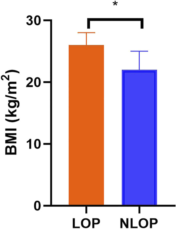
Nineteen patients (age 64 [52–70] years, 37% male) were included, 11 (55%) of them in the LOP group. Their main characteristics are reported in Table 1. The cause of ARDS was most frequently of pulmonary origin (74%), and five patients were treated with ECMO. The baseline scores of APACHE II and SOFA were 19 (16, 20) and 13 (11, 14) respectively. Patients in the LOP group had higher body mass index (26 [25-28] vs. 22 [17-25] kg/m2; p = 0.009) compared with the NLOP group (Figure 1). There was no difference between the two groups in other basic population characteristics. In the LOP group, no patient died during the ICU stay. However, 4 (50%) patients died in the NLOP group (p = 0.018). Hospital mortality was also lower in the LOP group (9% vs. 75%; p = 0.006) (Table 1).
TABLE 1.
Characteristics and outcomes.
| Characteristic | Total (n = 19) | NLOP (n = 8) | LOP (n = 11) | p Value |
|---|---|---|---|---|
| Age, year | 64 (52, 70) | 70 (53, 73) | 63 (52, 68) | 0.342 |
| Male, n (%) | 7 (37) | 3 (38) | 4 (36) | 1.000 |
| BMI (kg/m2) | 25 (21, 27) | 22 (17, 25) | 26 (25, 28) | 0.009 |
| BMI (kg/m2)-no ECMO | 24 (19, 26) | 21 (17.25) | 26 (23, 29) | 0.018 |
| APACHE II | 19 (16, 20) | 18 (18, 19) | 20 (14, 24) | 0.648 |
| SOFA | 13 (11, 14) | 14 (11, 14) | 12 (10, 14) | 0.560 |
| ARDS-risk factor | 0.338 | |||
| Extrapulmonary | 5 (26) | 1 (12) | 4 (36) | |
| Pulmonary | 14 (74) | 7 (88) | 7 (64) | |
| Lesion | 1.000 | |||
| Diffuse, n (%) | 8 (42) | 3 (38) | 5 (45) | |
| Focal, n (%) | 11 (58) | 5 (62) | 6 (55) | |
| Grade | 0.367 | |||
| Mild | 6 (32) | 4 (50) | 2 (18) | |
| Moderate | 12 (63) | 3 (38) | 9 (82) | |
| Severe | 1 (5) | 1 (12) | 0 (0) | |
| ECMO, n (%) | 5 (26) | 1 (12) | 4 (36) | 0.338 |
| ICU length of stay (d) | 32 (16, 44) | 32 (18, 77) | 32 (16, 44) | 0.756 |
| In-ICU mortality (%) | 4 (21) | 4 (50) | 0 (0) | 0.018 |
| Hospital mortality (%) | 7 (37) | 6 (75) | 1 (9) | 0.006 |
LOP, the patients whose optimal PEEP was lower in the prone than in the supine position; NLOP, the patients with the optimal PEEP, not lower in the prone position; BMI, body mass index; APACHE II, Acute Physiology and Chronic Health Evaluation II; SOFA, Sequential Organ-Failure Assessment; ARDS, acute respiratory distress syndrome; PEEP, positive end-expiratory pressure; ECMO, extracorporeal membrane oxygenation; ICU, intensive care unit.
FIGURE 1.

Body mass index of the two groups. Patients in the LOP group had higher body mass index compared with the NLOP group. *p < 0.05 compared with the LOP group.
Ventilator and Respiratory Parameters in Supine Position and Prone Positions
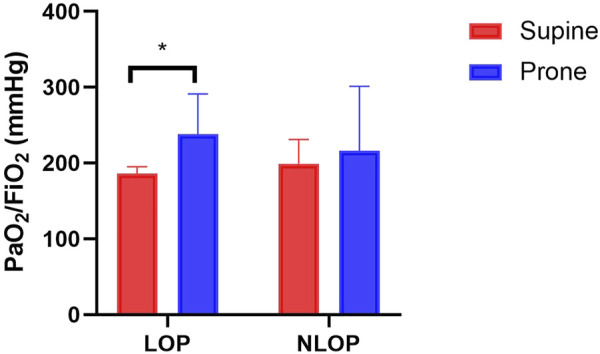
PaO2/FiO2 in the LOP group was significantly increased during PP (238 [170-291] vs. 186 [141-195] mmHg; p = 0.042) (Table 2 Figure 2). No significant difference was observed between the two positions regarding VT, RR, Cdyn, FiO2 and PaCO2 in both groups. The ventilator parameters at baseline did not differ between the two groups as well (Table 3).
TABLE 2.
Difference of ventilator parameters at supine position and prone position.
| Group | SP | PP | p Value | |
|---|---|---|---|---|
| VT (ml) | Total | 382 (235, 446) | 349 (224, 456) | 0.570 |
| LOP | 390 (276, 446) | 370 (213, 550) | 0.476 | |
| NLOP | 324 (208, 459) | 332 (272, 404) | 0.933 | |
| VT (ml/kg) | Total | 4.8 (3.5, 6.7) | 5.5 (3.1, 7.6) | 0.639 |
| LOP | 4.7 (3.5, 5.9) | 5.5 (3.0, 7.6) | 0.811 | |
| NLOP | 6.0 (3.1, 8.5) | 6.0 (3.9, 8.2) | 0.985 | |
| VT (ml/kg)-no ECMO | Total | 6.0 (4.4, 7.4) | 6.6 (5.2, 7.9) | 0.358 |
| LOP | 5.7 (4.7, 6.7) | 6.4 (5.5, 7.6) | 0.383 | |
| NLOP | 6.1 (2.8, 8.9) | 6.7 (4.9, 8.5) | 0.934 | |
| RR (bpm) | Total | 17 (16, 20) | 19 (16, 23) | 0.440 |
| LOP | 16 (14, 21) | 16 (14, 20) | 0.462 | |
| NLOP | 18 (17, 22) | 22 (18, 29) | 0.104 | |
| Crs (ml/cmH2O) | Total | 16.3 (14.0, 32.4) | 18.4 (16.7, 35.7) | 0.066 |
| LOP | 20.0 (14.3, 32.4) | 24.7 (17.2, 39.2) | 0.147 | |
| NLOP | 15.6 (12.7, 25.4) | 17.9 (14.5, 33.0) | 0.313 | |
| Crs (ml/cmH2O)-no ECMO | Total | 19.4 (13.9.41.5) | 28.8 (17.0.42.0) | 0.456 |
| LOP | 29.4 (16.3.40.1) | 38.24 (17.3.47.7) | 0.535 | |
| NLOP | 15.8 (7.5, 45.6) | 18.4 (16.3.33.1) | 0.205 | |
| FiO2 (%) | Total | 45 (40, 50) | 40 (38, 50) | 0.035 |
| LOP | 45 (40, 50) | 40 (38, 48) | 0.181 | |
| NLOP | 48 (40, 52) | 48 (39, 50) | 0.181 | |
| PaO2/FiO2 (mmHg) | Total | 186 (141, 218) | 238 (160, 298) | 0.023 |
| LOP | 186 (141, 195) | 238 (170, 291) | 0.042 | |
| NLOP | 199 (145, 231) | 216 (151, 301) | 0.313 | |
| PaO2/FiO2 (mmHg)-no ECMO | Total | 185 (132,217) | 250 (143,359) | 0.165 |
| LOP | 185 (132,195) | 250 (149,365) | 0.204 | |
| NLOP | 186 (132,224) | 162 (126,352) | 0.620 | |
| PaCO2 (mmHg) | Total | 43 (39, 48) | 43 (41, 50) | 0.732 |
| LOP | 42 (39, 47) | 42 (41, 51) | 0.286 | |
| NLOP | 47 (39, 54) | 45 (43, 49) | 0.575 | |
| PaCO2 (mmHg)-no ECMO | Total | 46 (41.53) | 46 (42.57) | 0.692 |
| LOP | 43 (42.47) | 45 (42.59) | 0.477 | |
| NLOP | 48 (40.58) | 47 (41.53) | 0.710 | |
| Pplat (cmH2O) | Total | 24 (21.28) | 24 (21.26) | 0.491 |
| LOP | 23 (21.28) | 23 (21.26) | 0.529 | |
| NLOP | 25 (18.33) | 25 (19.28) | 0.779 | |
| Baseline PEEP (cmH2O) | Total | 8 (5, 10) | 8 (6, 11) | 0.959 |
| LOP | 10 (6, 11) | 10 (6, 11) | 0.787 | |
| Optimal PEEP (cmH2O) | NLOP | 8 (5, 8) | 7 (6, 9) | 0.787 |
| Total | 9 (6, 12) | 9 (6, 9) | 0.116 | |
| LOP | 12 (10, 15) | 9 (8, 9) | 0.002 | |
| NLOP | 5 (3, 8) | 6 (6, 9) | 0.089 | |
| Optimal PEEP-no ECMO (cmH2O) | Total | 9 (3, 13) | 9 (6, 10) | 0.886 |
| LOP | 12 (12, 15) | 9 (9.12) | 0.084 | |
| NLOP | 3 (3, 6) | 6 (6, 9) | 0.139 |
LOP, the patients whose optimal PEEP, was lower in the prone than in the supine position; NLOP, the patients with the optimal PEEP, not lower in the prone position; SP, supine position; PP, prone position; EIT, electrical impedance tomography; VT, tidal volume; RR, respiratory rate; bpm, breaths per minute; Crs, respiratory system compliance; FiO2, fraction of inspired oxygen; PaO2=arterial partial pressure of oxygen; PaCO2, arterial partial pressure of carbon dioxide.
FIGURE 2.

Ratio between the arterial partial pressure of oxygen and fraction of inspired oxygen (PaO2/FiO2) in different positions of the two groups. PaO2/FiO2 in the LOP group was significantly increased during prone position compared with supine position. No significant difference was observed in the NLOP group. *p < 0.05 compared with supine position.
TABLE 3.
Difference of baseline ventilator parameters in LOP and NLOP.
| LOP | NLOP | p Value | |
|---|---|---|---|
| VT (ml) | 390 (276, 446) | 324 (208, 459) | 0.700 |
| VT (ml/kg) | 4.7 (3.5, 5.9) | 6.0 (3.1, 8.5) | 0.395 |
| RR (bpm) | 16 (14, 21) | 18 (17, 22) | 0.261 |
| Crs (ml/cmH2O) | 20.0 (14.3, 32.4) | 15.6 (12.7, 25.4) | 0.657 |
| PaO2/FiO2 (mmHg) | 186 (141, 195) | 199 (145, 231) | 0.717 |
| PaCO2 (mmHg) | 43 (42.47) | 48 (40.58) | 0.351 |
| Pplat (cmH2O) | 23 (21.28) | 25 (18.33) | 0.951 |
| Baseline PEEP (cmH2O) | 10 (6, 11) | 8 (5, 8) | 0.498 |
| Optimal PEEP (cmH2O) | 12 (10, 15) | 5 (3, 8) | 0.001 |
LOP, the patients whose optimal PEEP, was lower in the prone than in the supine position; NLOP, the patients with the optimal PEEP, not lower in the prone position; SP, supine position; PP, prone position; EIT, electrical impedance tomography; VT, tidal volume; RR, respiratory rate; bpm, breaths per minute; Crs, respiratory system compliance; FiO2, fraction of inspired oxygen; PaO2, arterial partial pressure of oxygen; PaCO2, arterial partial pressure of carbon dioxide.
Effect of Prone Position on EIT-Related Parameters at Different PEEP Levels
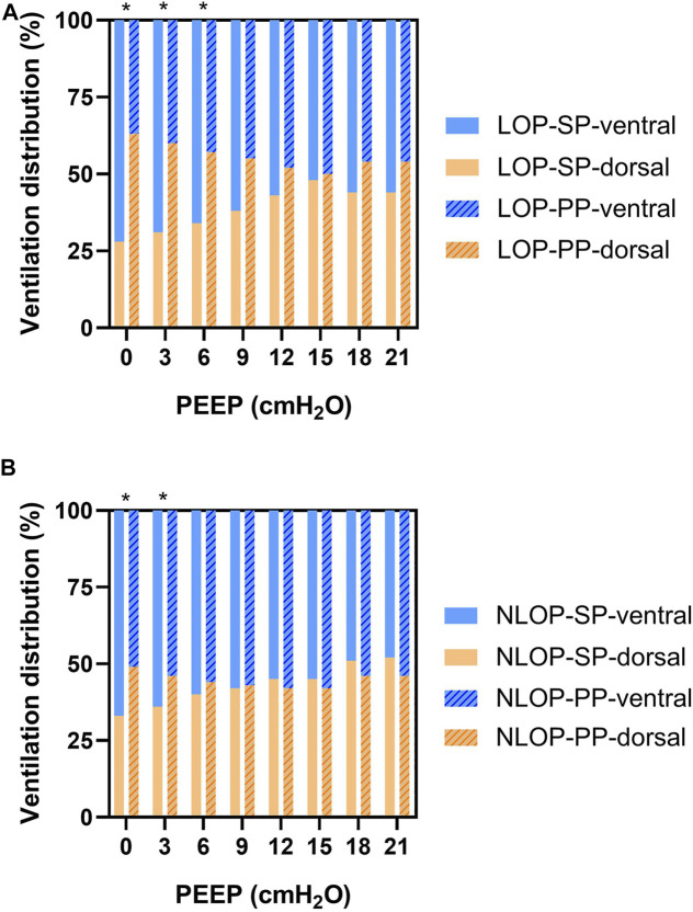
In the LOP group, dorsal ventilation was significantly increased during prone positioning at lower PEEP levels (PEEP = 0, 3, 6 cmH2O). The significant improvement of dorsal ventilation was also seen at 0 and 3 cmH2O of PEEP in the NLOP group (Figure 3). CoV was improved in the LOP group during PP at zero PEEP (41.7 ± 7.3% vs. 54.0 ± 6.3%; p = 0.01), PEEP = 3 cmH2O (42.6 ± 6.5% vs. 53.6 ± 5.2%; p = 0.01) and PEEP = 6 cmH2O (44.4 ± 7.4% vs. 52.9 ± 5.3%; p = 0.03); RVD was lower in the LOP group during PP at zero PEEP (6.54 ± 5.22 vs. 2.98 ± 1.36; p = 0.01) and PEEP = 3 cmH2O (4.01 ± 2.09 vs. 2.67 ± 1.10; p = 0.01). Similar effects were not found in the NLOP group (Table 4).
FIGURE 3.
Dorsal ventilation was significantly higher during prone position at lower PEEP in the LOP group (PEEP = 0, 3, 6 cmH2O) (A). The significant improvement of dorsal ventilation was also present at PEEP of 0 and 3 cmH2O in the NLOP group (B). *p < 0.05 compared with supine position.
TABLE 4.
Effect of prone position on EIT-related parameters at different PEEP levels.
| Baseline | PEEP = 21 | PEEP = 18 | PEEP = 15 | PEEP = 12 | PEEP = 9 | PEEP = 6 | PEEP = 3 | PEEP = 0 | |||
|---|---|---|---|---|---|---|---|---|---|---|---|
| GI | LOP | SP | 0.40 ± 0.11 | 0.40 ± 0.03 | 0.37 ± 0.02 | 0.42 ± 0.09 | 0.40 ± 0.08 | 0.44 ± 0.08 | 0.50 ± 0.11 | 0.56 ± 0.16 | 0.64 ± 0.22 |
| PP | 0.42 ± 0.24 | 0.40 ± 0.06 | 0.37 ± 0.02 | 0.43 ± 0.18 | 0.42 ± 0.18 | 0.43 ± 0.23 | 0.51 ± 0.28 | 0.50 ± 0.27 | 0.60 ± 0.35 | ||
| NLOP | SP | 0.44 ± 0.11 | 0.41 ± 0.05 | 0.40 ± 0.05 | 0.43 ± 0.07 | 0.43 ± 0.07 | 0.44 ± 0.09 | 0.45 ± 0.11 | 0.47 ± 0.12 | 0.50 ± 0.13 | |
| PP | 0.42 ± 0.18 | 0.38 ± 0.01 | 0.44 ± 0.07 | 0.47 ± 0.16 | 0.46 ± 0.15 | 0.45 ± 0.17 | 0.48 ± 0.18 | 0.72 ± 0.63 | 0.57 ± 0.22 | ||
| CoV | LOP | SP | 46.0 ± 6.6 | 44.9 ± 8.2 | 44.4 ± 6.8 | 47.5 ± 7.1 | 47.1 ± 7.0 | 45.9 ± 7.1 | 44.4 ± 7.4 | 42.6 ± 6.5 | 41.7 ± 7.3 |
| PP | 51.5 ± 6.4 | 51.3 ± 6.3 | 51.2 ± 5.4 | 50.2 ± 6.5 | 51.0 ± 5.9 | 52.2 ± 5.4 | 52.9 ± 5.3* | 53.6 ± 5.2* | 54.0 ± 6.3* | ||
| NLOP | SP | 48.0 ± 5.5 | 48.5 ± 2.8 | 48.1 ± 2.8 | 47.3 ± 4.4 | 47.8 ± 5.9 | 46.9 ± 5.7 | 46.1 ± 5.8 | 45.0 ± 6.2 | 44.1 ± 6.4 | |
| PP | 54.5 ± 8.6 | 49.0 ± 4.2 | 48.9 ± 4.0 | 50.4 ± 10.7 | 48.2 ± 12.1 | 48.7 ± 11.7 | 49.2 ± 11.4 | 49.9 ± 10.9 | 50.7 ± 10.5 | ||
| RVD | LOP | SP | 3.88 ± 1.70 | 1.74 ± 0.64 | 2.49 ± 1.52 | 3.02 ± 1.60 | 4.02 ± 2.87 | 4.01 ± 2.26 | 4.02 ± 2.37 | 4.01 ± 2.09 | 6.54 ± 5.22 |
| PP | 3.33 ± 1.85 | 1.59 ± 0.60 | 2.54 ± 0.82 | 3.04 ± 1.45 | 2.88 ± 1.32 | 2.86 ± 1.40 | 2.81 ± 1.19 | 2.67 ± 1.10* | 2.98 ± 1.36* | ||
| NLOP | SP | 4.55 ± 3.05 | 1.84 ± 1.01 | 1.81 ± 2.16 | 2.74 ± 1.25 | 3.24 ± 1.49 | 3.80 ± 1.63 | 3.90 ± 3.06 | 3.19 ± 0.93 | 3.78 ± 1.10 | |
| PP | 4.00 ± 2.19 | 1.70 ± 0.82 | 1.60 ± 1.03 | 3.59 ± 3.31 | 3.96 ± 3.12 | 4.02 ± 3.69 | 4.32 ± 3.40 | 3.92 ± 2.37 | 4.26 ± 2.07 | ||
PEEP, positive end-expiratory pressure; GI, global inhomogeneity index; CoV, center of ventilation; RVD, regional ventilation delay; *p < 0.05 compared with supine position.
EIT-Titrated Optimal PEEP Between Supine and Prone Positions
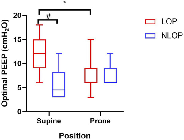
In 7 cases PEEP was titrated from 21 cmH2O and in twelve cases from 15 cmH2O. Broad variability in optimal PEEP between supine and prone positions was observed in the studied patients with ARDS. There were eleven patients whose EIT-based optimal PEEP was reduced in prone position in the LOP group, the optimal PEEP shifting from 12 (10, 15) to 9 (8, 9) cmH2O; However, in the NLOP group, PEEP was elevated during PP in four patients and unchanged in the other four patients (Figure 4).
FIGURE 4.

Electrical impedance tomography (EIT)-estimated optimal positive end-expiratory pressure (PEEP) in supine and prone positions. #p < 0.05 compared with the LOP group. *p < 0.05 compared with supine position.
Discussion
The main findings of our study can be summarized as follows: 1) Broad variability in optimal PEEP between supine and prone position was observed in the ARDS patients. Not all patients showed decreased optimal PEEP during PP. 2) Patients with lower optimal PEEP in prone position had higher body mass index and led to better oxygenation and ventilation homogeneity.
PP is currently widely applied in moderate-to-severe ARDS patients. Lung density redistributes from dorsal to ventral regions due to recruitment in dorsal lung regions and collapse of ventral ones when patients are shifted into the prone position. And some studies have shown that prone positioning can also improve transpulmonary pressure and lung stress. But the overall effect of PP is the decrease in chest wall compliance, which results in an increase in plateau pressure during volume-controlled ventilation or a decrease in VT during pressure-controlled ventilation (Guerin et al., 2004; Taccone et al., 2009; Gattinoni et al., 2013; Iftikhar et al., 2015; Guérin et al., 2020; Lai et al., 2021). Recent studies suggested that EIT-based optimal PEEP was significantly lower in prone than in supine position (Kotani et al., 2018; Martinsson et al., 2021). On the contrary, a study showed that in most patients a PEEP value above commonly used settings was necessary to avoid alveolar collapse in the prone position (Spaeth et al., 2016). Therefore, the choice of PEEP in the PP is still controversial. In our study we analyzed nineteen ARDS patients whose optimal PEEP values were titrated by EIT in both supine and prone positions, and we were able to show that not all of the patients had lower optimal PEEP in prone position. The optimal PEEP was decreased in 11 patients during PP, while it increased in four patients and remained unchanged in the other four patients. Due to the broad variability in optimal PEEP between supine and prone position observed in these patients, our research suggests that an individual PEEP for prone position might not be derived from the optimal PEEP for supine position in ARDS.
A recent study suggested that optimal PEEP was significantly lower in prone than in supine position (Franchineau et al., 2020). This does not correspond with our results. Thus, the patients were separated into two groups, one of which was named the LOP group where optimal PEEP was lower in the prone than in the supine group, the remaining patients were allocated into the NLOP group. We found that the BMI of the patients in the LOP group was significantly higher than that in the NLOP group. The median BMI of patients was 29 kg/m2 in the study from Franchineau et al. (Franchineau et al., 2020), which was also higher than the normal range. As previous study showed, fat accumulation in chest wall and abdomen of obese patients in supine position restricted the diaphragm movement and decreased lung compliance. These factors decrease lung compliance, functional residual capacity, and increase work of breathing and airway resistance in obese patients. Furthermore, PP could be more important in obese patients because of their decreased functional residual capacity and increased atelectasis. PP can partly offset these adverse effects, and obese patients have better responsiveness and prognosis to prone position as reported in recent studies (Chergui et al., 2007; De Jong et al., 2013).
Interestingly, PaO2/FiO2 in the LOP group was significantly increased during PP. EIT-derived parameters in the LOP group, including CoV and RVD, were also improved after PP at lower PEEP levels. Similar effects were not found in patients without decrease of optimal PEEP during prone positioning. Besides, the outcomes of the two groups were quite different, the ICU and hospital mortality in the NLOP group was significantly higher than that in the LOP group. This may be related to the poor responsiveness of the latter patients to prone position. There is evidence that PaO2/FiO2 after the PP differed significantly between ICU survivors and non-survivors (Lee et al., 2020). Although we observed improvement in oxygenation and better prognosis after PP in overweight patients, the small sample size of this retrospective study does not allow to draw the conclusion that the prone position is not required in lean patients.
Our study has several limitations. First, this is a retrospective study, but the PEEP titration method in the supine and prone positions were consistent. Optimal PEEP results were thus comparable. Although the time interval between the two examinations was different, the change in posture occurred within 1 day, approximately after 16 h on average. Second, the sample size was relatively small, and only 19 patients were included. Third, there were some potential confounding factors, e, g, five patients were treated with ECMO. However, no effect on the EIT-derived parameters is presumed because of identical evaluation of EIT data. Therefore, only some preliminary conclusions have been drawn so far, and prospective studies with larger sample sizes can be conducted in the future to explore and verify the current results.
Conclusion
Broad variability in optimal PEEP between supine and prone positions was observed in ARDS patients. Not all patients showed decreased optimal PEEP during prone positioning. Patients with lower optimal PEEP in prone position had higher body mass index and led to better oxygenation and ventilation homogeneity.
Key Messages
1) Broad variability in optimal PEEP between supine and prone position was observed in ARDS patients. Not all patients exhibited decreased optimal PEEP during prone positioning.
2) Patients with lower optimal PEEP in prone position had higher body mass index and led to better oxygenation and ventilation homogeneity.
Data Availability Statement
The original contributions presented in the study are included in the article, further inquiries can be directed to the corresponding authors.
Ethics Statement
The studies involving human participants were reviewed and approved by The Institutional Research and Ethics Committee of Peking Union Medical College Hospital. Written informed consent for participation was not required for this study in accordance with the national legislation and the institutional requirements.
Author Contributions
LM, YC, HH, and YL designed and planned the study. LM and HH were responsible for collection and assembly of data. YC, SY, YY, IF, and ZZ was responsible for data analysis and interpretation. LM, YC, HH wrote the initial manuscript draft, and the other authors were involved in critical revision of the final manuscript. All authors read and approved the final manuscript.
Funding
This study was supported by Capital’s Funds for Health Improvement and Research (NO. 2020-2-40111) and Excellence Program of Key Clinical Specialty of Beijing in 2020 for Critical Care Medicine, Beijing Municipal Science and Technology Commission (No. Z201100005520051).
Conflict of Interest
The authors declare that the research was conducted in the absence of any commercial or financial relationships that could be construed as a potential conflict of interest.
Publisher’s Note
All claims expressed in this article are solely those of the authors and do not necessarily represent those of their affiliated organizations, or those of the publisher, the editors and the reviewers. Any product that may be evaluated in this article, or claim that may be made by its manufacturer, is not guaranteed or endorsed by the publisher.
Abbreviations
APACHE, Acute Physiology and Chronic Health Evaluation; ARDS, acute respiratory distress syndrome; BMI, body mass index; CoV, center of ventilation; Cdyn, dynamic respiratory system compliance; ECMO, extracorporeal membrane oxygenation; EIT, electrical impedance tomography; FiO2, fraction of inspiratory oxygen; GI, global inhomogeneity; ICU, intensive care unit; PaO2, arterial partial pressure of oxygen; PaCO2, arterial partial pressure of carbon dioxide; PEEP, positive end-expiratory pressure; PP, prone positioning; ROI, region of interest; RR, respiratory rate; RVD, regional ventilation delay; SOFA, Sequential Organ-Failure Assessment; SP, supine position; VT, tidal volume.
References
- Aguirre-Bermeo H., Turella M., Bitondo M., Grandjean J., Italiano S., Festa O., et al. (2018). Lung Volumes and Lung Volume Recruitment in ARDS: a Comparison between Supine and Prone Position. Ann. Intensive Care 8, 25. 10.1186/s13613-018-0371-0 [DOI] [PMC free article] [PubMed] [Google Scholar]
- Becher T., Buchholz V., Hassel D., Meinel T., Schädler D., Frerichs I., et al. (2021). Individualization of PEEP and Tidal Volume in ARDS Patients with Electrical Impedance Tomography: a Pilot Feasibility Study. Ann. Intensive Care 11, 89. 10.1186/s13613-021-00877-7 [DOI] [PMC free article] [PubMed] [Google Scholar]
- Bellani G., Laffey J. G., Pham T., Fan E., Brochard L., Esteban A., et al. (2016). Epidemiology, Patterns of Care, and Mortality for Patients with Acute Respiratory Distress Syndrome in Intensive Care Units in 50 Countries. JAMA 315, 788–800. 10.1001/jama.2016.0291 [DOI] [PubMed] [Google Scholar]
- Chergui K., Choukroun G., Meyer P., Caen D. (2007). Prone Positioning for a Morbidly Obese Patient with Acute Respiratory Distress Syndrome: an Opportunity to Explore Intrinsic Positive End-Expiratory Pressure-Lower Inflexion Point Interdependence. Anesthesiology 106, 1237–1239. 10.1097/01.anes.0000265419.50900.f4 [DOI] [PubMed] [Google Scholar]
- Costa E. L. V., Borges J. B., Melo A., Suarez-Sipmann F., Toufen C., Bohm S. H., et al. (2009). Bedside Estimation of Recruitable Alveolar Collapse and Hyperdistension by Electrical Impedance Tomography. Intensive Care Med. 35, 1132–1137. 10.1007/s00134-009-1447-y [DOI] [PubMed] [Google Scholar]
- Dalla Corte F., Mauri T., Spinelli E., Lazzeri M., Turrini C., Albanese M., et al. (2020). Dynamic Bedside Assessment of the Physiologic Effects of Prone Position in Acute Respiratory Distress Syndrome Patients by Electrical Impedance Tomography. Minerva Anestesiol. 86, 1057–1064. 10.23736/s0375-9393.20.14130-0 [DOI] [PubMed] [Google Scholar]
- De Jong A., Molinari N., Sebbane M., Prades A., Futier E., Jung B., et al. (2013). Feasibility and Effectiveness of Prone Position in Morbidly Obese Patients with ARDS. Chest 143, 1554–1561. 10.1378/chest.12-2115 [DOI] [PubMed] [Google Scholar]
- Franchineau G., Bréchot N., Hekimian G., Lebreton G., Bourcier S., Demondion P., et al. (2020). Prone Positioning Monitored by Electrical Impedance Tomography in Patients with Severe Acute Respiratory Distress Syndrome on Veno-Venous ECMO. Ann. Intensive Care 10, 12. 10.1186/s13613-020-0633-5 [DOI] [PMC free article] [PubMed] [Google Scholar]
- Frerichs I., Hahn G., Golisch W., Kurpitz M., Burchardi H., Hellige G. (1998). Monitoring Perioperative Changes in Distribution of Pulmonary Ventilation by Functional Electrical Impedance Tomography. Acta Anaesthesiol. Scand. 42 (6), 721–726. 10.1111/j.1399-6576.1998.tb05308.x [DOI] [PubMed] [Google Scholar]
- Gattinoni L., Vagginelli F., Carlesso E., Taccone P., Conte V., Chiumello D., et al. (2003). Decrease in Paco2 with Prone Position Is Predictive of Improved Outcome in Acute Respiratory Distress Syndrome*. Crit. care Med. 31, 2727–2733. 10.1097/01.Ccm.0000098032.34052.F9 [DOI] [PubMed] [Google Scholar]
- Gattinoni L., Taccone P., Carlesso E., Marini J. J. (2013). Prone Position in Acute Respiratory Distress Syndrome. Rationale, Indications, and Limits. Am. J. Respir. Crit. Care Med. 188, 1286–1293. 10.1164/rccm.201308-1532CI [DOI] [PubMed] [Google Scholar]
- Gibot S., Conrad M., Courte G., Cravoisy A. (2021). Positive End-Expiratory Pressure Setting in COVID-19-Related Acute Respiratory Distress Syndrome: Comparison Between Electrical Impedance Tomography, PEEP/FiO2 Tables, and Transpulmonary Pressure. Front. Med. 8, 720920. 10.3389/fmed.2021.720920 [DOI] [PMC free article] [PubMed] [Google Scholar]
- Griffiths M. J. D., Mcauley D. F., Perkins G. D., Barrett N., Blackwood B., Boyle A., et al. (2019). Guidelines on the Management of Acute Respiratory Distress Syndrome. BMJ Open Resp. Res. 6, e000420. 10.1136/bmjresp-2019-000420 [DOI] [PMC free article] [PubMed] [Google Scholar]
- Guerin C., Gaillard S., Lemasson S., Ayzac L., Girard R., Beuret P., et al. (2004). Effects of Systematic Prone Positioning in Hypoxemic Acute Respiratory Failure. JAMA 292, 2379–2387. 10.1001/jama.292.19.2379 [DOI] [PubMed] [Google Scholar]
- Guérin C., Albert R. K., Beitler J., Gattinoni L., Jaber S., Marini J. J., et al. (2020). Prone Position in ARDS Patients: Why, when, How and for Whom. Intensive Care Med. 46, 2385–2396. 10.1007/s00134-020-06306-w [DOI] [PMC free article] [PubMed] [Google Scholar]
- He H., Chi Y., Long Y., Yuan S., Frerichs I., Möller K., et al. (2020). Influence of Overdistension/recruitment Induced by High Positive End-Expiratory Pressure on Ventilation-Perfusion Matching Assessed by Electrical Impedance Tomography with Saline Bolus. Crit. Care 24, 586. 10.1186/s13054-020-03301-x [DOI] [PMC free article] [PubMed] [Google Scholar]
- He H., Chi Y., Yang Y., Yuan S., Long Y., Zhao P., et al. (2021). Early Individualized Positive End-Expiratory Pressure Guided by Electrical Impedance Tomography in Acute Respiratory Distress Syndrome: a Randomized Controlled Clinical Trial. Crit. Care 25, 230. 10.1186/s13054-021-03645-y [DOI] [PMC free article] [PubMed] [Google Scholar]
- Hinz J., Hahn G., Neumann P., Sydow M., Mohrenweiser P., Hellige G., et al. (2003). End-expiratory Lung Impedance Change Enables Bedside Monitoring of End-Expiratory Lung Volume Change. Intensive Care Med. 29, 37–43. 10.1007/s00134-002-1555-4 [DOI] [PubMed] [Google Scholar]
- Holanda M. A., Sousa N. P. d., Melo L. T., Marinho L. S., Ribeiro-Filho H. V., Troncon L. E. d. A., et al. (2018). Helping Students to Understand Physiological Aspects of Regional Distribution of Ventilation in Humans: a Experience from the Electrical Impedance Tomography. Adv. physiology Educ. 42, 655–660. 10.1152/advan.00086.2018 [DOI] [PubMed] [Google Scholar]
- Hsu H.-J., Chang H.-T., Zhao Z., Wang P.-H., Zhang J.-H., Chen Y.-S., et al. (2021). Positive End-Expiratory Pressure Titration with Electrical Impedance Tomography and Pressure-Volume Curve: a Randomized Trial in Moderate to Severe ARDS. Physiol. Meas. 42, 014002. 10.1088/1361-6579/abd679 [DOI] [PubMed] [Google Scholar]
- Iftikhar I. H., Donley M. A., Owens W. B. (2015). Prone Positioning in Acute Respiratory Distress Syndrome. Crit. care Med. 43, e55–e56. 10.1097/ccm.0000000000000761 [DOI] [PubMed] [Google Scholar]
- Kotani T., Tanabe H., Yusa H., Saito S., Yamazaki K., Ozaki M. (2016). Electrical Impedance Tomography-Guided Prone Positioning in a Patient with Acute Cor Pulmonale Associated with Severe Acute Respiratory Distress Syndrome. J. Anesth. 30, 161–165. 10.1007/s00540-015-2084-y [DOI] [PMC free article] [PubMed] [Google Scholar]
- Kotani T., Hanaoka M., Hirahara S., Yamanaka H., Teschner E., Shono A. (2018). Regional Overdistension during Prone Positioning in a Patient with Acute Respiratory Failure Who Was Ventilated with a Low Tidal Volume: a Case Report. J. intensive care 6, 18. 10.1186/s40560-018-0290-z [DOI] [PMC free article] [PubMed] [Google Scholar]
- Kumaresan A., Gerber R., Mueller A., Loring S. H., Talmor D. (2018). Effects of Prone Positioning on Transpulmonary Pressures and End-Expiratory Volumes in Patients without Lung Disease. Anesthesiology 128, 1187–1192. 10.1097/aln.0000000000002159 [DOI] [PubMed] [Google Scholar]
- Lai C., Adda I., Teboul J.-L., Persichini R., Gavelli F., Guérin L., et al. (2021). Effects of Prone Positioning on Venous Return in Patients with Acute Respiratory Distress Syndrome*. Crit. care Med. 49, 781–789. 10.1097/ccm.0000000000004849 [DOI] [PubMed] [Google Scholar]
- Lee H. Y., Cho J., Kwak N., Choi S. M., Lee J., Park Y. S., et al. (2020). Improved Oxygenation after Prone Positioning May Be a Predictor of Survival in Patients with Acute Respiratory Distress Syndrome*. Crit. care Med. 48, 1729–1736. 10.1097/ccm.0000000000004611 [DOI] [PubMed] [Google Scholar]
- Martinsson A., Houltz E., Wallinder A., Lindgren S., Thorén A. (2021). Lung Recruitment in the Prone Position after Cardiac Surgery: a Randomised Controlled Study. Br. J. Anaesth. 126, 1067–1074. 10.1016/j.bja.2020.12.039 [DOI] [PubMed] [Google Scholar]
- Menk M., Estenssoro E., Sahetya S. K., Neto A. S., Sinha P., Slutsky A. S., et al. (2020). Current and Evolving Standards of Care for Patients with ARDS. Intensive Care Med. 46, 2157–2167. 10.1007/s00134-020-06299-6 [DOI] [PMC free article] [PubMed] [Google Scholar]
- Mezidi M., Parrilla F. J., Yonis H., Riad Z., Böhm S. H., Waldmann A. D., et al. (2018). Effects of Positive End-Expiratory Pressure Strategy in Supine and Prone Position on Lung and Chest Wall Mechanics in Acute Respiratory Distress Syndrome. Ann. Intensive Care 8, 86. 10.1186/s13613-018-0434-2 [DOI] [PMC free article] [PubMed] [Google Scholar]
- Muders T., Hentze B., Kreyer S., Wodack K. H., Leonhardt S., Hedenstierna G., et al. (2021). Measurement of Electrical Impedance Tomography-Based Regional Ventilation Delay for Individualized Titration of End-Expiratory Pressure. J. Clin. Med. 10, 2933. 10.3390/jcm10132933 [DOI] [PMC free article] [PubMed] [Google Scholar]
- Ranieri V., Rubenfeld G., Thompson B., Ferguson N., Caldwell E., Fan E., et al. (2012). Acute Respiratory Distress Syndrome. JAMA 307, 2526–2533. 10.1001/jama.2012.5669 [DOI] [PubMed] [Google Scholar]
- Scaramuzzo G., Ball L., Pino F., Ricci L., Larsson A., Guérin C., et al. (2020a). Influence of Positive End-Expiratory Pressure Titration on the Effects of Pronation in Acute Respiratory Distress Syndrome: A Comprehensive Experimental Study. Front. Physiol. 11, 179. 10.3389/fphys.2020.00179 [DOI] [PMC free article] [PubMed] [Google Scholar]
- Scaramuzzo G., Spadaro S., Dalla Corte F., Waldmann A. D., Böhm S. H., Ragazzi R., et al. (2020b). Personalized Positive End-Expiratory Pressure in Acute Respiratory Distress Syndrome: Comparison Between Optimal Distribution of Regional Ventilation and Positive Transpulmonary Pressure. Crit. care Med. 48, 1148–1156. 10.1097/ccm.0000000000004439 [DOI] [PubMed] [Google Scholar]
- Sella N., Boscolo A., Zarantonello F., Bonvecchio I., Andreatta G., Pettenuzzo T., et al. (2021). Electrical Impedance Tomography for Positive End-Expiratory Pressure Setting after Bilateral Lung Transplantation. J. Heart Lung Transplant. 40, S317. 10.1016/j.healun.2021.01.897 [DOI] [Google Scholar]
- Spaeth J., Daume K., Goebel U., Wirth S., Schumann S. (2016). Increasing Positive End-Expiratory Pressure (Re-)improves Intraoperative Respiratory Mechanics and Lung Ventilation after Prone Positioning. Br. J. Anaesth. 116, 838–846. 10.1093/bja/aew115 [DOI] [PubMed] [Google Scholar]
- Sweemey R. M., Mcauley D. F. (2016). Acute Respiratory Distress Syndrome. Lancet 388, 2416–2430. 10.1016/s0140-6736(16)00578-x [DOI] [PMC free article] [PubMed] [Google Scholar]
- Taccone P., Pesenti A., Latini R., Polli F., Vagginelli F., Mietto C., et al. (2009). Prone Positioning in Patients with Moderate and Severe Acute Respiratory Distress Syndrome. JAMA 302, 1977–1984. 10.1001/jama.2009.1614 [DOI] [PubMed] [Google Scholar]
- Wrigge H., Zinserling J., Muders T., Varelmann D., Günther U., von der Groeben C., et al. (2008). Electrical Impedance Tomography Compared with Thoracic Computed Tomography during a Slow Inflation Maneuver in Experimental Models of Lung Injury*. Crit. Care Med. 36, 903–909. 10.1097/ccm.0b013e3181652edd [DOI] [PubMed] [Google Scholar]
- Yun L., He H.-w., Möller K., Frerichs I., Liu D., Zhao Z. (2016). Assessment of Lung Recruitment by Electrical Impedance Tomography and Oxygenation in ARDS Patients. Medicine 95, e3820. 10.1097/md.0000000000003820 [DOI] [PMC free article] [PubMed] [Google Scholar]
- Zhao Z., Steinmann D., Frerichs I., Guttmann J., Möller K. (2010). PEEP Titration Guided by Ventilation Homogeneity: a Feasibility Study Using Electrical Impedance Tomography. Crit. Care 14, R8. 10.1186/cc8860 [DOI] [PMC free article] [PubMed] [Google Scholar]
- Zhao Z., Lee L.-C., Chang M.-Y., Frerichs I., Chang H.-T., Gow C.-H., et al. (2020). The Incidence and Interpretation of Large Differences in EIT-Based Measures for PEEP Titration in ARDS Patients. J. Clin. Monit. Comput. 34, 1005–1013. 10.1007/s10877-019-00396-8 [DOI] [PubMed] [Google Scholar]
Associated Data
This section collects any data citations, data availability statements, or supplementary materials included in this article.
Data Availability Statement
The original contributions presented in the study are included in the article, further inquiries can be directed to the corresponding authors.


