Abstract
To further develop genetic techniques for the enteropathogen Brachyspira hyodysenteriae, the gyrB gene of this spirochete was isolated from a λZAPII library of strain B204 genomic DNA and sequenced. The putative protein encoded by this gene exhibited up to 55% amino acid sequence identity with GyrB proteins of various bacterial species, including other spirochetes. B. hyodysenteriae coumermycin A1-resistant (Cnr) mutant strains, both spontaneous and UV induced, were isolated by plating B204 cells onto Trypticase soy blood agar plates containing 0.5 μg of coumermycin A1/ml. The coumermycin A1 MICs were 25 to 100 μg/ml for the resistant strains and 0.1 to 0.25 μg/ml for strain B204. Four Cnr strains had single nucleotide changes in their gyrB genes, corresponding to GyrB amino acid changes of Gly78 to Ser (two strains), Gly78 to Cys, and Thr166 to Ala. When Cnr strain 435A (Gly78 to Ser) and Cmr Kmr strain SH (ΔflaA1::cat Δnox::kan) were cultured together in brain heart infusion broth containing 10% (vol/vol) heat-treated (56°C, 30 min) calf serum, cells resistant to chloramphenicol, coumermycin A1, and kanamycin could be isolated from the cocultures after overnight incubation, but such cells could not be isolated from monocultures of either strain. Seven Cnr Kmr Cmr strains were tested and were determined to have resistance genotypes of both strain 435A and strain SH. Cnr Kmr Cmr cells could not be isolated when antiserum to the bacteriophage-like agent VSH-1 was added to cocultures, and the numbers of resistant cells increased fivefold when mitomycin C, an inducer of VSH-1 production, was added. These results indicate that coumermycin resistance associated with a gyrB mutation is a useful selection marker for monitoring gene exchange between B. hyodysenteriae cells. Gene transfer readily occurs between B. hyodysenteriae cells in broth culture, a finding with practical importance. VSH-1 is the likely mechanism for gene transfer.
A major impediment to investigations of the biology of spirochetes (members of the order Spirochaetales) has been an inability to genetically manipulate these bacteria. Complex culture requirements and the lack of common genetic tools, such as selection markers (e.g., antibiotic resistance genes), methods for mutagenesis, and natural gene transfer, have limited investigations of Borrelia burgdorferi, Treponema pallidum, Leptospira interrogans, and Treponema denticola (12). An ability to derive strains with specific mutations is important for identifying virulence-associated genes of these human pathogens. Recent reports of a shuttle vector plasmid for Leptospira biflexa, a nonpathogenic leptospire (9), of electrotransformation methods for B. burgdorferi (38), and of a heterologous plasmid that is stably maintained in noninfectious B. burgdorferi (30) are encouraging breakthroughs which will undoubtedly facilitate genetic investigations of pathogenic strains of Leptospira and B. burgdorferi.
Brachyspira (Serpulina) hyodysenteriae is an anaerobic spirochete and the etiologic agent of swine dysentery (11, 13). This enteropathogen offers several research advantages not currently available for other spirochete species. The cultural, nutritional, and metabolic properties of B. hyodysenteriae have been substantially characterized (32), and a serum-free, low-protein culture medium has been described (17). An experimental disease model featuring the natural host animal has been available for many years (20, 34). Most significantly, recent research has provided a basis for understanding and manipulating B. hyodysenteriae at the gene level.
A physical map of the B. hyodysenteriae chromosome has been created (42). A method for targeted mutagenesis of B. hyodysenteriae genes by electroporation-mediated allelic exchange using antibiotic resistance genes as selection markers was first reported by ter Huurne et al. (37) and has been used to identify virulence-associated traits of the spirochete (19, 27, 34, 37). VSH-1, a mitomycin C-inducible prophage which transduces B. hyodysenteriae genes, has been described (16, 17).
In previous studies, heterologous chloramphenicol and kanamycin resistance genes were used as selection markers for targeted mutagenesis of B. hyodysenteriae genes (18, 19, 27, 37) and investigations of gene transfer (17). It would be useful to have additional antibiotic resistance markers to investigate Brachyspira genetics.
Coumermycin A1 and other coumarins are fermentation-derived, broad-spectrum antibiotics targeting the GyrB subunit of DNA gyrase. DNA gyrase (EC 5.99.1.3; a type of DNA topoisomerase II) catalyzes ATP-dependent introduction of negative supercoils into DNA, which affects DNA topology, and is essential for DNA replication (5, 22, 23). The gyrA and gyrB genes encode DNA gyrase subunits that have conserved motifs in diverse bacterial species (15). Specific point mutations in gyrB result in single amino acid changes in the GyrB protein that confer coumermycin resistance (Cnr) in various bacterial species (1, 5–7, 14, 23, 36).
Coumarins are not used in the treatment of swine dysentery. Coumermycin A1-resistant strains of other spirochetes, B. burgdorferi (29) and T. denticola (10), have been isolated. Cnr has been used as a selection marker during mutagenesis of B. burgdorferi (26, 38). These features made coumermycin A1 resistance attractive as a selection marker for B. hyodysenteriae mutant strains. Consequently, the objectives of this study were to produce coumermycin A1-resistant B. hyodysenteriae strains with discernible mutations in their gyrB genes and to evaluate coumermycin A1 resistance as a selection marker for monitoring gene exchange among B. hyodysenteriae cells. In the course of accomplishing these objectives, we found that exchange of a mutant gyrB gene conferring Cnr readily occurs between B. hyodysenteriae cells in broth cultures and is likely to be mediated by the gene transfer agent VSH-1.
MATERIALS AND METHODS
Culture media and conditions.
B. hyodysenteriae B204 cells were routinely cultured at 38°C in stirred Difco brain heart infusion (BHI) broth containing 10% (vol/vol) heat-treated (56°C, 30 min) calf serum (BHIS broth) beneath an initial culture atmosphere containing 99% N2 and 1% O2 (33). BHI basal broth (no serum added) was used for diluting bacterial cultures. Trypticase soy blood (TSB) agar plates were prepared with 30 g of Trypticase soy broth plus dextrose (BBL, Becton Dickinson, Cockeysville, Md.), 950 ml of distilled water, and 9.0 g of Noble agar. After the medium was sterilized by autoclaving and cooled to 48°C, 50 ml of defibrinated bovine blood was added and the medium was dispensed (20 ml/plate). TSB agar plates were stored at 5°C in an air atmosphere but were placed in an anaerobic chamber 24 h before use.
For antibiotic-containing media, a 5-mg/ml stock solution of coumermycin A1 (Sigma) in dimethyl sulfoxide was diluted 1/100 in tubes containing anaerobic BHIS broth. While the coumermycin A1 was added, the tubes were flushed with sterile O2-free N2 gas to maintain anaerobic conditions and mixed vigorously in order to prevent precipitation of the antibiotic. This solution was diluted in BHIS broth and added to both TSB agar plates and BHIS broth. Media containing kanamycin and chloramphenicol have been described previously (17).
Sequencing and cloning the B. hyodysenteriae gyrB gene.
Based on consensus amino acid sequences of the GyrB proteins of Borrelia, Treponema, and other bacteria in the GenBank database, a degenerate PCR primer pair was designed to amplify the B. hyodysenteriae gyrB gene. Primer 1FOR (5′-CCT/AGGT/AATGTATATA/TGGT/ATC) corresponds to coding sequence base positions 73 to 92 in the B. hyodysenteriae gyrB gene and primer 1REV (5′-ATAGTAGTTTCCCAA/TAG/AT/CTG) is complementary to coding sequence base positions 1760 to 1741. This primer combination was used to PCR amplify an internal region of the gyrB gene by using purified B204 DNA as the template. The amplified product (1688 bp) was purified by ultrafiltration (Microcon 100 column) and directly sequenced. A single, unambiguous sequence was obtained. The entire gyrB gene and regions upstream and downstream of the coding region were obtained from a λ clone of B. hyodysenteriae DNA. A λZAPII library of B. hyodysenteriae B204 genomic DNA was screened for clones carrying the gyrB gene by plaque lift hybridization with 32P-labeled oligonucleotide probe GR (5′-TCTAATTCAAGTTTTTTAGC) by using standard techniques (28) and methods recommended by the library manufacturer (Stratagene). Probe GR is complementary to coding sequence base positions 895 to 914 of the B. hyodysenteriae gyrB gene and was designed based on the sequence of the amplified gyrB internal region. A λ clone containing the gyrB gene near the middle of a 6-kb insert was isolated and sequenced. Every base position on both strands of gyrB DNA was determined at least once in cycle sequencing reactions (8) at the Iowa State University Nucleic Acid Facility.
UV mutagenesis.
B. hyodysenteriae B204 cells in the exponential phase of growth (optical density at 620 nm [OD620], 0.7 [18-mm-path-length culture tubes]; approximately 8 × 10⁷ CFU/ml) were harvested from 15 ml of BHIS broth. The bacteria were harvested by centrifugation (5 min, 2900 × g) resuspended and washed once in 15 ml of ice-cold phosphate-buffered saline (PBS) (28) and resuspended in 45 ml of PBS (final cell density, approximately 2.5 × 10⁷ CFU/ml). Five-milliliter samples of the cell suspension were placed in sterile petri plates 17.5 cm beneath a single UV lamp bulb (the other bulbs were removed) in a Stratalinker 1800 UV box (Stratagene). The cells were exposed to a UV dose of 3,500 μJ as measured by the Stratalinker sensor. The UV box was placed on a rotating platform, and the cell suspensions were mixed (50 rpm) during UV exposure. Preliminary studies established that this exposure killed 99 to 99.5% of the cells. Control cultures used for determining cell killing and for isolating spontaneous coumermycin A1-resistant mutants were handled in the same way but were not exposed to UV light.
Six 5-ml suspensions of UV-exposed cells were pooled, and two 5-ml suspensions of control (unexposed) cells were combined. Each pooled suspension was harvested by centrifugation as described above, the supernatants were discarded, and the cell pellets in the centrifuge tubes were placed on ice and transferred into a Coy anaerobic chamber. The chamber was inflated with a mixture containing 85% N2, 10% H2, and 5% CO2. Inside the Coy chamber, the cells were resuspended in anaerobic BHI broth (37 ml for UV-exposed cells and 12.6 ml for control cells). Serial 10-fold dilutions of the cell suspensions (0.1 ml) were plated onto TSB agar plates to count the surviving bacteria. Separate cultures were created by dispensing samples of the suspensions into sterile 18-mm glass tubes (6.3 ml per tube). Heat-treated calf serum (0.7 ml) and a sterile magnetic stirring flea were added to each tube. The tubes were sealed with sterile rubber stoppers and removed from the Coy chamber. The culture atmosphere inside the tubes was replaced with 99% N2–1% O2, and the cultures were incubated with stirring in the dark at 38°C until the culture OD620 reached 0.4 to 0.6 (approximately 8 to 10 h for control cultures and 24 to 26 h for UV-treated cultures). Throughout these procedures, the bacteria were shielded from light by wrapping the glass vessels and culture tubes with aluminum foil.
Selection of coumermycin A1-resistant strains.
Six-milliliter cultures of UV-treated and control cells were concentrated 10-fold by centrifugation and transferred into an anaerobic chamber, and serial 10-fold dilutions were made in BHI basal broth. Samples of the dilutions were plated onto TSB agar plates to determine viable population densities and onto TSB agar plates containing coumermycin A1 at a final concentration of 0.5 μg/ml to select for Cnr mutants. This concentration of coumermycin A1 was approximately five-fold higher than the MIC of the antibiotic for B. hyodysenteriae B204 cells (wild type) and was based upon preliminary studies which demonstrated that higher concentrations of the antibiotic (2.0 and 5.0 μg/ml) inhibited colony formation during the initial isolation of both spontaneous and UV-induced Cnr strains. We do not understand the basis for this inhibition, since after multiple subcultures the strains formed colonies on agar medium containing substantially higher coumermycin A1 concentrations.
Cnr strains were cloned by subculturing single isolated colonies three times on TSB agar plates containing coumermycin A1. The strains were then cultured in BHIS broth supplemented with coumermycin A1 (0.5 μg/ml), harvested, and stored at −70°C. Each Cnr strain was considered an independent isolate, since only one strain was selected from each culture.
Coumermycin A1 MIC determination.
Cultures and cell suspensions of wild-type and Cnr strains were vortexed to disrupt cell aggregations. B. hyodysenteriae cells (2 ml) in the exponential phase of growth (3 × 10⁸ cells/ml, as determined by direct microscope counting) were harvested by centrifugation (10 min, 3,000 × g, Beckman GPR benchtop centrifuge). The cells were washed once in 2 ml of cold, sterile PBS and resuspended in 6 ml of PBS. Ten-microliter samples (approximately 5 × 10⁵ bacterial cells) of the suspension were spotted onto TSB agar plates containing various coumermycin A1 concentrations (spot plate MIC test). Additionally, the suspension was serially diluted 10-fold in PBS, and 100-μl samples of the 10−3, 10−4, and 10−5 dilutions were spread on the surfaces of TSB agar plates (spread plate MIC test). All plates were inoculated in laboratory air and immediately transferred into a Coy chamber for incubation. After 4 days of incubation, the coumermycin A1 MIC was identified as the lowest concentration at which growth was inhibited (no or very faint hemolysis on spot plates and no colony growth on spread plates). MICs were based on two to four separate determinations for each strain. The final coumermycin A1 concentrations were 0, 0.1, 0.25, 0.5, 1.0, 2.5, 5.0, 10, 25, 50, and 100 μg/ml.
Sequencing gyrB genes of coumermycin A1-resistant strains.
PCR primers 2FOR (5′-GATTTGTAATATTTAGTTATTC) and 2REV (5′-GTATTTATTATCCATAATTTG) complementary to regions 56 bp upstream and 82 bp downstream of the gyrB stop codon, respectively, were used to amplify the gyrB genes of nine Cnr strains isolated in this study. The amplified products were sequenced (8). Base differences between the gyrB genes of Cnr strains and wild-type strain B204 were confirmed by a second round of PCR amplification and sequencing.
Sequence analyses and computer software.
PCR primers were designed by using Oligo V5.0 for Windows (National Biosciences, Inc.). Gene and protein sequences were analyzed by using Vector NTI Suite V5.5 (InforMax, Inc.) and DNASIS v.1.0 (Hitachi Software Engineering Co.). The predicted amino acid sequence of the B. hyodysenteriae GyrB protein was compared to amino acid sequences in the GenBank nr peptide database by using BLASTP, version 2.0.13.
Gene exchange studies.
To detect genetic exchange among B. hyodysenteriae cells, coumermycin A1-resistant strain 435A (Table 1) was cocultured with kanamycin- and chloramphenicol-resistant strain SH. Strain SH was generated previously by allelic exchange and generalized transduction with purified VSH-1 particles (17). Strain SH cells contain a gene for chloramphenicol resistance inserted into the flaA1 gene (ΔflaA1 593–762::cat) and a gene for kanamycin resistance inserted into the nox gene (Δnox 438–760::kan). Exponential-growth-phase cells (0.1 ml; approximately 3 × 10⁷ bacteria, as determined by direct cell counting) of each strain were inoculated into the same culture tubes containing BHIS broth. When the OD620 of the cocultures reached 1.0, they were serially diluted 10-fold, and 0.1- or 0.2-ml portions of the dilutions were spread onto the surfaces of TSB agar plates prepared either without or with coumermycin A1 (final concentration 10 μg/ml), kanamycin (200 μg/ml), and chloramphenicol (10 μg/ml). Duplicate cultures were analyzed in three experiments. Two or three triply resistant colonies from each experiment (total, seven colonies) were selected in order to analyze their resistance genotypes. For kanamycin and chloramphenicol resistance, each strain was analyzed by PCR amplification of the Δnox::kan genetic construct with primers ForNK (5′-AATGCCAATATTTTATAATATAA-3′) (35) and REVKM (5′-CGCGGCCTCGAGCAAGACG-3′) (34) and by amplification of the ΔflaA1::cat genetic construct with primers ERL10 (5′-GGGGATCCTATGAAAAAGTTATTCGTAGT-ATTAACTTTCC-3′) and ERL16 (5′-GATTAAAGATCTCTTTTCTCTTCC-3′) (17). The amplifications yielded 610- and 750-bp products, respectively, for parent strain SH. The coumermycin-resistant genotype was identified by amplifying a 702-bp region of gyrB with PCR primers ForGyr (5′-GATTTGTAATATTTAGTTATTC-3′) and RevGyr (5′-CTCATCTTTAAGAGTAATCC-3′). The amplified product was sequenced to detect substitution of A for G at base position 232, a mutation associated with coumermycin resistance in parent strain 435A (Table 1).
TABLE 1.
Characteristics of coumermycin A1-resistant B. hyodysenteriae strains
| Strain | Coumermycin A1 MIC (μg/ml)a | Mutationb
|
|
|---|---|---|---|
| gyrB codon | GyrB amino acid | ||
| 120B | 25–100 | T232GT | Cys78 |
| 235C | 50–100 | A232GT | Ser78 |
| 435A | 50–100 | A232GT | Ser78 |
| 235E | 50–100 | G496CT | Ala166 |
| 235F | 25–50 | NC | NC |
MICs (in micrograms of coumermycin A1/per milliliter of TSB agar) are based on two to four determinations for each strain. B. hyodysenteriae wild-type strain B204 had a coumermycin A1 MIC of 0.1 to 0.25 μg/ml.
The subscript numbers refer either to nucleotide base positions in the gyrB coding sequences or to the corresponding amino acid positions in the predicted GyrB proteins. The corresponding wild-type strain B204 base positions are G232GT (Gly78) and A496CT (Thr166). NC, no change. Strain 235F and four other coumermycin-resistant strains (235C, 235D, 635F, and 420A) had gyrB sequences identical to that of wild-type strain B204.
VSH-1 antiserum.
Antiserum to bacteriophage VSH-1 was produced by injecting a rabbit with virion particles purified by density gradient ultracentrifugation (17). Six VSH-1 preparations were combined for immunization. The purified virions were mixed with RIBI adjuvant in 0.05 M sodium phosphate buffer (pH 7.0), as recommended by the manufacturer (RIBI ImmunoChem Research Inc.). The rabbit was given a primary injection (100 μg of total VSH-1 protein/0.4 ml) in the thigh muscle. This was followed by two consecutive monthly booster injections, each containing 50 μg of VSH-1 protein. The rabbit was anesthetized and exsanguinated 10 weeks after the primary injection. A 1:2,000 dilution of the antiserum has been used for Western immunoblot detection of VSH-1 proteins in B. hyodysenteriae cultures and to screen plaques of λ clones made from a B. hyodysenteriae genomic library (M. G. Thompson and T. B. Stanton, unpublished data). In the present study antiserum to VSH-1 was added to cultures at a final concentration of 0.5% (vol/vol) to examine its effect on gene exchange. A control blood sample (2 ml) was taken before immunization. All animal protocols were approved and conducted according to the guidelines of the National Animal Disease Center Animal Care and Use Committee.
Nucleotide sequence accession number.
The sequence of the B. hyodysenteriae gyrB gene has been deposited in the GenBank database under accession no. AF288224.
RESULTS
B. hyodysenteriae B204 GyrB sequence.
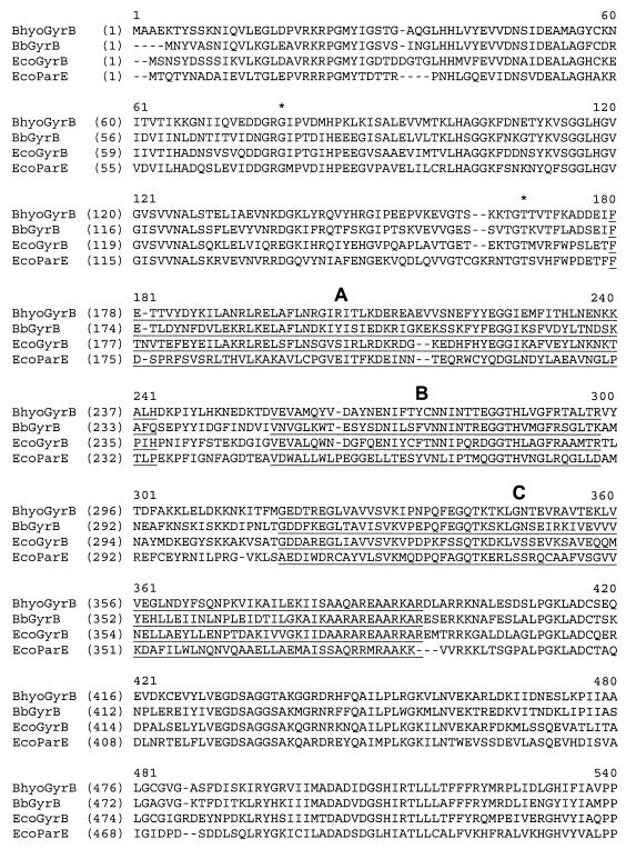
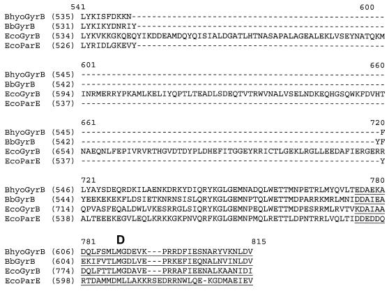
The B. hyodysenteriae gyrB gene sequence was determined from the sequences of a PCR amplicon of strain B204 DNA and a λZAPII clone containing DNA that hybridized with oligonucleotide probe GR, designed from the amplicon. The predicted amino acid sequence encoded by a 1908-bp open reading frame within the λ clone is shown in Fig. 1. This sequence exhibits significant similarity (BLAST Expect “E” values, e−113 to 0) over its entire length with 91 GenBank sequences that are identified or putative DNA gyrase subunit B proteins. The B. hyodysenteriae protein exhibits the highest sequence identities (50 to 56%) with GyrB proteins of bacteria that include other spirochetes, B. burgdorferi (Fig. 1), T. denticola, and T. pallidum. Four regions of the B. hyodysenteriae protein (Fig. 1, regions A to D) exhibit high sequence similarities (48 to 60%) with Escherichia coli GyrB. Three of these regions are conserved domains that differentiate bacterial GyrB proteins from homologous ParE proteins, which are subunits of DNA topoisomerase IV (31).
FIG. 1.
Comparison of amino acid sequences of the GyrB proteins of B. hyodysenteriae (BhyoGyrB), B. burgdorferi (BbGyrB), and E. coli (EcoGyrB) and the ParE protein of E. coli (EcoParE). B. hyodysenteriae GyrB exhibits overall sequence identities of 56, 42, and 37% with B. burgdorferi GyrB, E. coli GyrB, and E. coli ParE, respectively. In regions A to D, the B. hyodysenteriae GyrB protein exhibits the following sequence identities with B. burgdorferi GyrB, E. coli GyrB, and E. coli ParE, respectively: region A, 44, 48, and 22%; region B, 50, 58, and 34%; region C, 60, 54, and 33%; and region D, (D) 58, 60, and 21%. B. hyodysenteriae Cnr strains isolated in this study have amino acid mutations at positions Gly78 and Thr166, (indicated by asterisks).
Based on the considerations described above, we concluded that the cloned 1,908-bp open reading frame is the B. hyodysenteriae gyrB gene. Furthermore, as discussed below, coumermycin A1-resistant B. hyodysenteriae strains have amino acid substitutions in the GyrB sequence, and these substitutions parallel changes in the Cnr GyrB proteins of other bacterial species.
The GyrB protein of B. hyodysenteriae has an estimated molecular mass of 71,160 Da and is composed of 636 amino acids (Fig. 1). This protein is smaller than the E. coli GyrB protein, which has an additional 170-amino-acid insert at its C-terminal end (Fig. 1). Based on GenBank sequence comparisons performed with ClustalW (data not shown), this smaller sequence is characteristic of GyrB proteins of diverse bacterial species, including all spirochetes that have been examined, gram-positive and related bacteria (Mycoplasma pneumoniae, Staphylococcus aureus, Bacillus subtilis), the archaebacterium Thermotoga maritima, and the gram-negative anaerobe Bacteroides fragilis. In contrast, larger, E. coli-like GyrB proteins have been reported for the gram-negative facultative anaerobes Myxococcus xanthus, Salmonella enterica serovar Typhimurium, Pseudomonas aeruginosa, and Haemophilus influenzae.
UV mutagenesis and coumermycin A1 resistance.
Exposing B. hyodysenteriae cell suspensions to a UV dose of 3,500 μJ resulted in death of more than 99% of the bacteria (the bacterial viable counts decreased from 1.9 × 10⁷ to 1.8 × 10⁵ CFU/ml). This exposure resulted in a 10-fold increase in the number of cells forming colonies on solid medium containing 0.5 μg of coumermycin A1/ml (the number increased from 1.7 × 10⁻⁷ to 1.8 × 10⁻⁶ coumermycin-resistant CFU/total CFU). After 5 days of incubation at 38°C, coumermycin A1-resistant colonies appeared as discrete 1- to 4-mm hemolytic zones against a background of faint hemolysis caused by plating high densities of hemolytic cells of the spirochete.
Nine mutant strains able to grow on TSB agar plates containing coumermycin A1 were independently isolated (Table 1). Seven of these strains came from cell suspensions that had been exposed to UV light. Cnr strains 120B and 420A were isolated from cultures not exposed to UV light and thus were spontaneous mutants.
Coumermycin A1 MICs for the resistant strains were determined by both a spot plate test based on standard antibiotic MIC assays for Brachyspira species (21, 40) and a spread plate test. The spread plate test was used to determine antibiotic concentrations for subsequent genetic exchange studies. Results obtained from both techniques indicated that the coumermycin A1 MICs were 25 to 100 μg/ml for all of the resistant B. hyodysenteriae strains (Table 2). Under the same assay conditions, the MIC for wild-type strain B204 was 0.1 to 0.25 μg of coumermycin/ml. Dimethyl sulfoxide, the initial solvent used for the antibiotic, at final concentrations up to 1% (vol/vol) did not affect growth of the spirochete strains on TSB agar plates.
TABLE 2.
Exchange of antibiotic resistance genes between B. hyodysenteriae strains 435A (coumermycin A1 resistant) and SH (kanamycin and chloramphenicol resistant)a
| BHIS broth culture | CFU/ml of culture
|
|
|---|---|---|
| TSB agar | TSB agar + antibioticsb | |
| Strain 435A (Cnr) | 7.3 × 10⁷ | Und |
| Strain SH (Kmr Cmr) | 3.0 × 10⁸ | Und |
| Coculture (435A + SH) | 1.7 × 10⁸ | 357 |
| Coculture + antisera to VSH-1 | 2.4 × 10⁸ | Und |
| Coculture + preimmune sera | 2.2 × 10⁸ | 362 |
| Coculture + mitomycin Cc | 1.6 × 10⁸ | 1,810 |
The values are averages for cultures in three experiments, except for cocultures to which antiserum and mitomycin C were added (averages of two experiments). Strain 435H contains a coumermycin A1-resistant gyrB gene (Table 2). Strain SH (ΔflaA1 593–762::cat Δnox 438–760::kan) was constructed previously (17).
TSB agar containing kanamycin (200 μg/ml), chloramphenicol (10 μg/ml), and coumermycin A1 (10 μg/ml). Und, undetected (the incidence of spontaneous triply resistant mutants was less than 2 × 10⁻⁸ or less than 1 CFU per 5 × 10⁷ CFU plated).
Mitomycin C was added at a final concentration of 0.2 μg/ml to cocultures after inoculation of bacteria.
Coumermycin-resistant strains 120B, 235C, 435A, and 235E each had a single base mutation in gyrB, which provided three discernible genotypes (Table 1). Five other resistant strains had gyrB gene sequences identical to that of wild-type strain B204 and, due to their indiscernible genotypes, were not studied further.
Coumermycin A1 resistance gene exchange between B. hyodysenteriae strains.
To investigate the possibility of coumermycin A1 resistance transfer in B. hyodysenteriae broth cultures, cells of Cnr strain 435A (Table 1) were cultured with cells of Kmr Cmr strain SH in BHIS broth, and samples of the cocultures were plated onto TSB agar containing coumermycin A1, kanamycin, and chloramphenicol (Table 2). Based on MIC results (Table 1), a coumermycin A1 concentration of 10 μg/ml was used.
Cells resistant to all three antibiotics were isolated from the cocultures after overnight incubation and were present at levels of approximately 360 CFU/ml (Table 2). Strains resistant to all three antibiotics were not detected in (control) monocultures of either strain SH or strain 435A (Table 2). These results suggested that the triply resistant mutants resulted from gene transfer and were not the result of spontaneous mutations.
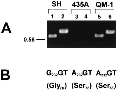
Triply resistant strains resulting from gene exchange in cocultures should have possessed the genotypes of both parent strains, SH and 435A. In three experiments, a total of seven B. hyodysenteriae strains (designated QM-1 to QM-7) were isolated by subculturing randomly chosen colonies from TSB agar plates containing coumermycin A1, chloramphenicol, and kanamycin. Based on PCR analyses, each triply resistant strain had kan and cat genes, like strain SH (Fig. 2A), and contained a gyrB gene mutated at base position 232 (A232 to G, resulting in a change from Gly78 to Ser), like strain 435A (Fig. 2B).
FIG. 2.
Genotype analyses of antibiotic resistance determinants of strains SH (Kmr Cmr), 435A (Cnr), and QM-1 (Cnr Kmr Cmr). Six other triply resistant strains (QM-2 to QM-7) were examined and gave results identical to those obtained with strain QM-1. (A) PCR amplification to detect the kan gene (lanes 1, 3, and 5) and the cat gene (lanes 2, 4, and 6). (B) DNA sequence analysis to detect the gyrB nucleotide change (corresponding amino acid change) associated with coumermycin resistance.
Cnr Kmr Cmr strains produced by gene exchange were not isolated from cocultures to which rabbit anti-VSH-1 antiserum (final concentration, 0.5% [vol/vol]) had been added (Table 2). The antiserum had no detectable effect on growth of either strain. Preimmunization serum from the same rabbit did not affect gene transfer (Table 2). An explanation for these findings is that VSH-1 (17) was produced spontaneously in B. hyodysenteriae cultures and was transfer agent for coumermycin-resistant gyrB in the cocultures. Also supporting this conclusion is the observation that addition of mitomycin C to cocultures resulted in a five-fold increase in the number of triply resistant cells obtained after incubation (Table 2). Mitomycin C induces production of VSH-1 particles in B. hyodysenteriae cultures (16, 17). In separate experiments, triply resistant B. hyodysenteriae cells were isolated from strain SH cultures to which VSH-1 particles purified from mitomycin C-treated 435A cultures had been added (Stanton and Humphrey, unpublished data).
DISCUSSION
The results of our investigations, as elaborated below, led to the following conclusions. Discernible mutations, both spontaneous and UV induced, in the B. hyodysenteriae gyrB gene result in coumermycin A1 resistance. Coumermycin A1 resistance due to a gyrB mutation is readily transferred between B. hyodysenteriae strains in broth cultures. VSH-1 is the most likely mechanism for Cnr gyrB gene transfer between B. hyodysenteriae cells. These findings have implications both for in vitro investigations of B. hyodysenteriae and for understanding aspects of the ecology and evolution of this spirochete.
The N-terminal domain of bacterial GyrB subunits contains the ATP binding site of DNA gyrase and is the site for mutations conferring coumarin resistance (23, 24). Amino acid substitutions in B. hyodysenteriae GyrB proteins occurred in the N-terminal domain, and these substitutions are comparable to changes associated with coumermycin resistance in other species. Mutations comparable to the GyrB Gly78-to-Ser change in B. hyodysenteriae 235C and 435A confer coumermycin A1 resistance in other species; such mutations include Gly74 to Ser in B. burgdorferi (D. S. Samuels, personal communication), Gly124 to Ser in Bartonella bacilliformis (1), and Gly85 to Ser in S. aureus (36). To our knowledge, the Gly78-to-Cys modification of GyrB in B. hyodysenteriae spontaneous mutant strain 120B is the only example of a mutation of this Gly residue to an amino acid other than Ser. The substitution in the coumermycin-resistant GyrB subunit of strain 235E (Thr166 to Ala) parallels amino acid substitutions observed in the coumermycin-resistant GyrB of B. bacilliformis (Thr214 to Ala or Ile), B. burgdorferi (Thr162 to Ile), and S. aureus (Thr173 to Asn) (1, 36; Samuels, personal communication).
Cnr strains of bacterial species other than B. hyodysenteriae commonly have homologous mutations corresponding to substitutions for Arg136 in E. coli (1, 5, 6, 14, 29, 36). T. denticola has a wild-type GyrB protein with Lys136 in place of Arg, and spontaneous coumermycin A1-resistant strains of this spirochete have modifications of Lys136 to Thr or Gln in GyrB (10). B. hyodysenteriae has a comparable Lys137 in wild-type GyrB (Fig. 1); however, none of the Cnr strains which we isolated had amino acid modifications at that residue. Characterization of additional Cnr strains may permit evaluation of whether Lys137 is a stable residue and thus whether there is a possible functional difference between the GyrB protein of B. hyodysenteriae and those of other bacteria.
The basis for coumermycin resistance in five B. hyodysenteriae strains remains unknown, since the gyrB sequences of these strains are identical to that of wild-type strain B204. Mutations in genes other than gyrB have been associated with coumermycin resistance in other bacteria (24).
B. hyodysenteriae Cnr Kmr Cmr strains most likely resulted from unidirectional transfer of gyrB from strain 435A to strain SH. The alternative explanation, that strain SH was the donor of both Kmr and Cmr, would require independent transfer of both flaA1::cat and nox::kan genes to the same 435A cell. This scenario seems improbable since the flaA1 and nox genes are unlinked and are located on opposite sides of the B. hyodysenteriae B78T chromosome (42).
VSH-1 is a bacteriophage-like element that packages random 7.5-kb fragments of B. hyodysenteriae genomic DNA (17). VSH-1 is the only known mechanism for gene transfer in B. hyodysenteriae. In this study, anti-VSH-1 antiserum inhibited Cnr gyrB transfer (Table 2). Mitomycin C, an inducer of VSH-1 (17), enhanced gene transfer. Purified VSH-1 particles transmit coumermycin resistance to strain SH cells. Based on these considerations, VSH-1 is the likely agent for Cnr gene transfer in cultures of this spirochete.
Previous investigators either have reported spontaneous appearance of bacteriophage particles resembling VSH-1 (25) or have described extrachromosomal DNA that is the size of VSH-1 DNA (7.5 kb) in cultures of B. hyodysenteriae (3, 4, 41; L. A. Joens, A. B. Margolin, and M. J. Hewlett, Abstr. 86th Annu. Meet. Am. Soc. Microbiol. 1986, abstr. H-173, 1986). VSH-1-like bacteriophages have recently been detected in other Brachyspira species (2). In our laboratory we have been unable to confirm reports of other investigators since we have been unable to directly detect VSH-1 particles or 7.5-kb DNA in B. hyodysenteriae cultures (16, 17). Based on a frequency of 1.5 × 10⁻⁶ transductant per phage particle for VSH-1 (17), a DNA content of 7.5 kb per phage particle (17), and production of 360 triply resistant CFU/ml due to the transfer of Cnr, we conservatively estimate that VSH-1 particles are produced in B. hyodysenteriae cultures at levels that are at least 5- to 10-fold lower than the limit of detection of our previous assays for VSH-1 DNA (limit of detection, 40 ng of VSH-1 DNA/ml of culture). The results of this study suggest that monitoring Cnr gene transfer in cocultures of strains 435A and SH is a more sensitive assay for VSH-1 production than either analysis for 7.5-kb extrachromosomal DNA fragments or (not surprisingly) electron microscopy to detect phage particles. We are currently using increases in the incidence of triply resistant strains in cocultures of the two strains as an assay for chemicals or conditions that induce VSH-1 production (Matson, unpublished data).
The observation that gene transfer readily occurs between B. hyodysenteriae strains in broth cultures has practical importance. Recently, by using the broth coculture method described in this paper, other investigators have been able to produce B. hyodysenteriae strains with double mutations in separate fla genes, and they are using these strains to investigate spirochete motility (C. Li and N. W. Charon, personal communications). The use of Cnr as a selection marker should facilitate additional investigations of B. hyodysenteriae involving gene exchange. Among the spirochetes, B. hyodysenteriae stands out as a good, practical choice for studying spirochete genetics and investigating aspects of spirochete biology through the use of mutant strains.
The findings of this study may also have ecological significance. In a comparative analysis of the genetic diversity of 231 B. hyodysenteriae isolates by multilocus enzyme electrophoresis, Trott and colleagues (39) concluded that substantial genetic recombination has shaped the overall population structure of this spirochete species. The gene transducing capability of VSH-1, especially since there are no other known mechanisms of gene transfer, leads to the hypothesis that VSH-1 has been a major factor in B. hyodysenteriae evolution. In view of this hypothesis, it would be worthwhile to examine natural factors that influence VSH-1 production and to assess VSH-1-mediated gene transfer between B. hyodysenteriae cells in their natural environment, the swine intestinal tract.
ACKNOWLEDGMENTS
We thank Tom Casey, Richard Zuerner, and Scott Samuels for comprehensive reviews of the manuscript. The insightful comments and enthusiastic advice of Scott Samuels regarding gyrB and coumermycin A1 resistance are both highly regarded and appreciated. We thank J. Hardham, E. Rosey, K. Tilly, A. F. Elias, J. L. Bono, P. Stewart, and P. Rosa for sharing prepublication manuscripts.
REFERENCES
- 1.Battisti J M, Smitherman L S, Samuels D S, Minnick M F. Mutations in Bartonella bacilliformis gyrB confer resistance to coumermycin A1. Antimicrob Agents Chemother. 1998;42:2906–2913. doi: 10.1128/aac.42.11.2906. [DOI] [PMC free article] [PubMed] [Google Scholar]
- 2.Calderaro A, Dettori G, Collini L, Ragni P, Grillo R, Cattani P, Fadda G, Chezzi C. Bacteriophages induced from weakly beta-haemolytic human intestinal spirochaetes by mitomycin C. J Basic Microbiol. 1998;38:323–335. [PubMed] [Google Scholar]
- 3.Calderaro A, Dettori G, Grillo R, Plaisant P, Amalfitano G, Chezzi C. Search for bacteriophages spontaneously occurring in cultures of haemolytic intestinal spirochaetes of human and animal origin. J Basic Microbiol. 1998;38:313–322. [PubMed] [Google Scholar]
- 4.Combs B G, Hampson D J, Harders S J. Typing of Australian isolates of Treponema hyodysenteriae by serology and by DNA restriction endonuclease analysis. Vet Microbiol. 1992;31:273–285. doi: 10.1016/0378-1135(92)90085-8. [DOI] [PubMed] [Google Scholar]
- 5.Contreras A, Maxwell A. gyrB mutations which confer coumarin resistance also affect DNA supercoiling and ATP hydrolysis by Escherichia coli DNA gyrase. Mol Microbiol. 1992;6:1617–1624. doi: 10.1111/j.1365-2958.1992.tb00886.x. [DOI] [PubMed] [Google Scholar]
- 6.del Castillo L, Vizan J L, Rodriguez-Sainz M C, Moreno F. An unusual mechanism for resistance to the antibiotic coumermycin A1. Proc Natl Acad Sci USA. 1991;88:8860–8864. doi: 10.1073/pnas.88.19.8860. [DOI] [PMC free article] [PubMed] [Google Scholar]
- 7.Fournier B, Hooper D C. Mutations in topoisomerase IV and DNA gyrase of Staphylococcus aureus: novel pleiotropic effects on quinolone and coumarin activity. Antimicrob Agents Chemother. 1998;42:121–128. doi: 10.1128/aac.42.1.121. [DOI] [PMC free article] [PubMed] [Google Scholar]
- 8.Frothingham R, Hills H G, Wilson K H. Extensive DNA sequence conservation throughout the Mycobacterium tuberculosis complex. J Clin Microbiol. 1994;32:1639–1643. doi: 10.1128/jcm.32.7.1639-1643.1994. [DOI] [PMC free article] [PubMed] [Google Scholar]
- 9.Girons I S, Bourhy P, Ottone C, Picardeau M, Yelton D, Hendrix R W, Glaser P, Charon N. The LE1 bacteriophage replicates as a plasmid within Leptospira biflexa: construction of an L. biflexa-Escherichia coli shuttle vector. J Bacteriol. 2000;182:5700–5705. doi: 10.1128/jb.182.20.5700-5705.2000. [DOI] [PMC free article] [PubMed] [Google Scholar]
- 10.Greene S R, Stamm L V. Molecular characterization of the gyrB region of the oral spirochete, Treponema denticola. Gene. 2000;253:259–269. doi: 10.1016/s0378-1119(00)00254-7. [DOI] [PubMed] [Google Scholar]
- 11.Hampson D J, Atyeo R F, Combs B G. Swine dysentery. In: Hampson D J, Stanton T B, editors. Intestinal spirochaetes in domestic animals and humans. Wallingford, United Kingdom: CAB International; 1997. pp. 175–209. [Google Scholar]
- 12.Hardham J M, Rosey E L. Antibiotic selective markers and spirochete genetics. J Mol Microbiol Biotechnol. 2000;2:425–432. [PubMed] [Google Scholar]
- 13.Harris D L, Hampson D J, Glock R D. Swine dysentery. In: Straw B E, D'Allaire S, Mengeling W L, Taylor D L, editors. Diseases of swine. 8th ed. Ames: Iowa State University Press; 1999. pp. 579–600. [Google Scholar]
- 14.Holmes M L, Dyall-Smith M L. Mutations in DNA gyrase result in novobiocin resistance in halophilic archaebacteria. J Bacteriol. 1991;173:642–648. doi: 10.1128/jb.173.2.642-648.1991. [DOI] [PMC free article] [PubMed] [Google Scholar]
- 15.Huang W M. Bacterial diversity based on type II DNA topoisomerase genes. Annu Rev Genet. 1996;30:79–107. doi: 10.1146/annurev.genet.30.1.79. [DOI] [PubMed] [Google Scholar]
- 16.Humphrey S B, Stanton T B, Jensen N S. Mitomycin C induction of bacteriophages from Serpulina hyodysenteriae and Serpulina innocens. FEMS Microbiol Lett. 1995;134:97–101. doi: 10.1111/j.1574-6968.1995.tb07921.x. [DOI] [PubMed] [Google Scholar]
- 17.Humphrey S B, Stanton T B, Jensen N S, Zuerner R L. Purification and characterization of VSH-1, a generalized transducing bacteriophage of Serpulina hyodysenteriae J. Bacteriol. 1997;179:323–329. doi: 10.1128/jb.179.2.323-329.1997. [DOI] [PMC free article] [PubMed] [Google Scholar]
- 18.Hyatt D R, ter Huurne A A H M, van der Zeijst B A M, Joens L A. Reduced virulence of Serpulina hyodysenteriae hemolysin-negative mutants in pigs and their potential to protect pigs against challenge with a virulent strain. Infect Immun. 1994;62:2244–2248. doi: 10.1128/iai.62.6.2244-2248.1994. [DOI] [PMC free article] [PubMed] [Google Scholar]
- 19.Kennedy M J, Rosey E L, Yancey R J., Jr Characterization of flaA- and flaB-mutants of Serpulina hyodysenteriae: both flagellin subunits, FlaA and FlaB, are necessary for full motility and intestinal colonization. FEMS Microbiol Lett. 1997;153:119–128. doi: 10.1111/j.1574-6968.1997.tb10472.x. [DOI] [PubMed] [Google Scholar]
- 20.Kinyon J M, Harris D L, Glock R D. Enteropathogenicity of various isolates of Treponema hyodysenteriae. Infect Immun. 1977;15:638–646. doi: 10.1128/iai.15.2.638-646.1977. [DOI] [PMC free article] [PubMed] [Google Scholar]
- 21.Kitai K, Kashiwazaki M, Adachi Y, Kume T, Arakawa A. In vitro activity of 39 antimicrobial agents against Treponema hyodysenteriae. Antimicrob Agents Chemother. 1979;15:392–395. doi: 10.1128/aac.15.3.392. [DOI] [PMC free article] [PubMed] [Google Scholar]
- 22.Levine C, Hiasa H, Marians K J. DNA gyrase and topoisomerase IV: biochemical activities, physiological roles during chromosome replication, and drug sensitivities. Biochim Biophys Acta. 1998;1400:29–43. doi: 10.1016/s0167-4781(98)00126-2. [DOI] [PubMed] [Google Scholar]
- 23.Maxwell A. DNA gyrase as a drug target. Biochem Soc Trans. 1999;27:48–53. doi: 10.1042/bst0270048. [DOI] [PubMed] [Google Scholar]
- 24.Maxwell A. The interaction between coumarin drugs and DNA gyrase. Mol Microbiol. 1993;9:681–686. doi: 10.1111/j.1365-2958.1993.tb01728.x. [DOI] [PubMed] [Google Scholar]
- 25.Ritchie A E, Robinson I M, Joens L A, Kinyon J M. A bacteriophage for Treponema hyodysenteriae. Vet Rec. 1978;102:34–35. doi: 10.1136/vr.103.2.34. [DOI] [PubMed] [Google Scholar]
- 26.Rosa P, Samuels D S, Hogan D, Stevenson B, Casjens S, Tilly K. Directed insertion of a selectable marker into a circular plasmid of Borrelia burgdorferi. J Bacteriol. 1996;178:5946–5953. doi: 10.1128/jb.178.20.5946-5953.1996. [DOI] [PMC free article] [PubMed] [Google Scholar]
- 27.Rosey E L, Kennedy M J, Yancey R J. Dual flaA1 flaB1 mutant of Serpulina hyodysenteriae expressing periplasmic flagella is severely attenuated in a murine model of swine dysentery. Infect Immun. 1996;64:4154–4162. doi: 10.1128/iai.64.10.4154-4162.1996. [DOI] [PMC free article] [PubMed] [Google Scholar]
- 28.Sambrook J, Fritsch E F, Maniatis T. Molecular cloning. 2nd ed. Cold Spring Harbor, N.Y: Cold Spring Harbor Laboratory Press; 1989. [Google Scholar]
- 29.Samuels D S, Marconi R T, Huang W M, Garon C F. gyrB mutations in coumermycin A1-resistant Borrelia burgdorferi. J Bacteriol. 1994;176:3072–3075. doi: 10.1128/jb.176.10.3072-3075.1994. [DOI] [PMC free article] [PubMed] [Google Scholar]
- 30.Sartakova M, Dobrikova E, Cabello F C. Development of an extrachromosomal cloning vector system for use in Borrelia burgdorferi. Proc Natl Acad Sci USA. 2000;97:4850–4855. doi: 10.1073/pnas.080068797. [DOI] [PMC free article] [PubMed] [Google Scholar]
- 31.Springer A L, Schmid M B. Molecular characterization of the Salmonella typhimurium parE gene. Nucleic Acids Res. 1993;21:1805–1809. doi: 10.1093/nar/21.8.1805. [DOI] [PMC free article] [PubMed] [Google Scholar]
- 32.Stanton T B. Physiology of ruminal and intestinal spirochaetes. In: Hampson D J, Stanton T B, editors. Intestinal spirochaetes in domestic animals and humans. Wallingford, United Kingdom: CAB International; 1997. pp. 7–45. [Google Scholar]
- 33.Stanton T B, Cornell C P. Erythrocytes as a source of essential lipids for Treponema hyodysenteriae. Infect Immun. 1987;55:304–308. doi: 10.1128/iai.55.2.304-308.1987. [DOI] [PMC free article] [PubMed] [Google Scholar]
- 34.Stanton T B, Rosey E L, Kennedy M J, Jensen N S, Bosworth B T. Isolation, oxygen sensitivity, and virulence of NADH oxidase mutants of the anaerobic spirochete Brachyspira (Serpulina) hyodysenteriae, etiologic agent of swine dysentery. Appl Environ Microbiol. 1999;65:5028–5034. doi: 10.1128/aem.65.11.5028-5034.1999. [DOI] [PMC free article] [PubMed] [Google Scholar]
- 35.Stanton T B, Sellwood R. Cloning and characteristics of a gene encoding NADH oxidase, a major mechanism for oxygen metabolism by the anaerobic spirochete, Brachyspira (Serpulina) hyodysenteriae. Anaerobe. 1999;5:539–546. [Google Scholar]
- 36.Stieger M, Angehrn P, Wohlgensinger B, Gmünder H. GyrB mutations in Staphylococcus aureus strains resistant to cyclothialidine, coumermycin, and novobiocin. Antimicrob Agents Chemother. 1996;40:1060–1062. doi: 10.1128/aac.40.4.1060. [DOI] [PMC free article] [PubMed] [Google Scholar]
- 37.ter Huurne A A H M, van Houten M, Muir S, Kusters J G, van der Zeijst B A M, Gaastra W. Inactivation of a Serpula (Treponema) hyodysenteriae hemolysin gene by homologous recombination: importance of this hemolysin in pathogenesis of S. hyodysenteriae in mice. FEMS Microbiol Lett. 1992;92:109–114. doi: 10.1016/0378-1097(92)90550-8. [DOI] [PubMed] [Google Scholar]
- 38.Tilly K, Elias A F, Bono J L, Stewart P, Rosa P. DNA exchange and insertional inactivation in spirochetes. J Mol Microbiol Biotechnol. 2000;2:433–442. [PubMed] [Google Scholar]
- 39.Trott D J, Oxberry S L, Hampson D J. Evidence for Serpulina hyodysenteriae being recombinant, with an epidemic population structure. Microbiology. 1997;143:3357–3365. doi: 10.1099/00221287-143-10-3357. [DOI] [PubMed] [Google Scholar]
- 40.Trott D J, Stanton T B, Jensen N S, Duhamel G E, Johnson J L, Hampson D J. Serpulina pilosicoli sp. nov. the agent of porcine intestinal spirochetosis. Int J Syst Bacteriol. 1996;46:206–215. doi: 10.1099/00207713-46-1-206. [DOI] [PubMed] [Google Scholar]
- 41.Turner A K, Sellwood R. Extracellular DNA from Serpulina hyodysenteriae consists of 6.5 kbp random fragments of chrosomal DNA. FEMS Microbiol Lett. 1997;150:75–80. doi: 10.1111/j.1574-6968.1997.tb10352.x. [DOI] [PubMed] [Google Scholar]
- 42.Zuerner R L, Stanton T B. Physical and genetic map of the Serpulina hyodysenteriae B78T chromosome. J Bacteriol. 1994;176:1087–1092. doi: 10.1128/jb.176.4.1087-1092.1994. [DOI] [PMC free article] [PubMed] [Google Scholar]


