Abstract
Objective
This study aimed to determine the correlation between sealer penetration into dentinal tubules and interfacial adaptation to root canal walls using a hydraulic calcium silicate-based sealer, Endosequence Bioceramic Sealer (Brasseler USA, Savannah, GA, USA), and an epoxy resin-based sealer, AH Plus (Dentsply DeTrey, Konstanz, Germany).
Methods
Sixty-four maxillary central incisors were endodontically prepared with nickel-titanium rotary instruments and randomly assigned into two groups (n=32). Roots were filled with gutta-percha using a single-cone technique in conjunction with one of the two sealers, AHP or BCS. Sealers were mixed with Rhodamine B and analysed under a confocal laser scanning microscope. Transverse sections at 5 mm from root apex were obtained. The circumference of the root canal wall was first outlined and measured to determine the circumferential percentage of sealer penetration. The regions along the canal walls where the sealer had penetrated the dentinal tubules were delineated and measured. Then, the outlined distances were divided by the canal circumference. The width of each gap was measured and pooled for each specimen for comparison to determine the interfacial adaptation. The measurements were repeated twice to ensure reproducibility. Mann-Whitney tests were performed to compare continuous variables between AHP and BCS groups. The correlation between gap width and percentage of sealer penetration was investigated using the Spearman correlation coefficient.
Results
No significant difference was observed between groups regarding the percentage of sealer penetration (P>0.05) and the gap width (P>0.05). Also, there was no significant correlation between the two variables analysed for AHP (r=0.165; P>0.05) and BCS (r=-0.147; P>0.05) and in the overall sample (r=0.061; P>0.05).
Conclusion
The present results show no correlation between interfacial adaptation and sealer penetration in dentinal tubules in the total sample and among sealer subgroups. The ability of root canal sealers to penetrate dentinal tubules cannot be considered a sign of better interfacial adaptation.
Keywords: Hydraulic calcium silicate-based sealer, interfacial adaptation, resin-based sealer, tubule penetration
HIGHLIGHTS
No correlation between interfacial adaptation and the penetration of sealer in dentinal tubules.
The capacity of root canal sealers to penetrate dentinal tubules cannot be considered a sign of better interfacial adaptation.
The absence of correlation reported is of significance and might be considered during the introduction of a new root canal sealer.
INTRODUCTION
The sealer characteristics play an essential role in the success of root canal obturation. Root canal sealers are used in combination with gutta-percha as the core material to obturate the root canal system (1). Sealers fill the gap between gutta-percha points and between gutta-percha and root canal wall and therefore assume high adaptability to the root canal wall and better interfacial adaptation (2). An increased contact surface area between the dentine and the sealer ensures good interfacial adaptation to dentine. The occurrence of voids and gaps at the interface sealer-canal walls reduces the interfacial adaptation to dentine (3). One of the main concerns of endodontic treatment is the control of infections caused by microorganisms in the root canal system (4). Studies have focused on the importance of sealer penetration into dentinal tubules as a prerequisite for improving the quality of root fillings; this idea has sparked considerable scientific debate (3-6). Sealer penetration into dentinal tubules can inactivate microorganisms within tubules, enhance retention of materials and improve the quality of the seals by increasing the contact surface of the sealers with dentine walls (7). Moreover, the high adaptability of root canal filling to root dentine will minimise microorganism infiltration since gaps usually serve as hubs for microbial housing (8). Contrary to common beliefs, no research has yet identified a causal link between the penetration of a root canal sealer into dentinal tubules and interfacial adaptation of the sealer (gap) (4). Scientific advances introduced hydraulic calcium silicate-based materials that are distinct from other materials, mainly because of their hydraulic characteristics and their interaction with the environment (9). Among a wide range of commercially available hydraulic calcium silicate sealers, EndoSequence BC Sealer (Brasseler USA, Savannah, GA, USA) (BCS) has gained popularity in recent years (10). In addition to its excellent physical properties, it adapts well to the canal walls and can form a chemical bond with the inorganic phase of dentine (11). The epoxy resin-based sealer AH Plus (Dentsply DeTrey, Konstanz, Germany) (AHP) has been considered the “gold standard” due to its physicochemical properties compared to other sealers and has been widely studied in terms of interfacial adaptation and dentinal tubule penetration (12).
Sealer penetration and interfacial adaptation to dentine walls can be assessed using several instruments, including the Confocal Laser Scanning Microscope (CLSM) (7, 13, 14-16). It allows sealer visualisation inside dentinal tubules and interfacial adaptation with few artefacts and clearly shows the amount of sealer in the entire cross-sectional specimen. It maintains the integrity of the sealer, as it does not require any special sample preparation (4). Moreover, with the CLSM, interfacial gaps can be distinguished from technique gaps that may result from the loss of material after high vacuum dehydration under a scanning electron microscope (4). The hypothesis formulated is that there is a positive correlation between sealer penetration into dentinal tubules and interfacial adaptation.
This laboratory study aimed to determine the correlation between sealer penetration into dentinal tubules and interfacial adaptation to root canal walls using a hydraulic calcium silicate-based sealer, BCS, and an epoxy resin-based sealer, AHP.
MATERIALS AND METHODS
Materials
The sealers used in this study were:
AHP, mixed and manipulated according to the manufacturers’ instructions.
BCS, a premixed root canal sealer available in ready-to-use syringes.
Ethical aspects
This study was approved by the ethics committee of Saint Joseph University, Beirut, Lebanon (registration no. 2016-61).
Minimal sample size calculation
The G*Power software (Version 3.0.10) was used to calculate the minimum sample size, considering a two-sided test to detect a simple correlation r (r=0.5), a 5% significance level test (α=0.05), and a power of 85% (β=0.15). The minimum sample required was 32 to determine whether a correlation coefficient differs from zero.
Specimen selection
A total of 95 maxillary central incisors were obtained from several dental offices. Teeth with fully formed roots were collected from patients aged between 50 and 60 and stored in distilled water after disinfection, and processed electronically to identify teeth with comparable internal anatomy. Teeth with intracanal calcifications and/or internal resorption were excluded. A single operator classified the category of radiographs according to similar root canal anatomy. The widths BL and MD of the root canals were measured at a point 5 mm from the root apex. Out of the 95 teeth, 70 had a BL/MD ratio ≤1.5, thus radiographically similar. Crowns were trimmed using a low-speed cutting steel disk, leaving all the roots 16 mm long, then root canals were irrigated with 5 mL of 2.5% NaOCl. A size 10 K-file (Dentsply Sirona, Ballaigues, Switzerland) was inserted in all root canals to achieve apical patency. A 25 K-file (Dentsply Sirona) was introduced to the apical foramen of these teeth, corresponding to an anatomic diameter of 250 μm, to standardise the apical foramen scale. Roots with wider foramina were removed. As a result, just 64 teeth fit the selection criteria.
Root canal preparation
One specialist in endodontics performed all the endodontic procedures. A 10 K-file was introduced into the canal until it was visible at the apical foramen and the length measured. The working length (WL) was calculated by subtracting 0.5 mm from this length. Root canals were prepared with the ProTaper Universal system (Dentsply Maillefer, Ballaigues, Switzerland), using a crown-down technique, according to the manufacturer’s instructions, up to the F4 instrument, which has an ISO 040 tip size, a 6% apical third taper, and then a progressively decreasing taper in the body. A size 10 K-file was inserted after each file to maintain apical patency. Canals were irrigated between each instrument with 0.5 mL of freshly prepared NaOCl 2.5%, and after instrumentation, with 10 mL EDTA 17% followed by 3 mL of 5.25% NaOCl for 3 minutes using a 30 G Navi Tip needle (Ultradent, South Jordan, UT, USA) and introduced 3 mm shorter than the WL (17). After all the procedures, each canal was irrigated with 10 mL of distilled water then dried using paper points (Dentsply Maillefer).
Specimen grouping
A block randomisation procedure was applied to assign teeth into two groups of equal numbers. A block size of 4 was taken, and possible balanced configurations were 1 for AHP and 2 for BCS (1122, 1212, 1221, 2112, 2121, 2211). Blocks were selected randomly to ensure the assignment of all 64 teeth with 32 teeth in each group. The comparability between groups, in terms of root canal length, BL/MD ratio (≤1.5), and apical diameter, was assessed and confirmed statistically (P>0.05).
Root canal obturation
All instrumented canals were filled with pre-fitted size gutta-percha F4 cone (Dentsply Maillefer) and sealers, using the single-cone filling technique. Each sealer was mixed with 0.1% Rhodamine B (Sigma-Aldrich, St Louis, MO, USA). To use the same amount of sealers, we put each sealer onto a glass slab, and we measured 3 mm of each sealer with a millimetre ruler. Sealers were placed inside the root canals 1-mm short of the WL with a size 30 lentulo spiral (Dentsply Maillefer) mounted on a slow-speed handpiece (18). The F4 master cone was then lightly coated with the sealer and placed in the root canal to the WL. The coronal excess filling material was sheared off with an electrical instrument. The quality of the filling in terms of length and density was confirmed by BL and MD digital radiographs. Access cavities were filled with a temporary filling material (Cavit, 3M; ESPE, St. Paul, MN, USA). All teeth were stored at 37°C, with fully saturated humidity (100%), for 14 days to allow the endodontic sealers to set, then placed into self-cure acrylic resin moulds.
Confocal microscopy examination
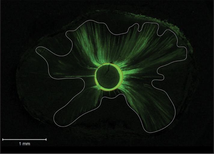
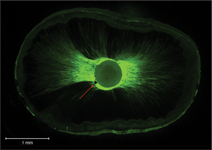
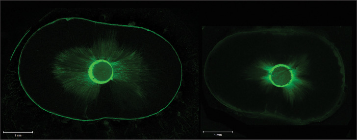
The sealer penetration and root dentine-sealer interface were assessed using CLSM and ImageJ software (National Institutes of Health, Bethesda, MD, USA). The roots were sectioned perpendicular to their long axis, and a 1.0-mm-thick slice was obtained 5 mm from the root apex using a diamond disk (Buehler, Lake Bluff, IL, USA) with a slow speed (25,000 rpm) handpiece. The coronal surface was polished using sandpaper numbers 500, 700, and 1200 under continuous water and polishing paste, then mounted onto glass slides. The Rhodamine B was visible at an excitation/emission wavelength of 514/543 nm. Specimens were assessed by CLSM (Zeiss, LSM 780, Germany) at ×5 and ×10 magnifications, 10 µm below the surface (Fig. 1). Areas into which the sealer penetrated the dentinal tubules and the circumference of the root canal wall were outlined and measured with ImageJ software. The outlined distances were divided by the canal circumference to calculate the circumferential percentage of sealer penetration (18) (Fig. 2), and the width of each gap was measured and pooled for each specimen to evaluate and compare the interfacial adaptation (19). The measurements were repeated twice to ensure reproducibility (Fig. 3).
Figure 1.
Representative CLSM images. On the left, an image from the AHP group, and on the right, an image from the BCS group. Note the sealer inside the dentine
CLSM: Confocal Laser Scanning Microscope, AHP: AH Plus, BCS: Endosequence Bioceramic sealer
Figure 2.

Representative CLSM image from AHP group. Calculation of the circumferential percentage of sealer penetration: example of the outline of areas into which the sealer penetrated the dentinal tubules (white line). The outlined distance was divided by the canal circumference (red line)
CLSM: Confocal Laser Scanning Microscope, AHP: AH Plus
Figure 3.

A representative CLSM image from the BCS group. Measurement of gap width: the width of each gap was measured and pooled for each specimen for comparison. The red arrow shows the gap-containing region at the interfacial adaptation.
CLSM: Confocal Laser Scanning Microscope, BCS: Endosequence Bioceramic sealer
Statistical analysis
Statistical analyses were performed using IBM SPSS Statistics (version 25.0, Chicago, IL, USA). The level of significance level was set at P≤0.05. Kolmogorov Smirnov tests were used to assess the normality distribution of the continuous variables. Since variables were not normally distributed, the correlation between gap width and percentage of sealer penetration was investigated using the Spearman correlation coefficient. Mann-Whitney tests were also performed to compare continuous variables between AHP and BCS groups.
RESULTS
Correlation analyses
There was no statistically significant correlation between circumferential percentage of sealer penetration and gap width in the overall sample (r=0.061; P=0.673); similar results were shown for AHP (r=0.165; P=0.442) and BCS (r=-0.147; P=0.475) (Table 1).
TABLE 1.
Correlation between circumferential sealer penetration and gap width
| Sealers | Correlation coefficient | P |
|---|---|---|
| AHP | 0.165 | 0.442 |
| BCS | -0.147 | 0.475 |
| AHP and BCS | 0.061 | 0.673 |
AHP: AH Plus, BCS: Endosequence Bioceramic sealer
Percentage of sealer penetration into dentinal tubules
The mean circumferential percentage of sealer penetration into dentinal tubules was 20.17±5.46% for AHP and 17.94±4.13% for BCS. The circumferential percentage of sealer penetration was not significantly different between the two groups P>0.05 (Table 2).
TABLE 2.
Percentage of sealer penetration in dentinal tubules in different groups
| Groups | Mean | Standard deviation | P |
|---|---|---|---|
| AHP, % | 20.17 | 5.46 | 0.156 |
| BCS, % | 17.94 | 4.13 |
AHP: AH Plus, BCS: Endosequence Bioceramic sealer
Gap width
The mean gap width (±SD) was 19.85±47.17 μm for AHP and 4.54±23.14 μm for BCS. The gap width was not significantly different among AHP and BCS (P>0.05) (Table 3).
TABLE 3.
Mean gap width (μm) in different groups
| Groups | Mean | Standard deviation | P |
|---|---|---|---|
| AHP | 19.85 | 47.17 | 0.136 |
| BCS | 4.54 | 23.14 |
AHP: AH Plus, BCS: Endosequence Bioceramic sealer
DISCUSSION
Several studies have assessed dentinal tubule penetration and interfacial adaptation of sealers based on the hypothesis that sealer penetration may enhance the interfacial adaptation of endodontic sealers, and the debate is still ongoing regarding its importance to improve the quality of root fillings (20, 21). Our results contradicted this hypothesis and showed that sealer penetration into dentinal tubules was not associated with an improvement in the interfacial adaptation, regardless of the type of sealer used.
Previous findings revealed higher sealer penetration in the middle third of the root (21, 22). In the coronal region, the number of dentinal tubules is higher and their diameter longer, making it easier for the sealer to penetrate the tubules. In that context, Mjör et al. (23) showed that some areas of the apical region might not contain tubules. Thus, our specimens were obtained 5 mm from the root apex.
In this study, transverse sections were assessed because longitudinal sections may enhance the potential to miss the sealer penetration area since it may not provide a total view of all the dentine that surrounds the canal (5). Also, to standardise the specimen, the study design considered only patients between 50 and 60 since tubular sclerosis is age-related.
From a statistical standpoint, the same samples were used to test two variables, a strategy considered necessary for the study of potential correlation between two or more variables (4). However, despite this objective, the study was designed to create two groups where roots were filled with gutta-percha and two endodontic sealers, the epoxy amine resin (AHP) and the calcium silicate (BCS). No correlation could be established with both sealers. The two groups were then brought together to increase the power of the study; even with a larger sample, our study could not demonstrate a correlation between sealer penetration and interfacial adaptation.
Although previous studies have assessed sealer penetration and interfacial adaptation (14, 21), none has addressed the possible correlation between these two variables. It has been hypothesised that sealer penetration into dentinal tubules may improve the quality of root filling (18). Mamootil and Messer (5) showed that sealer penetration into dentinal tubules might improve the interface quality between material and dentine, thus enhancing the sealing ability and retaining the sealer by mechanical locking. Other studies also investigated the link between tubule penetration and other variables, such as saleability, bond strength and apical infiltration but could not establish a correlation (1, 3, 4, 24). Sen et al. (1) reported a converse but not statistically significant correlation between dye leakage and tubular penetration, while Stevens et al. (24) found that it was not significantly correlated. To the best of our knowledge, our study is the first to investigate the correlation between the penetration of root canal sealers into dentinal tubules and interfacial adaptation to root canal walls.
Studies have suggested that different compositions of root canal sealers may affect the interaction of sealers with dentine (7). Also, the physical and chemical properties of sealers, such as viscosity, particle size, solubility, and surface tension, influence their penetration into dentinal tubules of the root canal (5). In that context, De-Deus et al. (19) showed that tubule penetration of AHP was mainly seen in gap-free regions. This result is in agreement with previous findings (10) reporting that AHP could not penetrate tubules in gap areas while gap formation was associated with calcium silicate based-sealers penetration into dentinal tubules. Previous studies had found that some calcium silicate based-sealers may contain carbon polymer susceptible to polymerisation shrinkage (25).
Our study could not establish a correlation between interfacial adaptation and intratubular penetration of both tested sealers. Although the sealer penetration and gaps occurred in both groups, these two variables do not seem to be correlated. A possible explanation could be that different properties interfere with the penetration of sealer into dentinal tubules and with the occurrence of gaps. Previous research comparing the interfacial characteristics with dentine of three hydraulic sealers versus AHP has shown dentinal tubule penetration for all sealers, particularly AHP. However, their interaction with the environment and the dentine was different (13). Viapiana et al. (26) have suggested that the dynamics of tubule penetration and gap formation probably follow different mechanisms, as sealers interact chemically and physically with dentine. The chemical interaction defines the adaptation of the sealer and its interaction with dentine, while the physical interaction is mainly the penetration of the sealer into tubules to create mechanical interlocking.
Our results revealed no significant differences regarding sealer penetration between the two groups. The high hydraulic conductance of calcium silicate-based sealers allowed them to penetrate the dentinal tubules similarly to resin-based sealers (27). Such penetration has clinical relevance since it may prevent reinfection via the antimicrobial effect of the sealer. This research could detect gaps with both sealers, despite sealer penetration, consistent with the findings of a micro-CT study (28), where BCS and AHP produced gaps in oval-shaped canals with the single-cone technique. Interestingly, round canals were selected in our research. Although the occurrence of gaps in our study could be related to the filling technique, gaps at the interface sealer-dentine were also detected with other methods (15, 29). Our study used the recommended single-cone technique for hydraulic calcium silicate-based sealers since the application of warm techniques could affect the proprieties of hydraulic calcium silicate-based sealers (9). Gaps after root canal filling are of great concern, as they cannot be visualised radiographically. Also, the infiltration of microorganisms from the oral cavity to these regions can be responsible for reinfection and endodontic failure.
Although this study is the first to investigate the correlation between interfacial adaptation and sealer penetration into dentinal tubules, it has some limitations. Using various anatomical configurations and a novel micro-CT imaging technique to select the sample would have eliminated the confounding impact of anatomical variations in root morphology and protocols (30). Another limitation is the sample size: the correlation obtained was below the one that was expected in the minimal sample size calculation, which precludes an appropriate conclusion of the absence of correlation between the variables of interest. The only conclusion that could be drawn is that the correlation was too weak to be detected by the current sample size. Further studies considering these limitations and using a larger sample and novel procedures are warranted to confirm our findings.
CONCLUSION
This study aimed to determine the potential correlation between interfacial adaptation and sealer penetration in dentinal tubules. Our results showed no correlation between these two variables in the total sample and among sealer subgroups. In the context of the current outcome, the capacity of root canal sealers to penetrate dentinal tubules cannot be considered a sign of better interfacial adaptation to root canal walls during the introduction of a new root canal sealer. Further studies are necessary to confirm our findings.
Footnotes
Please cite this article as: El Hachem R, El Osta N, Sacre H, Salameh P, Wassef E, Le Brun G, Pellen F, Le Jeune B, Daou M, Khalil I, Abboud M. Lack of Correlation Between the Penetration of Two Types of Sealers and Interfacial Adaptation to Root Dentine. Eur Endod J 2022; 7: 150-55
Disclosures
Conflict of interest:
The authors deny any conflict of interest.
Ethics Committee Approval:
This study was approved by The Saint Joseph University, Beirut, Lebanon Ethics Committee (Date: 22/09/2016, Number: 2016-61).
Peer-review:
Externally peer-reviewed.
Financial Disclosure
This study did not receive any financial support.
Authorship contributions
Concept – R.E.H., G.L.B., M.A.; Design – F.P., G.L.B.; Supervision – R.E.H., M.A., I.K.; Funding - None; Materials - R.E.H., E.W., M.A., M.D.; Data collection and/or processing – R.E.H., G.L.B., F.P., B.L.J.; Analysis and/or interpretation – N.E.O., P.S., M.A.; Literature search – R.E.H., M.A.; Writing – R.E.H., H.S.; Critical Review – R.E.H., M.A., P.S.
References
- 1.Sen BH, Pişkin B, Baran N. The effect of tubular penetration of root canal sealers on dye microleakage. Int Endod J. 1996;29(1):23–8. doi: 10.1111/j.1365-2591.1996.tb01355.x. [DOI] [PubMed] [Google Scholar]
- 2.Michaud RA, Burgess J, Barfield RD, Cakir D, McNeal SF, Eleazer PD. Volumetric expansion of gutta-percha in contact with eugenol. J Endod. 2008;34(12):1528–32. doi: 10.1016/j.joen.2008.08.025. [DOI] [PubMed] [Google Scholar]
- 3.Tedesco M, Chain MC, Felippe WT, Alves AMH, Garcia LDFR, Bortoluzzi EA, et al. Correlation between bond strength to dentin and sealers penetration by push-out test and CLSM analysis. Braz Dent J. 2019;30(6):555–62. doi: 10.1590/0103-6440201902766. [DOI] [PubMed] [Google Scholar]
- 4.De-Deus G, Brandão MC, Leal F, Reis C, Souza EM, Luna AS, et al. Lack of correlation between sealer penetration into dentinal tubules and sealability in nonbonded root fillings. Int Endod J. 2012;45(7):642–51. doi: 10.1111/j.1365-2591.2012.02023.x. [DOI] [PubMed] [Google Scholar]
- 5.Mamootil K, Messer HH. Penetration of dentinal tubules by endodontic sealer cements in extracted teeth and in vivo. Int Endod J. 2007;40(11):873–81. doi: 10.1111/j.1365-2591.2007.01307.x. [DOI] [PubMed] [Google Scholar]
- 6.Caceres C, Larrain MR, Monsalve M, Peña Bengoa F. Dentinal tubule penetration and adaptation of Bio-C Sealer and AH-Plus: A comparative SEM evaluation. Eur Endod J. 2021;6(2):216–20. doi: 10.14744/eej.2020.96658. [DOI] [PMC free article] [PubMed] [Google Scholar]
- 7.Balguerie E, van der Sluis L, Vallaeys K, Gurgel-Georgelin M, Diemer F. Sealer penetration and adaptation in the dentinal tubules: a scanning electron microscopic study. J Endod. 2011;37(11):1576–9. doi: 10.1016/j.joen.2011.07.005. [DOI] [PubMed] [Google Scholar]
- 8.DeLong C, He J, Woodmansey KF. The effect of obturation technique on the push-out bond strength of calcium silicate sealers. J Endod. 2015;41(3):385–8. doi: 10.1016/j.joen.2014.11.002. [DOI] [PubMed] [Google Scholar]
- 9.Camilleri J. Sealers and warm gutta-percha obturation techniques. J Endod. 2015;41(1):72–8. doi: 10.1016/j.joen.2014.06.007. [DOI] [PubMed] [Google Scholar]
- 10.Al-Haddad A, Abu Kasim NH, Che Ab Aziz ZA. Interfacial adaptation and thickness of bioceramic-based root canal sealers. Dent Mater J. 2015;34(4):516–21. doi: 10.4012/dmj.2015-049. [DOI] [PubMed] [Google Scholar]
- 11.Koch KA, Brave DG, Nasseh AA. Bioceramic technology: closing the endo-restorative circle, Part I. Dent Today. 2010;29(2):100–5. [PubMed] [Google Scholar]
- 12.Almeida LHS, Moraes RR, Morgental RD, Cava SS, Rosa WLO, Rodrigues P, et al. Synthesis of silver-containing calcium aluminate particles and their effects on a MTA-based endodontic sealer. Dent Mater. 2018;34(8):e214–23. doi: 10.1016/j.dental.2018.05.011. [DOI] [PubMed] [Google Scholar]
- 13.Kebudi Benezra M, Schembri Wismayer P, Camilleri J. Interfacial characteristics and cytocompatibility of hydraulic sealer cements. J Endod. 2018;44(6):1007–17. doi: 10.1016/j.joen.2017.11.011. [DOI] [PubMed] [Google Scholar]
- 14.El Hachem R, Le Brun G, Le Jeune B, Pellen F, Khalil I, Abboud M. Influence of the EndoActivator irrigation system on dentinal tubule penetration of a novel tricalcium silicate-based sealer. Dent J (Basel) 2018;6(3):45. doi: 10.3390/dj6030045. [DOI] [PMC free article] [PubMed] [Google Scholar]
- 15.Gandolfi MG, Parrilli AP, Fini M, Prati C, Dummer PM. 3D micro-CT analysis of the interface voids associated with Thermafil root fillings used with AH Plus or a flowable MTA sealer. Int Endod J. 2013;46(3):253–63. doi: 10.1111/j.1365-2591.2012.02124.x. [DOI] [PubMed] [Google Scholar]
- 16.Özlek E, Neelakantan P, Akkol E, Gündüz H, Uçar AY, Belli S. Dentinal tubule penetration and dislocation resistance of a new bioactive root canal sealer following root canal medicament removal using sonic agitation or laser-activated irrigation. Eur Endod J. 2020;5(3):264–70. doi: 10.14744/eej.2020.92905. [DOI] [PMC free article] [PubMed] [Google Scholar]
- 17.Jeong JW, DeGraft-Johnson A, Dorn SO, Di Fiore PM. Dentinal tubule penetration of a calcium silicate-based root canal sealer with different obturation methods. J Endod. 2017;43(4):633–7. doi: 10.1016/j.joen.2016.11.023. [DOI] [PubMed] [Google Scholar]
- 18.Moon YM, Shon WJ, Baek SH, Bae KS, Kum KY, Lee W. Effect of final irrigation regimen on sealer penetration in curved root canals. J Endod. 2010;36(4):732–6. doi: 10.1016/j.joen.2009.12.006. [DOI] [PubMed] [Google Scholar]
- 19.De-Deus G, Reis C, Di Giorgi K, Brandão MC, Audi C, Fidel RA. Interfacial adaptation of the Epiphany self-adhesive sealer to root dentin. Oral Surg Oral Med Oral Pathol Oral Radiol Endod. 2011;111(3):381–6. doi: 10.1016/j.tripleo.2010.08.020. [DOI] [PubMed] [Google Scholar]
- 20.Generali L, Prati C, Pirani C, Cavani F, Gatto MR, Gandolfi MG. Double dye technique and fluid filtration test to evaluate early sealing ability of an endodontic sealer. Clin Oral Investig. 2017;21(4):1267–76. doi: 10.1007/s00784-016-1878-0. [DOI] [PubMed] [Google Scholar]
- 21.Pirani C, Generali L, Iacono F, Cavani F, Prati C. Evaluation of the root filling quality with experimental carrier-based obturators: a CLSM and FEG-SEM analysis. Aust Endod J. 2021 Oct 8; doi: 10.1111/aej.12577. [Epub ahead of print] [DOI] [PubMed] [Google Scholar]
- 22.El Hachem R, Khalil I, Le Brun G, Pellen F, Le Jeune B, Daou M, et al. Dentinal tubule penetration of AH Plus, BC Sealer and a novel tricalcium silicate sealer: a confocal laser scanning microscopy study. Clin Oral Investig. 2019;23(4):1871–6. doi: 10.1007/s00784-018-2632-6. [DOI] [PubMed] [Google Scholar]
- 23.Mjör IA, Smith MR, Ferrari M, Mannocci F. The structure of dentine in the apical region of human teeth. Int Endod J. 2001;34(5):346–53. doi: 10.1046/j.1365-2591.2001.00393.x. [DOI] [PubMed] [Google Scholar]
- 24.Stevens RW, Strother JM, McClanahan SB. Leakage and sealer penetration in smear-free dentin after a final rinse with 95% ethanol. J Endod. 2006;32(8):785–8. doi: 10.1016/j.joen.2006.02.027. [DOI] [PubMed] [Google Scholar]
- 25.Borges RP, Sousa-Neto MD, Versiani MA, Rached-Júnior FA, De-Deus G, Miranda CE, et al. Changes in the surface of four calcium silicate-containing endodontic materials and an epoxy resin-based sealer after a solubility test. Int Endod J. 2012;45(5):419–28. doi: 10.1111/j.1365-2591.2011.01992.x. [DOI] [PubMed] [Google Scholar]
- 26.Viapiana R, Guerreiro-Tanomaru J, Tanomaru-Filho M, Camilleri J. Interface of dentine to root canal sealers. J Dent. 2014;42(3):336–50. doi: 10.1016/j.jdent.2013.11.013. [DOI] [PubMed] [Google Scholar]
- 27.Ersahan S, Aydin C. Solubility and apical sealing characteristics of a new calcium silicate-based root canal sealer in comparison to calcium hydroxide-, methacrylate resin-and epoxy resin-based sealers. Acta Odontol Scand. 2013;71(3-4):857–62. doi: 10.3109/00016357.2012.734410. [DOI] [PubMed] [Google Scholar]
- 28.Celikten B, Uzuntas CF, Orhan AI, Orhan K, Tufenkci P, Kursun S, et al. Evaluation of root canal sealer filling quality using a single-cone technique in oval shaped canals: An In vitro Micro-CT study. Scanning. 2016;38(2):133–40. doi: 10.1002/sca.21249. [DOI] [PubMed] [Google Scholar]
- 29.Kierklo A, Tabor Z, Pawińska M, Jaworska M. A microcomputed tomography-based comparison of root canal filling quality following different instrumentation and obturation techniques. Med Princ Pract. 2015;24(1):84–91. doi: 10.1159/000368307. [DOI] [PMC free article] [PubMed] [Google Scholar]
- 30.De-Deus G, Simões-Carvalho M, Belladonna FG, Versiani MA, Silva EJNL, Cavalcante DM, et al. Creation of well-balanced experimental groups for comparative endodontic laboratory studies: a new proposal based on micro-CT and in silico methods. Int Endod J. 2020;53(7):974–85. doi: 10.1111/iej.13288. [DOI] [PubMed] [Google Scholar]


