PLOS Neglected Tropical Diseases logoLink to PLOS Neglected Tropical Diseases
. 2022 Jul 5;16(7):e0009995. doi: 10.1371/journal.pntd.0009995

Cervical lesion proportion measure using a digital gridded imaging technique to assess cervical pathology in women with genital schistosomiasis

Louise Thomsen Schmidt Arenholt 1,2,3, Katrina Kaestel Aaroe 1, Kanutte Norderud 1, Mads Lumholdt 1, Bodo Sahondra Randrianasolo 4, Charles Emile Ramarokoto 4, Oliva Rabozakandraina 4, Dorthe Broennum 1, Hermann Feldmeier 5, Peter Derek Christian Leutscher 1,3,*
Editor: Jennifer A Downs6
PMCID: PMC9286223  PMID: 35788749

Abstract

Female genital schistosomiasis (FGS) is characterized by a pattern of lesions which manifest at the cervix and the vagina, such as homogeneous and grainy sandy patches, rubbery papules in addition to neovascularization. A tool for quantification of the lesions is needed to improve FGS research and control programs. Hitherto, no tools are available to quantify clinical pathology at the cervix in a standardized and reproducible manner. This study aimed to develop and validate a cervical lesion proportion (CLP) measure for quantification of cervical pathology in FGS. A digital imaging technique was applied in which a grid containing 424 identical squares was positioned on high resolution digital images from the cervix of 70 women with FGS. CLP was measured for each image by observers counting the total number of squares containing at least one type of FGS associated lesion. For assessment of inter- and intra-observer reliability, three different observers measured CLP independently. In addition, a rubbery papule count (RPC) was determined in a similar manner. The intraclass correlation coefficient was 0.94 (excellent) for the CLP inter-rater reliability and 0.90 (good) for intra-rater reliability and the coefficients for the RPC were 0.88 and 0.80 (good), respectively. The CLP facilitated a reliable and reproducible quantification of FGS associated lesions of the cervix. In the future, grading of cervical pathology by CLP may provide insight into the natural course of schistosome egg-induced pathology of the cervix and may have a role in assessing praziquantel treatment efficacy against FGS.

Trial Registration: ClinicalTrials.gov, trial number NCT04115072; trial URL https://clinicaltrials.gov/ct2/show/NCT04115072?term=Female+genital+schistosomiasis+AND+Madagascar&draw=2&rank=1.

Author summary

Female genital schistosomiasis (FGS) is characterized by development of egg-induced chronic inflammatory lesions of the cervix and the vagina. FGS causes various symptoms such vaginal discharge, genital itch, pelvic pain and post-coital bleeding, and the disease is further associated with reproductive complications such as ectopic pregnancy and infertility. Moreover, FGS is today hypothesized as a risk factor for transmission of HIV in Sub-Saharan Africa. General prevention directed against Schistosoma infection and use of praziquantel as anthelmintic drug therapy are cornerstones in the FGS control strategy. The aim of the study was to test inter- and intra-rater reliability using a cervical lesion proportion (CLP) measure in a series of digital images from women living in a Schistosoma haematobium hyperendemic area in Madagascar”. In that overall context, we have developed a digital image-based tool for quantitative assessment of FGS associated cervical lesions, which enables evaluation of treatment outcome at individual as well as community level with particular focus on resolution of cervical pathology, but also on risk of recurrence. The tool will also provide new information in understanding the natural history of FGS including development of clinical pathology.

Introduction

Schistosoma haematobium, a trematode worm, occurs in Africa and in the Middle East [1]. In women S. haematobium affects particularly the external and internal genital organs in addition to the urinary tract. The adult worm pairs migrate through the venous plexus anastomoses in the pelvic region. Eggs are then trapped in the genital tissue evoking egg-induced inflammatory chronic changes with granuloma formation followed by fibrotic transformation. blood vessel distortion and calcifications of the cervico-vaginal epithelium [2]. These pathological manifestations occur in the disease entity of female genital schistosomiasis (FGS) [3]. Millions of women suffer from FGS, which causes a spectrum of genital signs and symptoms, including vaginal discharge, genital itch and pelvic discomfort [26] Moreover, FGS may lead to infertility and ectopic pregnancy [7,8]. In an overall context, FGS is associated with a reduced health-related quality of life and may cause stigmatization [9,10]. FGS is hypothesized to be contributing risk factor for transmission of human immunodeficiency virus (HIV) in Schistoma endemic areas of Sub-Saharan Africa [11,12].

A pocket atlas of the characteristic clinical pathology has been published by World Health Organization (WHO) to assist health-care workers in the diagnosis of FGS in Schistosoma endemic areas [13]. At the cervix, four types of lesions are associated with FGS: grainy sandy patches, yellow homogeneous sandy patches, rubbery papules and neovascularization [14,15]. According to a consensus recommendation, at least one of the four types of lesions should be present to establish the clinical diagnosis of FGS [16].

Furthermore, women should undergo colposcopy to ensure proper visualization and identification of the cervical lesions. This examination requires a colposcope, which is an expensive diagnostic instrument requiring maintenance and usually not available in schistosomiasis-endemic areas [17]. Moreover, colposcopes with an inbuilt digital camera are virtually non-existent. Thus, the interpretation of clinical pathology of the cervix is provided in a descriptive written form at best. Finally, proper use of a colposcope for diagnostic purposes requires extensive training. This makes comparison of changes in cervical pathology difficult. If the clinical pathology of the cervix is not available in a standard digital format, assessment of diagnostic accuracy will be impossible. A digital method to document clinical pathology in women with FGS in a standardized and reproducible manner is needed. Such a method would be extremely useful in understanding the natural history of the pathology as well as to assess the efficacy of chemotherapy with praziquantel.

We applied a camera equipped with a macro-lens generating high-resolution digital images of the cervix. The images were then reviewed by use of a digital gridded imaging technique enabling observers to quantify lesions on the cervical portio. The aim of the study was to test inter- and intra-rater reliability using a cervical lesion proportion (CLP) measure in a series of digital images from women living in a Schistosoma haematobium hyperendemic area in Madagascar.

Methods

Ethics statement

The study was carried out in accordance with the Declaration of Helsinki and the Guideline for Good Clinical Practice. Approval was obtained from the Committee of Ethics at the Ministry of Health in Antananarivo (Comité d’Ethique de la Recherche Bio-Médicale auprès du Ministère de la Santé Publique), Madagascar. Informed written consent was obtained from all the participating women, including gynecological examination, specimen sampling and image-documentation of clinical pathology. A written consent was obtained from the parent/guardian of participants below 18 years of age.

Study linkage and location

The gridded imaging technique study was performed in conjunction with a randomized controlled trial (RCT) in Madagascar assessing the efficacy of different dosages of praziquantel to reduce pathology of the cervix (ClinicalTrials.gov, trial number NCT04115072; trial URL https://clinicaltrials.gov/ct2/show/NCT04115072?term=Female+genital+schistosomiasis+AND+Madagascar&draw=2&rank=1). The trial participants lived in the district of Ambanja in the Northwest region of Madagascar, a highly endemic area for S. haematobium [18]. In total, 116 participants were recruited from two primary health centers in the municipalities of Antsakoamanondro and Antranokarany. Inclusion criteria for participation in the clinical trial were age 15 to 35 years and presence of cervical lesions associated to FGS. Baseline information on medical history and complaints was obtained from the study participants. Urine samples were collected, and 50 ml of urine were filtrated through a polycarbonate membrane [19]. After treatment at baseline with praziquantel the study participants were followed up after 5, 10 and 15 weeks involving a clinical re-assessment including photo documentation of the cervical pathology.

Collection of digital images

A gynecological examination using a speculum was performed at baseline and at each follow-up visit. Photographic documentation of the cervix was obtained using a Canon EOS M50 Camera equipped with a 100 mm macro lens and a circular LED light mounted on the lens. A polarization filter mounted to eliminate reflection of light from the surface of the cervix. The camera was placed on a tripod at 30 cm distance from the surface of the cervix and was mounted on a microscope sledge to allow precise adjustment of the distance. For each patient, the microscope sledge was adjusted to ensure the orifice of the cervix was localized in the center of camera display with a free fringe around the rims of the cervix of 3–5 mm. All digital images captured during the RCT were stored in the REDCap data management system (Research Electronic Data Capture Version REDCap 9.5.6).

Digital gridded imaging technique and cervical lesion proportion

Testing of the digital gridded imaging technique took place in the Centre for Clinical Research at North Denmark Regional Hospital after termination of the RCT. In the RCT 412 images were captured (at inclusion, at week 5, 10 and 15). Images from inclusion and week 15 were not used in the present study, since scoring of these images previously had been performed by two of the observers for another study. A portfolio of 70 digital images was established from the remaining 220 images captured at week 5 and 10. The computer software R studio (version 1.2.5033) was used to randomly select the images also allowing images from the same women (image from both visit 5 and 10) to be selected. Identifiers of the images were anonymized concerning RCT participant number and time of image capture.

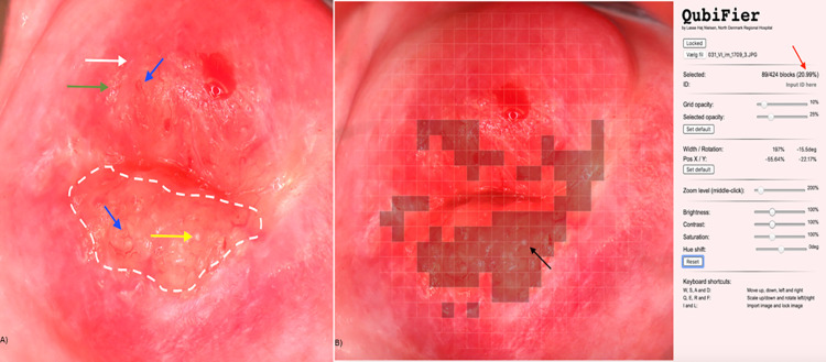
The JavaScript programming software was used to develop the digital gridded imaging technique, named FGS QubiFier, for quantitative measurement of CLP based upon the identification of cervical lesions on a digital image by an observer. With the software programme, a semi-transparent circularly formed grid is superimposed as an additional layer upon the digital image of the cervix. The grid covers the whole circumference of the cervix with the orifice appearing in the center of the grid by use of a zoom technique. When the grid is correctly placed it is locked and follows the image when zoom is applied for further analysis of the image. The grid consists of 424 equally sized squares enabling the observer to mark the squares containing any of the four FGS associated lesion types manually in a structured manner, hence leaving the squares without any lesions unmarked. (Fig 1).

Fig 1. Digital image showing female genital schistosomiasis associated pathognomonic lesions of the cervix (picture A) and the same lesions being digitally marked by QubiFier to determine the cervical lesion proportion (picture B).

Fig 1

The left picture (A) shows an area, which contains homogenous yellow sandy patches appearing as yellow colored area (indicated by white dashed line). The differently colored arrows indicate the other pathognomonic lesion types: a grainy sandy patch (white arrow), clustered grainy sandy patches or rice-grain shaped sandy patches (green arrow) and a rubbery papule colored in beige with an uneven surface (yellow arrow). Abnormal blood vessel (rounded, uneven-calibered, corkscrew or convoluted) are indicated by the blue arrow. The right picture (B) shows squares of the grid marked digitally containing any types of pathognomonic lesions. The red arrow shows the proportion of the cervix covered by any type of pathognomonic lesions.

During the review of the 424 squares the number of squares marked by the observer is counted automatically by the FGS Qubifier software program. To obtain the final CLP measure, as a result of the review, the total number of marked squares are divided automatically by 424, equal the total number of squares in the grid, and then multiplied by a factor 100. Hence, the estimated percentage constituted the CLP in the range of zero to 100 as a quantitative measure of lesions covering the cervical portio as identified by the observer. As an example, if 25 squares of the 424 squares are marked by an observer, the CLP will then be estimated to 5.8% (6.0%). Hence, measurement of CLP provides a mean for grading the cervical pathology as mild, moderate, or severe, which corresponds to intervals of low, intermediary, or high CLP measures, respectively. The number of rubbery papules are also countable by the observer and registered as a measure of rubbery papule count (RPC).

Types of lesions associated to FGS

The following four types of lesions are occurring in FGS [14,20] and displayed in the WHO FGS pocket atlas [13].

  1. Grainy sandy patches, defined as areas with distinct single or clusters of oblong grains (approximately 0.05 × 0.2 mm) in the cervicovaginal mucosa.

  2. Yellow homogenous sandy patches, defined as homogenous foci without detectable grains at 15 times the original magnification.

  3. Rubbery papules, previously described in the urinary bladder mucosa only, defined as spheroid, firm, beige, smooth papules (size, 0.3–1.2 mm) in the cervicovaginal mucosa.

  4. Neovascularization, indicating pathological changes of small venules of corkscrew, unevenly calibrated and convoluted appearance [21,22]. The distorted blood vessels are the result of eggs released by a female worm which did not achieve to penetrate the wall of the blood vessel and hence become trapped in a venule, thereby blocking the natural blood flow.

Study design

Three observers (A, B and C), all physicians trained and practicing medicine in Denmark, reviewed the 70 randomly selected images in the gridded imaging technique study (Fig 2). The observers had different levels of clinical experience regarding gynecology in general and FGS specifically: observer A (recently graduated from the medical school and just introduced to FGS at a textbook level), observer B (gynecology specialist with minor FGS field experience) and observer C (gynecology internship with major FGS field experience).

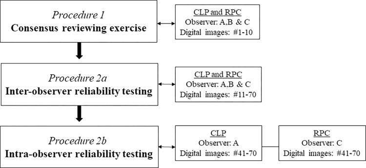
Fig 2. Flowchart displaying the different testing procedures in the FGS digital gridded imaging technique study in which 70 images of the cervical portio were reviewed for cervical lesion proportions (CLP) and rubbery papule counts (RPC), respectively.

Fig 2

In an initial consensus rating exercise (Procedure 1), each of the three observers independently examined ten randomly selected images (images 1 to 10) and rated the cervical lesions (Fig 2). The three observers then shared and discussed their findings to reach consensus on the uniform rating of the images. Subsequently, the remaining 60 images (images 11 to 70) were rated independently by the three observers testing inter-observer reliability (CLP and RPC) in Procedure 2a. Two weeks later intra-rater reliability was tested for images 41 to 70 in a similarly blinded manner by observers A (CLP) and C (RPC) in Procedure 2b. To ensure sufficient scoring, the images could be reviewed at all zoom levels determined by the reviewer.

Quality of the digital images

Unfortunately, the surface of the cervix could not always be depicted completely for anatomical reasons, e.g. when the labia majora were partially constricted or the cervix was anteverted or retroverted. The impact of the image quality on the CLP rating was therefore assessed between the three observers in separate sessions (A and B, A and C, and B and C, respectively). The quality of the digital images was stratified into whether the entire surface of the cervix was depicted optimally or partially.

Data management and statistical analysis

The collected data were entered into REDCap data management system. Data validity was ensured by two researchers independently. Data analysis was performed by use of the RStudio programming language (version 1.2.5033).

The CLP and RP determined by the three observers was compared using the Kruskal-Wallis rank-sum test, and CLP between raters were compared using the Wilcoxon rank-sum test. To test if the gridded imaging technique were able to distinguish between clinical pathology including rubbery papules and without rubbery papules, Fleiss kappa was calculated. The value of Fleiss kappa was interpreted as follows: <0.20 (poor), 0.21 to 0.40 (fair), 0.41 to 0.60 (moderate), 0.61 to 0.80 (good) and 0.81 to 1.00 (very good) [23].

To test the degree of agreement between the three observers (inter-rater reliability) and the degree of agreement among the same observer at two timepoints (intra-rater reliability) the intraclass correlation coefficient (ICC) was calculated by a two-way mixed-effects model based on average rating values and absolute agreement. Intra-observer variation was calculated by a two-way mixed-effects model based on a single-rating and absolute agreement. The ICC values were interpreted as follows: ≤ 0.5 (poor), 0.5 to 0.75 (moderate), 0.76 to 0.9 (good), and > 0.90 (excellent) [24].

Results

Age and parasitology

The 60 randomly selected images used for intra- and inter-observer reliability testing covered 50 different women: one image from 40 women (n = 40) obtained at either follow-up at week 5 or week 10 and two images from 10 women obtained at both follow-up visits (n = 20). Median age of the 50 women in the study was 26.5 years (interquartile range (IQR) 20.8–33.0). The median number of S. haematobium eggs per 50 ml of urine was 2.5 (IQR 0–62.0). In 13 women, the filtration of 50 ml urine did not reveal eggs.

Quality of digital images

In 33 (55%) of the 60 digital images used for intra- and interobserver reliability testing, the surface of the cervix was depicted completely, whereas in 27 images (45%) up to 20% of the cervical surface was not visible. However, the observers still found the quality of images to be adequate for CLP and RPC determination, and all images were included in the further analysis.

Cervical lesion proportion

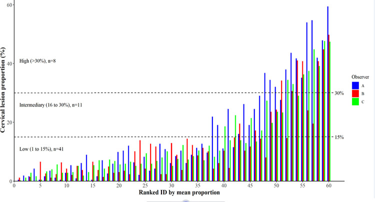
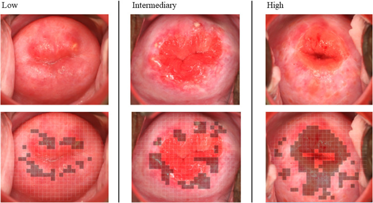
In Procedure 2a (60 images were individually scored by the three reviewers), the CLP ranged from 0.5% to 59.4% in observer A, from 0% to 49.8% in observer B, and from 0.2% to 47.6% in observer C. Fig 3 shows mean CLP by the three observers for each the 60 digital images. The mean proportions were divided into three interval levels defined arbitrarily as low (1 to 15%), intermediary (16 to 30%) and high (>30%) with a distribution as follows: 68% (n = 41), 18% (n = 11) and 13% (n = 8). Three randomly selected cases representing each of the three levels are displayed in Fig 4.

Fig 3. Distribution of cervical lesion proportions (%) for the 60 digital images as rated by the three observers and ranked in accordance with the respective individual mean values.

Fig 3

Fig 4. Three cases representing different levels of female genital schistosomiasis (FGS) associated pathology in accordance with the cervical lesion proportion (CLP) categories: low (1 to 15%), intermediary (16 to 30%), and high (>30%).

Fig 4

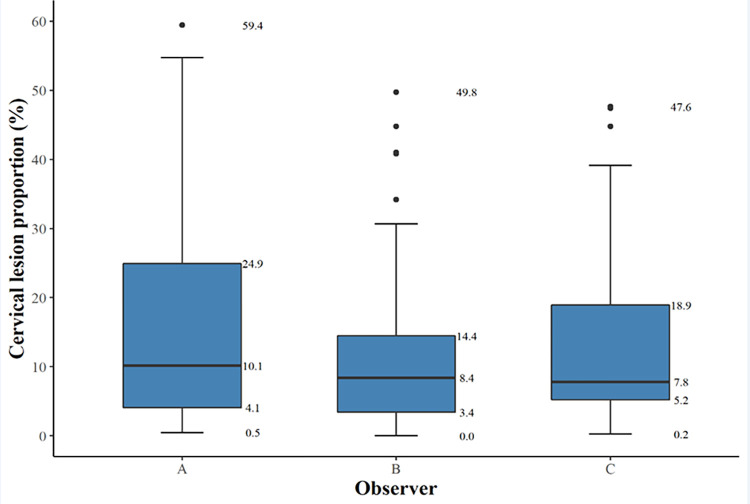
Median CLP (interquartile range, IQR) for observers A, B and C was 10.1% (4.1–24.9), 8.4% (3.4–14.5) and 7.8% (5.2–18.9), respectively (Fig 5). The difference in scores between all raters was not statistically significant (p = 0.094). Hence, inter-rater differences were not significant between the raters; A vs. B p = 0.53, A vs. C p = 0.53 and B vs. C p = 0.65. The scoring over time between the same reviewer (Procedure 2a versus Procedure 2b) the median CLP (IQR) was 6.3% (1.7–11.9) in Procedure 2a and 8.1% (3.4–14.0) in Procedure 2b. The difference was not statistically significant (p = 0.294).

Fig 5.

Fig 5

A box-and-whisker-plot of cervical lesion proportions (%) for each observer (A, B, and C) rating the 60 digital images. The bold horizontal line indicates the median, the upper and the lower line of the box the interquartile range.

Rubbery papule count

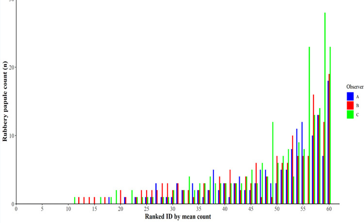
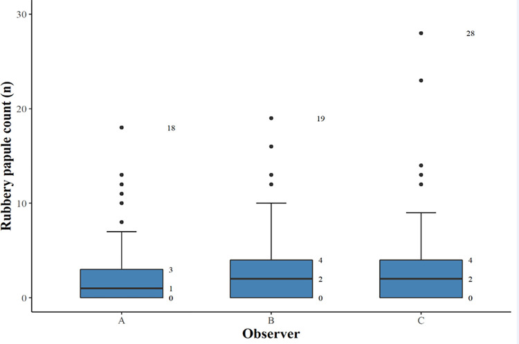
In Procedure 2a, the RPC ranged from 0 to 18 in observer A, from 0 to 19 in observer B, and from 0 to 28 in observer C. Fig 6 shows the distribution of RPC for each of the 60 digital images. Median RPC (IQR) for observers A, B and C were 1 (0–3), 2 (0–4) and 2 (0–4), respectively (Fig 7). Difference in scores between all raters was not statistically significant (p = 0.341). Inter-rater differences were not significant; A vs. B p = 0.41, A vs. C p = 0.41 and B vs. C p = 0.97. Median RPC (IQR) was 1 (0–3) in Procedure 2a and 1 (0–2) in Procedure 2b. The difference was not significant (p = 0.055). The Fleiss kappa value for the three observers in distinguishing rubbery papules was 0.55 (IC 0.54–0.55; p<0001), demonstrating a moderate agreement.

Fig 6.

Fig 6

A box-and-whisker plot of median rubbery papule counts (n) and interquartile range for each observer (A, B, and C) rating the 60 digital images.

Fig 7. Distribution of rubbery papule count (n) for the 60 digital images as rated by the three observers and ranked in accordance with the respective individual mean values.

Fig 7

Inter- and intra-observer reliability

The inter-observer reliability (A, B and C) for CLP measured by ICC was 0.93 (95% CI 0.90–0.96) indicating an excellent performance. The inter-observer reliability (A, B and C) for RPC measured by ICC was 0.88 (95% CI 0.82–0.92), indicating a good performance. The intra-observer reliability (A) for CLP measured by the intra-class correlation coefficient ICC was 0.90 (95% CI 0.79–0.95), indicating an excellent performance. The intra-observer reliability (C) for RPC measured by ICC was 0.80 (95% CI 0.59–0.90), indicating a good performance.

Factors influencing discrepancies

Data were further analyzed to identify whether certain characteristics of the surface of the cervix contributed to significant discrepancies in CLP between observers. In ten images with the most pronounced discrepancy in CLP between observers A and B, the differences ranged from 13% to 35%. When comparing observer A with observer C, the differences ranged from 11% to 17% for the ten images representing the most pronounced discrepancy between the two, and for observers B and C, differences ranged between 8% to 25%. Two major domains contributed to discrepancies between observers: 1. difference in zoom-level of the digital images resulting in a different coverage of the surface of the cervix by the grid, 2. disagreement when different cervical lesions co-existed and overlapped in the same square particularly in case of neovascularization. In addition, disagreement occurred also in a few cases due to reduced image quality and/or because the glandular lining of the endocervix was intense and categorized as yellow homogenous sandy patches.

Discussion

In this study, we have developed and tested a digital gridded imaging technique for assessment of cervical lesions occurring in FGS. Using this technique, we demonstrated that CLP and RPC are useful measures enabling quantification of cervical pathology. The four different lesion types (grainy sandy patches, yellow homogeneous sandy patches, rubbery papules, and neovascularization) searched for by the observers in the review of the digital images may not all be associated solely to FGS. Sandy patches and neovascularization are also associated to other conditions commonly affecting women living in Schistosoma endemic areas, including sexually transmitted infections (e.g. human papilloma virus and herpes simplex virus), likewise rubbery papules may resemble nabothian cysts [15]. In any case, the technique provides an opportunity to explore further into the natural history of FGS from a clinical as well as an epidemiological perspective. Moreover, the CLP and RPC measures offer an opportunity to assess pathological changes following treatment of FGS, in addition to measurement of subsequent re-occurrence of pathology after repeated exposure to Schistosoma infested water.

The proposed arbitrary grading of cervical pathology as low (1 to 15%), intermediary (16% to 30%) and high (>30%) according to the CLP, requires further evaluation in other groups of women with FGS. The CLP cut-off points were arbitrarily selected for purpose of the study, and these should be tested clinically to investigate if different levels of cervical pathology correlate with genital morbidity, including gynecological complaints.

The ICC values for the CLP and RPC indicated good to excellent inter- and intra-observer reliability. The difference in inter-observer reliability concerning CLP (excellent) and RPC (good) can be explained by the difficulty in evaluating a pronounced convex structure such as a rubbery papule on a 2D surface [15]. A moderate agreement between the three observers was found regarding identification of rubbery papule lesions, emphasizing another important aspect in the assessment of the gridded imaging technique. The kappa value demonstrated that the observers were capable of distinguishing between the presence versus absence of this FGS associated lesion. We believe that rubbery papule lesions represent the sign of recent eosinophil inflammatory response compared to the sandy patch lesions representing a late stage in the natural history of FGS. Thus, a validated RPC could be a good proxy for recent deposition of eggs in the epithelium.

The comparison of CLP and RPC median scores between observers showed no significant difference between any of the observers but for a few images a discrepancy up to 35% was noted. Two major domains were identified when evaluating the cause of discrepancy between observers. Firstly, different zoom-levels between the observers, and secondly, co-existence of lesions in same grid square. Reduced image quality also played a role in the discrepancy between observers. The possible causes of discrepancies between observers should be addressed in future studies to further optimize the gridded imaging technique protocol.

Use of digital images for the diagnosis and management of FGS provides different advantages in comparison to the colposcopy. FGS associated lesions are more easily identified on a computer screen as it is possible to enlarge portions of the cervix by increasing the zoom level, thus providing a more detailed visualization of the lesions. Digital images can be revaluated any time and shared with other observers or health care professionals for a second clinical opinion. The digital images of the cervix can be captured by use of the camera alone or by using a camera in conjunction with the colposcopy. A camera with proper technical features may potentially replace the colposcope. Introduction and use of the colposcope in FGS control programs is challenged by lack of supporting health care and staff training capacity as well as logistic and financial constraints. Previous studies by Holmen and colleagues have suggested use of colorimetric image analysis as a diagnostic tool in FGS [17,25]. However, the computer analysis is performed on colposcopy images, which are typically not available in district hospitals in sub-Saharan Africa. The digital camera image documentation approach overcomes the shortage of colposcopy equipment. However, access to cameras may also be limited in rural high endemic areas. Previous studies on visualization of cervical pre-cancer pathology have shown that image documentation could be obtained by mobile phone cameras [26,27], which seems also to have a major potential in a large-scale FGS diagnostic context.

Limitations

This study has some limitations. Firstly, underestimation of the CLS and RPC may have occurred because up to 20% of the cervical surface was not visible in half of the images selected for this study. Secondly, colposcopy is currently considered the standard procedure for identification of FGS associated lesions. In this study, the digital images used for the development and validation of the gridded imaging technique were obtained in women living in an S. haematobium endemic area enrolled in a therapeutic trial prior to this study. Main inclusion criteria for participation in this trial were presence of FGS associated lesions. However, the lesions were identified by digital camera images and not by colposcopy. A combined use of a colposcope and digital camera could have made it possible to study important comparative findings between the two diagnostic methods. As another limitation in this study, digital gridded imaging technique was not compared with real-time PCR for the detection of Schistosoma DNA in genital specimens as an accepted FGS diagnostic method. Moreover, other potential causes of homogeneous yellow sandy patches and neovascularization, such sexually transmitted infections (e.g. Chlamydia trachomatis, herpes simplex virus and human papilloma virus) and malignancy, respectively, were not included in a comparative analysis.

Conclusion

The digital gridded imaging technique provides an opportunity to quantify FGS associated cervical lesions. Furthermore, we found an excellent reliability for the CLP and a good reliability for RPC. Future studies need to explore to what extent Schistosoma DNA PCR as an FGS diagnostic biomarker and severity of disease as perceived by the patient correlate with CLP and RPC. If so, the digital gridded imaging technique provides new opportunities in future FGS research and control programs. The study has shown that the WHO FGS pocket atlas constitutes a very useful reference guide for physicians to identify FGS associated cervical lesions regardless of clinical experience level. Use of WHO FGS atlas together with the digital gridded imaging technique, allows for potential deployment in S. haematobium endemic countries in conjunction with future epidemiological and therapeutic studies aiming at improved FGS control in Africa.

Acknowledgments

We like to thank the field team for their dedicated work and the women in the district of Ambanja in the Northwest region of Madagascar for willing to participate in the study. Photographer Lasse Høj Nielsen contributed with expertise in development of the gridded clinical image technique,

Data Availability

Data are available in the RedCAP data management hosted by the Centre for Clinical Reseach, North Denmark Regional Hospital, 9800 Hjørring, Denmark (phone: +45 97 64 26 52; mail: forskning.rhn@rn.dk - contactperson: Signe Westmark.

Funding Statement

The study was funded by Merck KGaA (https://www.merckgroup.com/en). PDCL received the funding. The funder had no role in study design, data collection and analysis, decision to publish, or preparation of the manuscript.

References

  • 1.Chitsulo L., Engels D., Montresor A., and Savioli L. The global status of schistosomiasis and its control. Acta Trop. 2000. Oct 23; 77(1): 41–51. doi: 10.1016/s0001-706x(00)00122-4 [DOI] [PMC free article] [PubMed] [Google Scholar]
  • 2.Kjetland EF, Leutscher PDC, Ndhlovu PD. A review of female genital schistosomiasis. Trends in Parasitology February 2012; 28: 58–65. doi: 10.1016/j.pt.2011.10.008 [DOI] [PubMed] [Google Scholar]
  • 3.Feldmeier H, Krantz I. A synoptic inventory of needs for research on women and tropical parasitic diseases. I. Application to urinary and intestinal schistosomiasis. Acta Tropica. 1993;55:117–138. doi: 10.1016/0001-706x(93)90073-k [DOI] [PubMed] [Google Scholar]
  • 4.Feldmeier H, Poggensee G, I Krantz I, Helling-Giese G. Female genital schistosomiasis. New challenges from a gender perspective. Trop Geogr Med. 1995; 47(2 Suppl): S2–15. [PubMed] [Google Scholar]
  • 5.Helling-Giese G, Kjetland EF, Gundersen SG, Poggensee G, Richter J, Krantz I, et al. Schistosomiasis in women: manifestations in the upper reproductive tract. 1996;62:225–38. doi: 10.1016/s0001-706x(96)00025-3 [DOI] [PubMed] [Google Scholar]
  • 6.Swai B, Poggensee G, Mtweve S, Krantz I. Female genital schistosomiasis as an evidence of a neglected cause for reproductive ill-health: a retrospective histopathological study from Tanzania. BMC Infect Dis. 2006;6:134. doi: 10.1186/1471-2334-6-134 [DOI] [PMC free article] [PubMed] [Google Scholar]
  • 7.Odubamowo KH, Akinpelu OM, Lawal OO, Okolo CA, Odukogbe AA, Adekunle AO. Bilateral tubal gestation associated with schistosomiasis in an African Woman. Case Rep Obstet Gynecol. 2014; 2014: 674514. doi: 10.1155/2014/674514 [DOI] [PMC free article] [PubMed] [Google Scholar]
  • 8.Christinet V, Lazdins-Helds JK, Russell Stothard J, Jutta Reinhard-Rupp J. Female genital schistosomiasis (FGS): from case reports to a call for concerted action against this neglected gynaecological disease. Int J Parasitology. 2016,46:395–404. doi: 10.1016/j.ijpara.2016.02.006 [DOI] [PubMed] [Google Scholar]
  • 9.Trainor-Moss S, Mutapi F. Schistosomiasis therapeutics: whats in the pipeline? Expert Rev Clin Pharmacol. 2016;9:157–60. doi: 10.1586/17512433.2015.1102051 [DOI] [PubMed] [Google Scholar]
  • 10.Poggensee G, Feldmeier H. Female genital schistosomiasis: facts and hypotheses. Acta Trop. 2001;79:193–210. doi: 10.1016/s0001-706x(01)00086-9 [DOI] [PubMed] [Google Scholar]
  • 11.Feldmeier H, Krantz I, Poggensee G. Female genital schistosomiasis as a risk-factor for the transmission of HIV. Int J STD AIDS. 1994;5:368–72. doi: 10.1177/095646249400500517 [DOI] [PubMed] [Google Scholar]
  • 12.Patel P, Rose CE, Kjetland EF, Downs JA, Mbabazi PS, Sabin K, et al. Association of schistosomiasis and HIV infections: A systematic review and meta-analysis. Int J Infect Dis. 2021;102:544–553. [DOI] [PMC free article] [PubMed] [Google Scholar]
  • 13.World Health Organization. Female genital schistosomiasis. A pocket atlas for clinical health-care professionals. WHO/HTM/NTD/2015.4. Geneva 2015. [Google Scholar]
  • 14.Kjetland EF, Norsetha HM, Taylor M, Lillebø K, Kleppa E, Holmen SD, et al. Classification of the lesions observed in female genital schistosomiasis. International J Gynecology and Obstetrics. 2014;127:227–228. doi: 10.1016/j.ijgo.2014.07.014 [DOI] [PubMed] [Google Scholar]
  • 15.Norseth HM, Ndhlovu PD, Kleppa E, Randrianasolo BS, Jourdan PM, Roald B, et al. The colposcopic atlas of schistosomiasis in the lower female genital tract based on studies in Malawi, Zimbabwe, Madagascar and South Africa. PLoS Negl Trop Dis. 2014;8:e3229. doi: 10.1371/journal.pntd.0003229 [DOI] [PMC free article] [PubMed] [Google Scholar]
  • 16.Colley DG, Bustinduy AL, Secor WE, King CH. Human schistosomiasis. Lancet. 2014;383:2253–64. doi: 10.1016/S0140-6736(13)61949-2 [DOI] [PMC free article] [PubMed] [Google Scholar]
  • 17.Holmen SD, Kleppa E, Lillebø K, Pillay P, van Lieshout L, Taylor M, et al. The first step toward diagnosing female genital schistosomiasis by computer image analysis. Am J Trop Med Hyg. 2015. a;93: 80–86. doi: 10.4269/ajtmh.15-0071 [DOI] [PMC free article] [PubMed] [Google Scholar]
  • 18.Leutscher PDC, Ramarokoto CE, Hoffmann S, Jensen JS, Ramaniraka V, Randrianasolo B, et al. Coexistence of urogenital schistosomiasis and sexually transmitted infection in women and men living in an area where Schistosoma haematobium is endemic. Clin Infect Dis. 2008. 15;47:775–82. doi: 10.1086/591127 [DOI] [PubMed] [Google Scholar]
  • 19.Feldmeier H, Bienzle U, Dietrich M, Sievertsen HJ. Combination of a viability test and a quantification method for Schistosoma haematobium eggs (filtration—trypan blue-staining-technique). Tropenmed Parasitol. 1979;30:417–22. [PubMed] [Google Scholar]
  • 20.Randrianasolo BS, Jourdan PM, Ravoniarimbinina P, Ramarokoto CE, Rakotomanana F, Ravaoalimalala VE, et al. Gynecological manifestations, histopathological Findings, and schistosoma-specific polymerase chain reaction results among women with Schistosoma haematobium infection: a cross-sectional study in Madagascar. J Infect Dis. 2015; 212: 275–284. doi: 10.1093/infdis/jiv035 [DOI] [PMC free article] [PubMed] [Google Scholar]
  • 21.Jourdan PM, Randrianasolo BS, Feldmeier H, Chitsulo L, Ravoniarimbinina P, Roald B, et al. Pathologic mucosal blood vessels in active female genital schistosomiasis: new aspects of a neglected tropical disease. Int J Gynecol Pathol. 2013;32:137–40. doi: 10.1097/PGP.0b013e31824fe327 [DOI] [PubMed] [Google Scholar]
  • 22.Holmen S, Galappaththi-Arachchige HN, Kleppa E, Pillay P, Naicker T, Taylor M et al. Characteristics of blood vessels in female genital schistosomiasis: paving the way for objective diagnostics at the point of care. PLoS Negl Trop Dis. 2016;10: e0004628. doi: 10.1371/journal.pntd.0004628 [DOI] [PMC free article] [PubMed] [Google Scholar]
  • 23.Austin Nichols A. Causal inference with observational data. The Stata Journal. 2007;7, 507–541. [Google Scholar]
  • 24.Koo TK, Li MY. A Guideline of Selecting and Reporting Intraclass Correlation Coefficients for Reliability Research. J Chiropr Med. 2016;15:155–63. doi: 10.1016/j.jcm.2016.02.012 [DOI] [PMC free article] [PubMed] [Google Scholar]
  • 25.Holmen SD, Kjetland EF, Taylor M, Kleppa E, Lillebø K, Gundersen SG, et al. Colourimetric image analysis as a diagnostic tool in female genital schistosomiasis. Med Eng Phys. 2015. b;37: 309–14. doi: 10.1016/j.medengphy.2014.12.007 [DOI] [PubMed] [Google Scholar]
  • 26.Quinley KE, Gormley RH, Ratcliffe SJ, Shih T, Szep Z, Steiner A, et al. Use of mobile telemedicine for cervical cancer screening. J Telemed Telecare 2011;17:203–9. doi: 10.1258/jtt.2011.101008 [DOI] [PMC free article] [PubMed] [Google Scholar]
  • 27.Søfteland S, Sebitloane MH, Taylor M, Roald BB, Holmen S, Galappaththi-Arachchige HN, et al. A systematic review of handheld tools in lieu of colposcopy for cervical neoplasia and female genital schistosomiasis. Int J Gynaecol Obstet. 2021;153:190–199. doi: 10.1002/ijgo.13538 [DOI] [PMC free article] [PubMed] [Google Scholar]
PLoS Negl Trop Dis. doi: 10.1371/journal.pntd.0009995.r001

Decision Letter 0

Francesca Tamarozzi, Jennifer A Downs

22 Jan 2022

Dear Prof Leutscher,

Thank you very much for submitting your manuscript "Validation of cervical lesion proportion measure using a gridded imaging techique to assess cervical pathology in women with genital schistosomiasis" for consideration at PLOS Neglected Tropical Diseases. As with all papers reviewed by the journal, your manuscript was reviewed by members of the editorial board and by several independent reviewers. In light of the reviews (below this email), we would like to invite the resubmission of a significantly-revised version that takes into account the reviewers' comments.

We thank the authors for this important work, and agree that there is an urgent need to quantify FGS lesions. Please address reviewers' comments in order to strengthen this manuscript, which has some significant weaknesses at present. The manuscript could be made clearer particularly regarding whether the computer or the observers performed the actual counting, and precision of language throughout, as noted by Reviewer 1, is needed.

It will also be important to describe (1) rates and types of STIs that could confound this analysis, (2) the non-specificity of lesions (particularly neovascularization and homogeneous sandy patches), and to report the frequencies of the four different WHO atlas findings.

Finally, please double-check the whole text for typos and editing mistakes.

We cannot make any decision about publication until we have seen the revised manuscript and your response to the reviewers' comments. Your revised manuscript is also likely to be sent to reviewers for further evaluation.

When you are ready to resubmit, please upload the following:

[1] A letter containing a detailed list of your responses to the review comments and a description of the changes you have made in the manuscript. Please note while forming your response, if your article is accepted, you may have the opportunity to make the peer review history publicly available. The record will include editor decision letters (with reviews) and your responses to reviewer comments. If eligible, we will contact you to opt in or out.

[2] Two versions of the revised manuscript: one with either highlights or tracked changes denoting where the text has been changed; the other a clean version (uploaded as the manuscript file).

Important additional instructions are given below your reviewer comments.

Please prepare and submit your revised manuscript within 60 days. If you anticipate any delay, please let us know the expected resubmission date by replying to this email. Please note that revised manuscripts received after the 60-day due date may require evaluation and peer review similar to newly submitted manuscripts.

Thank you again for your submission. We hope that our editorial process has been constructive so far, and we welcome your feedback at any time. Please don't hesitate to contact us if you have any questions or comments.

Sincerely,

Jennifer A. Downs, M.D., Ph.D.

Associate Editor

PLOS Neglected Tropical Diseases

Francesca Tamarozzi

Deputy Editor

PLOS Neglected Tropical Diseases

***********************

We thank the authors for this important work, and agree that there is an urgent need to quantify FGS lesions. Please address reviewers' comments in order to strengthen this manuscript, which has some significant weaknesses at present. The manuscript could be made clearer particularly regarding whether the computer or the observers performed the actual counting, and precision of language throughout, as noted by Reviewer 1, is needed.

It will also be important to describe (1) rates and types of STIs that could confound this analysis, (2) the non-specificity of lesions (particularly neovascularization and homogeneous sandy patches), and to report the frequencies of the four different WHO atlas findings.

Reviewer's Responses to Questions

Key Review Criteria Required for Acceptance?

As you describe the new analyses required for acceptance, please consider the following:

Methods

-Are the objectives of the study clearly articulated with a clear testable hypothesis stated?

-Is the study design appropriate to address the stated objectives?

-Is the population clearly described and appropriate for the hypothesis being tested?

-Is the sample size sufficient to ensure adequate power to address the hypothesis being tested?

-Were correct statistical analysis used to support conclusions?

-Are there concerns about ethical or regulatory requirements being met?

Reviewer #1: Major Comments:

1) Validation: I believe a validation study would compare a new technology against a “gold standard” to confirm that the new technology accurately measures what it claims to measure. Admittedly, there is not a consensus reference standard for FGS. However, was there a comparison made with another accepted diagnostic method for FGS (biopsy, PCR, etc)? If there was no comparison with PCR or biopsy (or Holmen’s colorimetric image analysis), I’m not sure it is correct to call this a validation study. Please rephrase the title and line 301. Please also acknowledge this in the limitations.

2) What proportion of women in the study had STI? Please see PMID: 15772328. In Kjetland 2005, FGS-associated lesions (the homogeneous yellow sandy patch) were associated with STI (please see Table 4 in Kjetland 2005). There was strong evidence that chlamydia, HSV, and high risk HPV were associated with the homogeneous yellow sandy patches. Thus, to ensure that the lesions described were not associated with STI, please describe what STI testing was performed and what proportion of the women with sandy patches also had STI.

3) Please could the authors describe any assessment for dysplasia or malignancy? Neovascularization (see Kjetland 2005 Table 5) has been strongly associated with CIN I-III). Thus, in lines 114 & 116 these sentences could say “in women with FGS-associated lesions” or some other language to inform the reader that we do not know with certainty that these lesions are due to FGS, rather only that they look like lesions that are associated with FGS.

4) Given that FGS lesions lack specificity, I think it is challenging to call these “pathognomonic” lesions. Please rephrase throughout. Additionally, without an evaluation for STI + malignancy + other diagnostic methods for FGS, it seems appropriate to use less definitive language around causality of these lesions by FGS (see lines 36 (fgs-evoked), line 42-43 “women with FGS”, line 116 “women with FGS”, line 130). However, in some locations the authors are examples of more measured language, suggesting association rather than causality. For example, at line 35 the authors say that “Female genital schistosomiasis (FGS) is characterized by a pattern of lesions”. I think this type of language throughout would be more appropriate.

5) Abstract: (Line 38) You say that “no tools are able to quantify clinical pathology at the cervix in a standardized and reproducible manner”. However, in the discussion you reference Holmen’s colorimetric image analysis. Is that tool not standardized and reproducible? Also Holmen’s method can also be applied to digital images.

6) Methods:

a. Line 159 – 164. It seems to me from these lines that the computer algorithm automatically counted and marked the FGS associated lesions (so the computer algorithm calculates the CLP)? If this is true, what was the role of the human reviewers? In line 214 it appears the reviewers also calculate a CLP – how is this done? Without a clear understanding of the human component, it is challenging to understand how meaningful the inter-rater reliability is.

b. Lines 165-167 – to clarify, if there were 25 lesions/squares, this number 25/424 = 6% coverage of the cervical surface? These sentences might need to be rephrased slightly - this information was challenging to access.

c. Line 188 - What are the two components of the experience profile (1+1) – is one number for gynecology and one for FGS? This should be clarified, and how does one ‘achieve’ minor/moderate/major experience? This seems slightly arbitrary and should be further explained and disclosed as such (as you did very nicely with the grading of lesions into mild/mod/severe).

Minor Comments:

1) Lines 178 - Types of lesions. I think there is some debate regarding whether neovascularization should be included as an FGS associated lesion (in PMID: 25412334 the authors note that only homogeneous and grainy sandy patches, along with rubbery papules meet the criteria for FGS based the result of an expert meeting in 2010). I acknowledge that all 4 FGS-associated lesion the authors describe are displayed in the pocket atlas, thus making all 4 reasonable to include. However, it might be nice to have a sentence in the discussion outlining the controversy.

2) Referencing

a. Line 82 – epidemiology (Africa and the middle east) – please reference

b. Line 83 – how does Shaematobium affect the genital organs? Please expand and reference.

c. Line 64 – please reference these complaints. There is only one study I am aware of in which dyspareunia is associated with FGS (PMID: 21572820)

d. Line 66 (Author Summary). I think it is an overstatement to say that “FGS is considered as a major risk factor for transmission of HIV” – please rephrase

e. Line 89. I think it is an overstatement to say “FGS associated with increased risk of HIV acquisition”. There has only been one study suggesting an association with HIV acquisition and this was a retrospective study in women with Shaematobium antibodies (they did not have FGS evaluations) (PMID 30543654). Please rephrase.

f. Line 90 – While it is a compelling hypothesis, I am unsure that we have evidence to claim that FGS increases the risk of HPV infection. The references you have cited do not support this statement (#9-11). Please amend this statement or provide revised references.

g. Line 87-88. Please elaborate on which concrete symptoms or features of FGS (?dyspareunia, discharge, genital itching) might lead to a “poor quality of sexual life”.

3) Other minor comments:

Line 38 (abstract): “A tool for quantification of the lesions is needed to improve FGS research and control programs” – would the tool also be useful for evaluating treatment?

Line 70-72 – How does your tool allow for treatment at the community level? Would your tool also allow for monitoring of lesion resolution (as well as lesion development)?

Line 122 – should RCT stand for “randomized controlled trial”?

Line 127 – do you mean that the area highly endemic for S. haematobium (with a high prevalence of FGS?)?

Line 142 – what is the digital part of the study – do you mean the image review?

Line 346 – would there be a role for a mobile phone colposcope here as well?

Reviewer #2: The objectives are clear: This study aimed to develop and validate a cervical lesion proportion (CLP) measure for quantification of cervical pathology in FGS.

The RCT study design has been clearly explained.

The sample selected from the population needs further explanation. There were 116 women enrolled in the RCT between 15-35y with FGS pathognomonic lesions. How were 70 of the 412 images selected? For the comparison between the observers, why were 40 images selected from weeks 5 or 10 and two images from each of 10 women at both visits? Which women were selected, and why?

--------------------

Results

-Does the analysis presented match the analysis plan?

-Are the results clearly and completely presented?

-Are the figures (Tables, Images) of sufficient quality for clarity?

Reviewer #1: 1) Results:

a. What proportion of each of the FGS-associated lesions did the authors find? This is important given that neovascularization is not uniformly considered to be FGS-associated.

b. Please review for the reader again here which steps were 2a and 2b.

c. It might be helpful to define inter-observer and intra-observer reliability for those readers who do not have a background with these methods

Reviewer #2: The results are accompanied by figures showing the comparisons of the findings by the three different observers.

Please can the authors further elucidate the proportion of homogeneous sandy patches and grainy sandy patches found.

Since getting photographs of the cervix may be difficult it would be on interest to know if when only 20% pf the cervical surface was only partially depicted, when the similar results were found between the more and less experienced observers. Can the authors please comment?

--------------------

Conclusions

-Are the conclusions supported by the data presented?

-Are the limitations of analysis clearly described?

-Do the authors discuss how these data can be helpful to advance our understanding of the topic under study?

-Is public health relevance addressed?

Reviewer #1: -I'm afraid from the way the methods are written that I don't fully understand the role of the human reviewers if the computer algorithm is calculating the CLP (please see above).

-Please see above for additional items for the limitations

Reviewer #2: The conclusions are supported by the data and they describe the limitations, but since a colposcope is the recommended tool, the authors need to consider a way forward.

The authors do not suggest a comparison study between use of the the colposcope (a heavy and expensive piece of equipment) and the digital image documentation approach which overcomes the shortage of colposcopic equipment. We would need information comparing the accuracy of the colposcope and the camera, to determine the accuracy/effectiveness of this new approach.

Finding an effective solution to the diagnosis of FGS urgently needs such a study .

--------------------

Editorial and Data Presentation Modifications?

Use this section for editorial suggestions as well as relatively minor modifications of existing data that would enhance clarity. If the only modifications needed are minor and/or editorial, you may wish to recommend “Minor Revision” or “Accept”.

Reviewer #1: 1) Line 190-199 – The authors have explained that the 70 images were chosen randomly – was this from a total of 412 total images from the study? Overall, I find it the numbers (70 from 212 or 412) and timepoints (the majority from week 5 but some from week 10 but from 50 women) hard to follow. Taking one randomly selected image per woman makes sense (line 198) but why then 2 from 10 women? This needs clearer explanation and perhaps Figure 2 needs a flow diagram to show the total number of participants, the time points, and images. This would allow further understanding of the risk of bias. The explanation given from lines 237-239 is easier to follow than the explanation used in the methods.

2) Reducing stigma – I think we as scientists can do our part to reduce stigma through the language we choose in our manuscripts. Please consider rephrasing:

a. Line 93 “resource poor”

b. Line 102 “in rural Africa”

c. Line 308 “contaminated” water

d. Line 346 “in an African context”

Reviewer #2: (No Response)

--------------------

Summary and General Comments

Use this section to provide overall comments, discuss strengths/weaknesses of the study, novelty, significance, general execution and scholarship. You may also include additional comments for the author, including concerns about dual publication, research ethics, or publication ethics. If requesting major revision, please articulate the new experiments that are needed.

Reviewer #1: This important study evaluates the use of a clinical lesion proportion measure for quantification of cervical pathology in Madagascan women with FGS. The clinical lesion proportion allowed a reproducible quantification of the surface of the cervix affected by FGS lesions. I agree with the authors that a quantitative and reproducible means to evaluate FGS-associated cervical lesions would be beneficial to the field. I also agree with the authors that a quantifiable tool would improve research, control and also treatment. I also agree that having a quantifiable tool will be useful in investigating whether certain levels of genital pathology correlate with symptoms.

Reviewer #2: (No Response)

--------------------

PLOS authors have the option to publish the peer review history of their article (what does this mean?). If published, this will include your full peer review and any attached files.

If you choose “no”, your identity will remain anonymous but your review may still be made public.

Do you want your identity to be public for this peer review? For information about this choice, including consent withdrawal, please see our Privacy Policy.

Reviewer #1: No

Reviewer #2: No

Figure Files:

While revising your submission, please upload your figure files to the Preflight Analysis and Conversion Engine (PACE) digital diagnostic tool, https://pacev2.apexcovantage.com. PACE helps ensure that figures meet PLOS requirements. To use PACE, you must first register as a user. Then, login and navigate to the UPLOAD tab, where you will find detailed instructions on how to use the tool. If you encounter any issues or have any questions when using PACE, please email us at figures@plos.org.

Data Requirements:

Please note that, as a condition of publication, PLOS' data policy requires that you make available all data used to draw the conclusions outlined in your manuscript. Data must be deposited in an appropriate repository, included within the body of the manuscript, or uploaded as supporting information. This includes all numerical values that were used to generate graphs, histograms etc.. For an example see here: http://www.plosbiology.org/article/info%3Adoi%2F10.1371%2Fjournal.pbio.1001908#s5.

Reproducibility:

To enhance the reproducibility of your results, we recommend that you deposit your laboratory protocols in protocols.io, where a protocol can be assigned its own identifier (DOI) such that it can be cited independently in the future. Additionally, PLOS ONE offers an option to publish peer-reviewed clinical study protocols. Read more information on sharing protocols at https://plos.org/protocols?utm_medium=editorial-email&utm_source=authorletters&utm_campaign=protocols

PLoS Negl Trop Dis. doi: 10.1371/journal.pntd.0009995.r003

Decision Letter 1

Francesca Tamarozzi, Jennifer A Downs

2 Apr 2022

Dear Prof Leutscher,

Thank you very much for submitting your manuscript "Cervical lesion proportion measure using a digital gridded imaging techique to assess cervical pathology in women with genital schistosomiasis" for consideration at PLOS Neglected Tropical Diseases. As with all papers reviewed by the journal, your manuscript was reviewed by members of the editorial board and by several independent reviewers. The reviewers appreciated the attention to an important topic. Based on the reviews, we are likely to accept this manuscript for publication, providing that you modify the manuscript according to the review recommendations.

Thank you for your responsiveness to the prior reviews. The manuscript is substantially improved. However, there are still some items that need to be revised before this manuscript can be accepted. These are outlined by the reviewer. Those most critical for revision include:

1. Please improve image resolution for the figures, which are currently illegible.

2. Please be more precise in the use of the word "pathognomonic," which still remains in places in the text. This should either be removed, or should be better justified as pathognomonic based on other studies.

3. Please revise statements about FGS being a possible risk factor for HIV transmission so that they are more circumspect, as there are not sufficient data to conclude this strongly.

I believe that the use of the Wilcoxon rank-sum test to compare median CLP and median numbers of rubbery papules in the Methods section is acceptable and does not need further revision.

Please prepare and submit your revised manuscript within 30 days. If you anticipate any delay, please let us know the expected resubmission date by replying to this email.

When you are ready to resubmit, please upload the following:

[1] A letter containing a detailed list of your responses to all review comments, and a description of the changes you have made in the manuscript.

Please note while forming your response, if your article is accepted, you may have the opportunity to make the peer review history publicly available. The record will include editor decision letters (with reviews) and your responses to reviewer comments. If eligible, we will contact you to opt in or out

[2] Two versions of the revised manuscript: one with either highlights or tracked changes denoting where the text has been changed; the other a clean version (uploaded as the manuscript file).

Important additional instructions are given below your reviewer comments.

Thank you again for your submission to our journal. We hope that our editorial process has been constructive so far, and we welcome your feedback at any time. Please don't hesitate to contact us if you have any questions or comments.

Sincerely,

Jennifer A. Downs, M.D., Ph.D.

Associate Editor

PLOS Neglected Tropical Diseases

Francesca Tamarozzi

Deputy Editor

PLOS Neglected Tropical Diseases

***********************

Thank you for your responsiveness to the prior reviews. The manuscript is substantially improved. However, there are still some items that need to be revised before this manuscript can be accepted. These are outlined by the reviewer. Those most critical for revision include:

1. Please improve image resolution for the figures, which are currently illegible.

2. Please be more precise in the use of the word "pathognomonic," which still remains in places in the text. This should either be removed, or should be better justified as pathognomonic based on other studies.

3. Please revise statements about FGS being a possible risk factor for HIV transmission so that they are more circumspect, as there are not sufficient data to conclude this strongly.

I believe that the use of the Wilcoxon rank-sum test to compare median CLP and median numbers of rubbery papules in the Methods section is acceptable and does not need further revision.

Reviewer's Responses to Questions

Key Review Criteria Required for Acceptance?

As you describe the new analyses required for acceptance, please consider the following:

Methods

-Are the objectives of the study clearly articulated with a clear testable hypothesis stated?

-Is the study design appropriate to address the stated objectives?

-Is the population clearly described and appropriate for the hypothesis being tested?

-Is the sample size sufficient to ensure adequate power to address the hypothesis being tested?

-Were correct statistical analysis used to support conclusions?

-Are there concerns about ethical or regulatory requirements being met?

Reviewer #1: This important study has been very nicely revised. Technology that allows the visual findings associated with FGS to be quantified and reproduced is an important contribution to the field. This work evaluates the use of a clinical lesion proportion measure for quantification of cervical pathology in Madagascan women with FGS. The clinical lesion proportion allowed a reproducible quantification of the surface of the cervix affected by FGS lesions. I agree with the authors that a quantifiable tool would improve research, control and also treatment. I also agree that having a quantifiable tool will be useful in investigating whether certain levels of genital pathology correlate with symptoms and other diagnostics. This work is needed will have an impact on the field.

I do have a few questions about the methods and statistics:

1) Line 288 – 290. It seems interesting that the mean RPC count was statistically different between reviewers A and B and also A and C for the RPC count when the numbers were so small (0-3, 0-3, and 0-4). Since these numbers are so low, is there another way to compare them that would be more meaningful?

2) Line 291-292. I am not sure how the median RPC could be significantly different in step 2a and 2b can be different when they are both 1? “Median RPC (IQR) was 1 (0-3) in Step 2a and 1 (0-2) in Step 2b, thus statistically significant (p=0.002)”

3) Lines 218-223: When the impact of the image quality on the CLP rating was assessed between the reviewers in separate sessions , the authors say that the images were stratified to whether they were depicted optimally or suboptimally? I see later in line 264 – 265 that the reviewers felt that the reviewers felt that the quality of all the images was adequate for CLP and RPC determination? So could I clarify that ultimately no images were stratified to the group that was suboptimally depicted?

4) Could I clarify that while all observers had access to the same images, each observer could choose to rate/view the images at different zoom settings?

--------------------

Results

-Does the analysis presented match the analysis plan?

-Are the results clearly and completely presented?

-Are the figures (Tables, Images) of sufficient quality for clarity?

Reviewer #1: The Figures are a nice addition, but Figure 2 and 3 were challenging to access due to the text size

1) Figure 1 – This is a nice figure. Unfortunately the text under the “qubifier” heading is too small to read

2) Figure 3 – Unfortunately the labeling of the axes is too small and blurry to read

--------------------

Conclusions

-Are the conclusions supported by the data presented?

-Are the limitations of analysis clearly described?

-Do the authors discuss how these data can be helpful to advance our understanding of the topic under study?

-Is public health relevance addressed?

Reviewer #1: Yes, overall the conclusions are supported by the data presented. The limitations of the analysis are clearly described. The authors discuss how the data can be helpful to advance the field of FGS.

I do still have concerns about how two of the FGS lesions are still presented in the conclusion as being pathognomonic, without adequate data to support this conclusion. Given that FGS lesions lack specificity, I think it is challenging to call any of the four lesions “pathognomonic” (even rubbery papules or grainy sandy patches).

For example, in the discussion line – 326 – 327 the authors say “it seems reasonable to categorize grainy sandy patches and rubbery papules as FGS pathognomonic lesions.” Where in the manuscript have the authors provided evidence to support this statement? Since rubbery papules can look like Nabothian cysts, thus I think this position will be challenging to support (I do acknowledge that in Randrianosolo 2015 that 100% of the rubbery papules biopsied contained eggs [Table 2], but this is a small number). Additionally, in Kjetland’s 2005 “Simple clinical manifestations” paper, if I have read the tables correctly, didn’t only 30-60% of women with grainy sandy patches have eggs in any genital specimen vs biopsy (Table 3)? I am not sure this evidence is compelling enough to consider the grainy sandy patches pathognomonic either. Please consider either removing this statement, or providing additional evidence. If there is compelling evidence to cite, please could the authors consider adding this at line 185 where the four lesions are described.

I have still found a few other instances where FGS lesions are referred to as “pathognomonic”.

b. Discussion line 345 – 346 In this sentence the authors describe the rubbery papule as a “fgs pathognomonic lesion”. Please rephrase.

c. Line 133 – In the inclusion criteria – there is a reference to pathognomonic FGS lesions, please rephrase.

d. Line 185 – "types of pathognomonic lesions" please rename this section to be congruent with the changes made in the last revisions (Types of FGS associated cervical changes/lesions etc)

--------------------

Editorial and Data Presentation Modifications?

Use this section for editorial suggestions as well as relatively minor modifications of existing data that would enhance clarity. If the only modifications needed are minor and/or editorial, you may wish to recommend “Minor Revision” or “Accept”.

Reviewer #1: 1) Line 46 - one type of FGS associated lesions (Please check plurals, should this be lesion?)

2) Line 51-53 - I think the last few sentences of the abstract might need to be clarified. Currently, the way the abstract is worded (in regards to the natural history of egg-induced pathology and assessing praziquantel treatment efficacy) it almost seems like these measures will be evaluated in this manuscript. Perhaps adjusting the wording to clarify this will be a future endeavor would be helpful here, such as: “In the future, grading of cervical pathology by CLP may provide insight into the natural course of schistosome egg-induced pathology of the cervix and may have a role in assessing praziquantel treatment efficacy against FGS.”

3) Lines 62- 74 - Author Summary: This segment gives a nice summary about FGS and the future uses of this technology but I am not sure that this segment tells the reader what the manuscript is about. I think the main aim was “The aim of the study was to test inter- and intra-rater reliability using a cervical lesion proportion (CLP) measure in a series of digital images from women living in a Schistosoma haematobium hyperendemic area in Madagascar”. Perhaps this could be brought into the author summary?

4) Lines 85 – 89. The content of these sentences is challenging to access because the sentence is so long. Could it potentially be broken up with a period (potentially between the “anastomoses in the pelvic region” and “eggs”?

5) Please review lines 88 and 92 for sentence fragments.

6) Line 92-93. Could the sentence containing the “poor quality of sexual life” potentially be rephrased to express a more objective concept? I think the authors are saying that FGS may have a negative impact on the woman’s sexual experience, but the current phrasing feels somewhat subjective. The authors could also consider removing this phrase.

7) Schistosoma should be italicized throughout – please review lines 98, 324, 331, 393, 402

8) Line 113 – “imperatively needed” feels redundant to me – could one word or the other suffice?

9) Line 167: superimposed?

10) Line 344: validation (Since this is not a validation study, do the authors mean assessment?)

11) Lines 323-326 – please provide a reference.

12) Lines 378-379: I’m not sure what you mean by “as mobile phones with camera features have pioneered the African continent” and I’m wondering if this particular portion of the sentence is necessary? And if necessary, should this be referenced?

--------------------

Summary and General Comments

Use this section to provide overall comments, discuss strengths/weaknesses of the study, novelty, significance, general execution and scholarship. You may also include additional comments for the author, including concerns about dual publication, research ethics, or publication ethics. If requesting major revision, please articulate the new experiments that are needed.

Reviewer #1: The revised manuscript is much clearer and is easier to follow. Thank you for the helpful revisions.

I do think that the wording around the association between FGS and HIV could be clarified by the authors. I believe there has only been one study suggesting an association with HIV transmission and schistosomiasis in women and this was a retrospective study in participants with Schistosoma antibodies (the participants in this study did not have FGS evaluations) (PMID 30543654). Thus, I think it is not accurate to state that FGS is considered a risk factor for HIV transmission.

I think it is also important to be clear regarding the difference between the association between schistosomiasis/FGS and HIV. At line 94, the authors reference a meta-analysis by Patel et al. The abstract for the Patel article clearly says “A significant association of schistosomiasis with HIV was found, however, a specific summary estimate for FGS could not be generated.” If an association with HIV is discussed, based on the Patel reference (12), I am not sure that it is correct to make the leap to FGS and then again to HIV transmission. Reference 11 is a hypothesis piece and can't stand alone to support the association between FGS and HIV transmission.

a. Line 66 (Author Summary). I think it is still an overstatement to say that “FGS is considered as a risk factor for transmission of HIV” – please rephrase.

b. Line 94. I still think it is an overstatement to say “FGS is suspected to be contributing risk factor for transmission of human immunodeficiency virus (HIV) in Schistosoma endemic areas of Sub-Saharan Africa”. Please rephrase.

--------------------

PLOS authors have the option to publish the peer review history of their article (what does this mean?). If published, this will include your full peer review and any attached files.

If you choose “no”, your identity will remain anonymous but your review may still be made public.

Do you want your identity to be public for this peer review? For information about this choice, including consent withdrawal, please see our Privacy Policy.

Reviewer #1: No

Figure Files:

While revising your submission, please upload your figure files to the Preflight Analysis and Conversion Engine (PACE) digital diagnostic tool, https://pacev2.apexcovantage.com. PACE helps ensure that figures meet PLOS requirements. To use PACE, you must first register as a user. Then, login and navigate to the UPLOAD tab, where you will find detailed instructions on how to use the tool. If you encounter any issues or have any questions when using PACE, please email us at figures@plos.org.

Data Requirements:

Please note that, as a condition of publication, PLOS' data policy requires that you make available all data used to draw the conclusions outlined in your manuscript. Data must be deposited in an appropriate repository, included within the body of the manuscript, or uploaded as supporting information. This includes all numerical values that were used to generate graphs, histograms etc.. For an example see here: http://www.plosbiology.org/article/info%3Adoi%2F10.1371%2Fjournal.pbio.1001908#s5.

Reproducibility:

To enhance the reproducibility of your results, we recommend that you deposit your laboratory protocols in protocols.io, where a protocol can be assigned its own identifier (DOI) such that it can be cited independently in the future. Additionally, PLOS ONE offers an option to publish peer-reviewed clinical study protocols. Read more information on sharing protocols at https://plos.org/protocols?utm_medium=editorial-email&utm_source=authorletters&utm_campaign=protocols

References

Please review your reference list to ensure that it is complete and correct. If you have cited papers that have been retracted, please include the rationale for doing so in the manuscript text, or remove these references and replace them with relevant current references. Any changes to the reference list should be mentioned in the rebuttal letter that accompanies your revised manuscript. If you need to cite a retracted article, indicate the article's retracted status in the References list and also include a citation and full reference for the retraction notice.

PLoS Negl Trop Dis. doi: 10.1371/journal.pntd.0009995.r005

Decision Letter 2

Francesca Tamarozzi, Jennifer A Downs

19 May 2022

Dear Prof Leutscher,

We are pleased to inform you that your manuscript 'Cervical lesion proportion measure using a digital gridded imaging techique to assess cervical pathology in women with genital schistosomiasis' has been provisionally accepted for publication in PLOS Neglected Tropical Diseases.

Before your manuscript can be formally accepted you will need to complete some formatting changes, which you will receive in a follow up email. A member of our team will be in touch with a set of requests.

Please note that your manuscript will not be scheduled for publication until you have made the required changes, so a swift response is appreciated.

IMPORTANT: The editorial review process is now complete. PLOS will only permit corrections to spelling, formatting or significant scientific errors from this point onwards. Requests for major changes, or any which affect the scientific understanding of your work, will cause delays to the publication date of your manuscript.

Should you, your institution's press office or the journal office choose to press release your paper, you will automatically be opted out of early publication. We ask that you notify us now if you or your institution is planning to press release the article. All press must be co-ordinated with PLOS.

Thank you again for supporting Open Access publishing; we are looking forward to publishing your work in PLOS Neglected Tropical Diseases.

Best regards,

Jennifer A. Downs, M.D., Ph.D.

Associate Editor

PLOS Neglected Tropical Diseases

Francesca Tamarozzi

Deputy Editor

PLOS Neglected Tropical Diseases

***********************************************************

Please note that the figures are still blurry. Please be sure to improve resolution for publication.

PLoS Negl Trop Dis. doi: 10.1371/journal.pntd.0009995.r006

Acceptance letter

Francesca Tamarozzi, Jennifer A Downs

20 Jun 2022

Dear Prof Leutscher,

We are delighted to inform you that your manuscript, "Cervical lesion proportion measure using a digital gridded imaging techique to assess cervical pathology in women with genital schistosomiasis," has been formally accepted for publication in PLOS Neglected Tropical Diseases.

We have now passed your article onto the PLOS Production Department who will complete the rest of the publication process. All authors will receive a confirmation email upon publication.

The corresponding author will soon be receiving a typeset proof for review, to ensure errors have not been introduced during production. Please review the PDF proof of your manuscript carefully, as this is the last chance to correct any scientific or type-setting errors. Please note that major changes, or those which affect the scientific understanding of the work, will likely cause delays to the publication date of your manuscript. Note: Proofs for Front Matter articles (Editorial, Viewpoint, Symposium, Review, etc...) are generated on a different schedule and may not be made available as quickly.

Soon after your final files are uploaded, the early version of your manuscript will be published online unless you opted out of this process. The date of the early version will be your article's publication date. The final article will be published to the same URL, and all versions of the paper will be accessible to readers.

Thank you again for supporting open-access publishing; we are looking forward to publishing your work in PLOS Neglected Tropical Diseases.

Best regards,

Shaden Kamhawi

co-Editor-in-Chief

PLOS Neglected Tropical Diseases

Paul Brindley

co-Editor-in-Chief

PLOS Neglected Tropical Diseases

Associated Data

    This section collects any data citations, data availability statements, or supplementary materials included in this article.

    Supplementary Materials

    Attachment

    Submitted filename: Responses and description of changes.docx

    Attachment

    Submitted filename: Response to Reviewers 29.04.2022.docx

    Data Availability Statement

    Data are available in the RedCAP data management hosted by the Centre for Clinical Reseach, North Denmark Regional Hospital, 9800 Hjørring, Denmark (phone: +45 97 64 26 52; mail: forskning.rhn@rn.dk - contactperson: Signe Westmark.


    Articles from PLoS Neglected Tropical Diseases are provided here courtesy of PLOS

    RESOURCES