Abstract
Aims
The purpose of this study was to compare the natural head position (NHP) in the sitting position to the NHP in a standing position using inertial measurement unit (IMU) and lateral photographs.
Matierials & Methods
Twenty healthy young adult volunteers were asked to look at a mirror located at 1 metre in front of their eyes while being recorded with the IMU system. Lateral photographs were also taken. This procedure was undertaken for the standing and sitting positions, on two separate occasions within a one‐week interval.
Results
A strong correlation was found between the IMU system and the lateral photographs (r > .99) with regard to the pitch axis, the absolute mean difference was 0.4 ± 0.5 (p = .99) for both standing and sitting positions. We found that in the sitting position the head was elevated by 2.5 ± 2.4 (p < .05) more than in the standing position, but no significant differences were observed for the other two axes (roll and yaw).
Conclusion
The IMU system is comparable to lateral photographs for pitch assessment. Except for a slight elevation of the head in the sitting position, no clinical differences were observed for the NHP when comparing the standing and sitting positions.
Keywords: clinicians, head, position, sitting position, standing position, wearable electronic devices
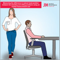
The differences in the natural head position between the standing and sitting positions were measured usinginertial measurement units. This system allows us to record the changes of the head position on the three axessimultaneously.

1. INTRODUCTION
The concept of natural head position (NHP) was first introduced in the 19th century by Broca, 1 who defined it as the most balanced head position, when a person is standing and when their visual axis is horizontal. The deviations of NHP have been correlated to different alterations in several fields such as craniofacial morphology, 2 malocclusions, 3 facial growth pattern, 4 physiology of respiration 5 , 6 and ophthalmology. 7 , 8
The analysis of head position is done on three axes (pitch, roll and yaw). 9 The NHP is usually measured when a patient is standing in front of a mirror (namely the mirror‐guided head position). 10 However, analysing the NHP in a sitting position may be important for certain populations such as individuals with a handicap, the elderly or young children for whom it is not always possible to stand up. Few studies have focused on this subject. Bjerin 11 found that the head in the sitting position is slightly more elevated 11 and the variations in head balance were slightly greater. 12 However, Huggare found no difference between the standing or sitting position, due to the methodology used, whereby the head position was corrected by the operator using a fluid level fixed on the head. 13 Those previous studies focused only on the pitch axis; thus, it would be interesting to search if there are differences in the head position between the sitting and standing position looking at all three axes.
There are different methods available to measure the NHP such as the operator's estimation, 14 , 15 lateral photographs 16 or radiographs, 10 head‐mounted equipment 17 and three‐dimensional scans. 18 The analysis can also be done with an electronic head‐mounted equipment using the inertial motion unit (IMU) 19 , 20 that can measure the three axes simultaneously. This system has already been tested in a previous study and showed excellent accuracy in measuring head position (M. Al‐Yassary, K. Billiaert, G. S. Antonarakis, & S. Kiliaridis, submitted for publication).
The aims of this study were to:
Evaluate the NHP (mirror‐guided head position) in all three axes comparing the standing and sitting positions;
Compare the observations on the pitch axis obtained from the electronic head‐mounted equipment sensors to those acquired by the lateral photographs;
Analyse the reliability and variation of the measurements of the NHP for both the standing and sitting positions on two separate occasions.
2. METHODS
2.1. Ethics statement
The present study was approved by the Swiss Association of Research Ethics Committees. The experimental procedures were conducted in conformity with the Declaration of Helsinki. Informed consent for participation in the study and publication in an open access format was obtained from the participants, with regard to their photographs and personal information. The procedures of the study were fully explained to the participants, and they provided their written informed consent before testing.
2.2. Participants
Twenty healthy young adult volunteers at the University clinics of dental medicine in Geneva, Switzerland (7 men, 13 women), aged from 20 to 30 years, were recruited for this study.
2.3. Experimental set‐up and protocol
To perform a reproducible analysis, a standardised position is needed. For the sitting position, the 90–90–90 position described by Engström 21 (corresponding to a 90° angle on the hips, the knees and the feet) is described as a reproducible and stable position. For the standing position, the orthoposition described by Molhave 22 is that most commonly used for research purposes. We used two methods to analyse the NHP, namely lateral photographs allowing an assessment of pitch and wearable sensors (IMU) allowing an assessment of pitch as well as roll and yaw.
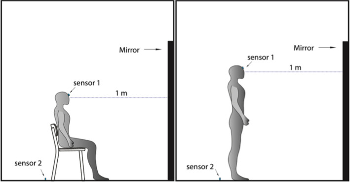
The participants were seated on a chair in the 90–90–90 position, facing a mirror which was one metre in front of their eyes. We calibrated the sensors on the ground to assess the neutral position (corresponding to 0° on each axis). The sensor was then placed on their forehead. We took a picture when the participants were gazing at their eyes in the mirror. Following this, the participants turned their heads to the photographer (for 5 s) then looked at the mirror for a second time (gazing at their eyes), when a second picture was taken. The same procedure was done with the participants standing up (Figure 1). In order to test the reliability of this method, the same procedure was performed on two separate occasions for each participant within a one‐week interval.
FIGURE 1.

Representative figure of the set‐up: The participants were seated on a chair in the 90–90–90 position 1 m in front of a mirror. After calibration of the inertial measurement unit on the floor to assess the reference, the first sensor was placed on the forehead to measure the natural head position. Following this, the participants stood up into a standing position at 1 m in front of the mirror and the natural head position was once again measured
2.4. Lateral photographic measurement
We displayed a laser giving us the horizontal plane behind the participant. The lateral photograph allowed us to assess the pitch by measuring the angle between the Frankfurt plane and the horizontal plane given by the laser.
2.5. IMU measurement
We used the MetaMotionR (Mbientlablnc., San Francisco, CA, USA) as the IMU, composed of two sensors. Before measuring the head position, this system needs to be calibrated. This is done by placing the first sensor on the ground in front of the participants and the second one behind them to assess the neutral position (corresponding to 0° on each axis). After calibration, the first sensor was placed on the forehead. Error of measurement could be caused by improper calibration, incorrect position of the sensor and the inclination of the forehead. To minimise the error caused by the calibration, we used the standardised procedure described above. To minimise the errors related to incorrect placement of the sensor on the forehead, we took a photograph in front of the participant to make sure that the sensor was parallel to the bipupillary line. Finally, for the possible sources of error in relation to forehead inclination, we took a lateral photograph and measured the inclination of the forehead. All angles are measured using Pixelstick (version 2.16.2, Plum Amazing Software LLC, USA).
2.6. Statistical analysis
All statistical analyses were performed using SPSS (version 24.0, SPSS Inc., Chicago, IL, USA). For each participant, we took two lateral photographs and two IMU recordings for the sitting position and standing position respectively, and this was done on two separate occasions.
To analyse the difference between the standing and sitting positions, linear regression analyses were performed with the standing data as the independent variable and sitting data as the dependent variable. A Bland‐Altman plot was used to visualise the spread of the differences between the two positions compared to their mean with a 95% confidence interval (95% CI).
To compare the pitch measured by the IMU system to the lateral photographs, Pearson correlation coefficients were calculated. Linear regression analysis was performed with the lateral photographic data as the independent variable and IMU data as the dependent variable. Intra‐class correlation (ICC) (based on a single rating, absolute‐agreement, 2‐way random‐effects model) estimates and their 95% CI were calculated. 23 ICC values less than 0.5 are indicative of poor reliability, those between 0.5 and 0.75 indicate moderate reliability, those between 0.75 and 0.9 indicate good reliability and those greater than 0.90 indicate excellent reliability. 24 The standard error measurement (SEM) of the difference between standing and sitting was calculated for each ICC (using the formula SEM = ).
We compared the two sessions, by calculating the paired t test for systematic differences between the two sessions. A Bland‐Altman plot was used to visualise the spread of the differences between the two sessions compared with their mean with a 95% CI.
3. RESULTS
3.1. Evaluating the NHP (mirror‐guided head position) in all three axes comparing the standing and sitting positions
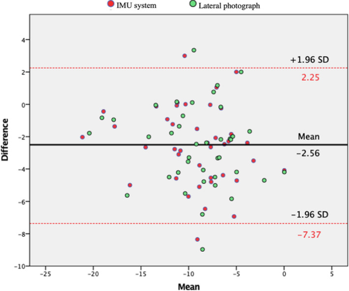
When comparing the two positions, we found that in the sitting position the head was more elevated (corresponding to the pitch axis) by 2.5 ± 2.4 (p < .05). We found no significant difference in the roll and yaw axes. The Bland‐Altman plot shows a bias line at −2.6°, with a 95% CI of −7.4 to +2.6. The data points are distributed equally below and above the bias line (Figure 2).
FIGURE 2.

Bland‐Altman plot comparing the sitting to the standing positions: The x‐axis represents the mean between the two and the y‐axis the differences. Red lines indicate the 95% confidence interval, and the black line indicates the bias (mean of the differences). Red points indicate the inertial measurement unit system, while green points indicate the lateral photographs
3.2. Comparing the observations on the pitch axis obtained from the electronic head‐mounted equipment sensors to those acquired by the lateral photographs
The absolute mean difference between the IMU and the lateral photographic data was 0.5 ± 0.5 for the pitch axis. A strong correlation was found between them (r = .99; p < .05). The ICCs were excellent (ICC = 0.99) with a 95% CI of 0.991–0.998 corresponding to an SEM = 0.03°.
3.3. Analysing the reliability of the measurements of the NHP for both the standing and sitting positions on two separate occasions
When comparing the two sessions, we observed no systematic differences for the three axes (p > .05) for the standing and sitting position. The correlation was strong for the pitch, moderate for the roll and poor for the yaw (Table 1). When comparing the differences between the two sessions, we observed that the differences in the sitting position were systematically lower than in the standing position for the roll axis (p = .01). No statistically significant differences were observed for the pitch and the yaw axes. (Table 1).
TABLE 1.
Difference in natural head position between the two sessions (T1‐T2), using the inertial measurement unit system
| Sitting | Standing | |||||
|---|---|---|---|---|---|---|
| Pitch | Roll | Yaw | Pitch | Roll | Yaw | |
| Mean ± SD |
−0.2 ± 2.1 (p = .55) |
0.4 ± 1.4 (p = .82) |
0 ± 2.3 (p = .36) |
0.3 ± 2.5 (p = .53) |
−0.4 ± 1.6 (p = .75) |
0.7 ± 2.8 (p = .78) |
| r (p‐value) | .89 (<.001) | .66 (<.001) | .27 (.25) | .87 (<.001) | .56 (.01) | .08 (.75) |
Means and standard deviations (SD) were calculated according to the differences between the first (corresponding to T1) and the second (corresponding to T2) sessions, done separately for each axis. The Pearson correlation (r) and p‐value between the first and second sessions was also calculated.
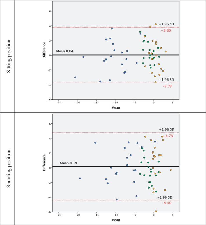
The Bland‐Altman plot shows the bias line at 0.04° for the sitting position and 0.19° for the standing position. The data points are distributed equally below and above the bias line. Interindividual variation for the roll and the yaw were approximately ±5°; however, for the pitch, the interindividual variation was higher (from 0° to −20°). (Figure 3).
FIGURE 3.

Bland‐Altman plot showing the relationship between the two sessions: The x‐axis represents the mean between the two and the y‐axis the differences. Red lines indicate the 95% confidence interval, and the black line indicates the bias (mean of the differences). Blue points indicate the pitch, while green and orange points indicate the roll and yaw, respectively
4. DISCUSSION
The present study, looking at differences in natural head position between the sitting and standing position, has shown that the head is slightly more elevated (pitch axis) in the sitting compared with the standing position. For the roll and yaw axis however, no differences were found when comparing the two positions. These results found on the pitch axis are similar to those found by Bjehin. 11
When comparing the measurement obtained by the electronic head‐mounted equipment to the lateral photographs, we found excellent correlations. With a precise and adequate protocol, the electronic head‐mounted equipment system can be used to measure the head position with great accuracy. The results found in our study were similar to the ones found by M. Al‐Yassary, K. Billiaert, G. S. Antonarakis, and S. Kiliaridis (submitted for publication) measuring the head position, Beange et al. 25 measuring the movement of lumbar spine and Fennema et al. 26 measuring the flexion of the knee. This method is easy to use, quick to set up and can be used on patients in the sitting or standing position. Moreover, the IMU system has an advantage over the other methods in that it allows an assessment of NHP not only in the pitch axis, but also in the roll and yaw axes.
The reliability of the measurements of the NHP on two separate occasions was different depending on the axis and the positions. The axis with the highest correlation was the pitch followed by the roll and yaw. With regard to the position, we found slightly greater correlation in the sitting position than the standing position in the three axes. The range of variations of the NHP on two separate occasions depends also on the axis and position. The axis with the least amount of variation was the roll, followed by the pitch and finally the yaw. Concerning the positions, we found no systematic difference in the range of variation for the pitch and the yaw. We did find however that the roll in the sitting position had systematically less variation compared to the standing positions. These results, in contrast to those found by Bjehin, 11 demonstrate that the sitting position is at least as reliable as the standing position and has the same range of variations or even less.
The elevation of the NHP found when sitting compared with standing may also be present in patients who are generally seated. However, it is difficult to demonstrate this phenomenon due to the significant interindividual variation. The interindividual variation for the pitch is 4 times greater than for the roll or the yaw. This observation was also made by Huh et al. 27 who analysed the angle between the Frankfort plane and Sella‐Nasion plane and found an individual variation ranging from 1.82° to 16.59°. If this elevation exists, it may have different implications on areas such as craniofacial growth and occlusion.
The results of the present study must be interpreted with caution because the participants included in our study were exclusively healthy young adults. The methodology used allows us to evaluate the short‐term reproducibility. A one‐week interval most likely prevents the participants from remembering the initial position, but it does not allow us to observe any changes in the head position over time. The methodology used allows us to evaluate the head position based on a one‐shot procedure but the NHP is known to be a balance of several positions with a certain range of variations. 14 It would be interesting to analyse the NHP for a long period of time in order to record the full range of variations and the tendencies for each axis.
5. CONCLUSION
When comparing the NHP of the sitting to the standing position, no significant differences were found for the roll and yaw axes. For the pitch axis however, the NHP in the sitting position is slightly more elevated. The stability of the sitting position is comparable to the standing position for the pitch and yaw axes. However, the roll axis is more stable in the sitting position. The electronic head‐mounted equipment, when used correctly, is comparable to lateral photographs for the pitch axis, with the advantage of recording the three axes simultaneously.
CONFLICT OF INTEREST
The authors declare no competing interests.
AUTHOR CONTRIBUTIONS
K.B. and M.A. contributed to the data collection and data analysis; G.S.A. and S.K. contributed to the study design and drafting of the manuscript. Figure 1 has been drawn by K.B., M.A. using Adobe Illustrator (version 24.3, cc 2020) (https://www.adobe.com
PEER REVIEW
The peer review history for this article is available at https://publons.com/publon/10.1111/joor.13233
ACKNOWLEDGEMENTS
The authors received no specific funding for this work. Open Access Funding provided by Universite de Geneve.
Billiaert K, Al‐Yassary M, Antonarakis GS, Kiliaridis S. Measuring the difference in natural head position between the standing and sitting positions using an inertial measurement unit. J Oral Rehabil. 2021;48:1144–1149. 10.1111/joor.13233
Billiaert and Al‐Yassary authors contributed equally to this work.
Contributor Information
Kelly Billiaert, Email: Kelly.Billiaert@unige.ch.
Gregory S. Antonarakis, Email: Gregory.Antonarakis@unige.ch.
DATA AVAILABILITY STATEMENT
All data included in this study are available upon reasonable request from the corresponding author.
REFERENCES
- 1. Broca M. Sur les projections de la tète, et sur un nouveau procède de cephalometrié. Bull Soc Anthropol Paris. 1862;3:514‐544. [Google Scholar]
- 2. Solow B, Tallgren A. Head posture and craniofacial morphology. Am J Phys Anthropol. 1976;44:417‐435. 10.1002/ajpa.1330440306 [DOI] [PubMed] [Google Scholar]
- 3. Solow B, Sonnesen L. Head posture and malocclusions. Eur J Orthod. 1998;20:685‐693. 10.1093/ejo/20.6.685 [DOI] [PubMed] [Google Scholar]
- 4. Solow B, Siersbaek‐Nielsen S. Growth changes in head posture related to craniofacial development. Am J Orthod. 1986;89:132‐140. 10.1016/0002-9416(86)90089-8 [DOI] [PubMed] [Google Scholar]
- 5. Cuccia AM, Lotti M, Caradonna D. Oral breathing and head posture. Angle Orthod. 2008;78:77‐82. 10.2319/011507-18.1 [DOI] [PubMed] [Google Scholar]
- 6. Neiva PD, Kirkwood RN, Mendes PL, Zabjek K, Becker HG, Mathur S. Postural disorders in mouth breathing children: a systematic review. Braz J Phys Ther. 2018;22:7‐19. 10.1016/j.bjpt.2017.06.011 [DOI] [PMC free article] [PubMed] [Google Scholar]
- 7. Nucci P, Curiel B, Lembo A, Serafino M. Anomalous head posture related to visual problems. Int Ophthalmol. 2015;35:241‐248. 10.1007/s10792-014-9943-7 [DOI] [PubMed] [Google Scholar]
- 8. Nucci P, Kushner BJ, Serafino M, Orzalesi N. A multi‐disciplinary study of the ocular, orthopedic, and neurologic causes of abnormal head postures in children. Am J Ophthalmol. 2005;140:65‐68. 10.1016/j.ajo.2005.01.037 [DOI] [PubMed] [Google Scholar]
- 9. Leung MY, Lo J, Leung YY. Accuracy of different modalities to record natural head position in 3 dimensions: a systematic review. J Oral Maxillofac Surg. 2016;74:2261‐2284. 10.1016/j.joms.2016.04.022 [DOI] [PubMed] [Google Scholar]
- 10. Solow B, Tallgren A. Natural head position in standing subjects. Acta Odontol Scand. 1971;29:591‐607. 10.3109/00016357109026337 [DOI] [PubMed] [Google Scholar]
- 11. Bjehin R. A Comparison between the Frankfort horizontal and the Sella Turcica ‐Nasion as reference planes in cephalometric analysis. Acta Odontol Scand. 1957;15:1‐12. 10.3109/00016355709041090 [DOI] [Google Scholar]
- 12. Moorrees CFA, Kean MR. Natural head position, a basic consideration in the interpretation of cephalometric radiographs. Am J Phys Anthropol. 1958;16:213‐234. 10.1002/ajpa.1330160206 [DOI] [Google Scholar]
- 13. Huggare JA. A natural head position technique for radiographic cephalometry. Dentomaxillofac Radiol. 1993;22:74‐76. 10.1259/dmfr.22.2.8375558 [DOI] [PubMed] [Google Scholar]
- 14. Lundstrom A, Forsberg CM, Westergren H, Lundstrom F. A comparison between estimated and registered natural head posture. Eur J Orthod. 1991;13:59‐64. 10.1093/ejo/13.1.59 [DOI] [PubMed] [Google Scholar]
- 15. Bass NM. The aesthetic analysis of the face. Eur J Orthod. 1991;13:343‐350. 10.1093/ejo/13.5.343 [DOI] [PubMed] [Google Scholar]
- 16. Lundstrom F, Lundstrom A. Natural head position as a basis for cephalometric analysis. Am J Orthod Dentofacial Orthop. 1992;101:244‐247. 10.1016/0889-5406(92)70093-P [DOI] [PubMed] [Google Scholar]
- 17. Audette I, Dumas JP, Cote JN, De Serres SJ. Validity and between‐day reliability of the cervical range of motion (CROM) device. J Orthop Sports Phys Ther. 2010;40:318‐323. 10.2519/jospt.2010.3180 [DOI] [PubMed] [Google Scholar]
- 18. Cassi D, De Biase C, Tonni I, Gandolfini M, Di Blasio A, Piancino MG. Natural position of the head: review of two‐dimensional and three‐dimensional methods of recording. Br J Oral Maxillofac Surg. 2016;54:233‐240. 10.1016/j.bjoms.2016.01.025 [DOI] [PubMed] [Google Scholar]
- 19. Patel S, Park H, Bonato P, Chan L, Rodgers M. A review of wearable sensors and systems with application in rehabilitation. J Neuroeng Rehabil. 2012;9:21. 10.1186/1743-0003-9-21 [DOI] [PMC free article] [PubMed] [Google Scholar]
- 20. Ghislieri M, Gastaldi L, Pastorelli S, Tadano S, Agostini V. Wearable inertial sensors to assess standing balance: a systematic review. Sensors (Basel). 2019;19(19):1–25. 10.3390/s19194075 [DOI] [PMC free article] [PubMed] [Google Scholar]
- 21. Engström B. Ergonomic seating: a true challenge: seating and mobility for the physically challenged: risks & possibilities when using wheelchairs. Stockholm: Posturalis Books; 2002. [Google Scholar]
- 22. Molhave A. The erect position in man; theoretical and statometrical aspects: a biostatic study. Nord Med. 1959;62:1148‐1150. [PubMed] [Google Scholar]
- 23. Portney LG, Watkins MP. Foundations of Clinical Research: Applications to Practice. Upper Saddle River, NJ: Pearson/Prentice Hall; 2015. [Google Scholar]
- 24. Koo TK, Li MY. A guideline of selecting and reporting intraclass correlation coefficients for reliability research. J Chiropr Med. 2016;15:155‐163. 10.1016/j.jcm.2016.02.012 [DOI] [PMC free article] [PubMed] [Google Scholar]
- 25. Beange KHE, Chan ADC, Beaudette SM, Graham RB. Concurrent validity of a wearable IMU for objective assessments of functional movement quality and control of the lumbar spine. J Biomech. 2019;97:109356. 10.1016/j.jbiomech.2019.109356 [DOI] [PubMed] [Google Scholar]
- 26. Fennema MC, Bloomfield RA, Lanting BA, Birmingham TB, Teeter MG. Repeatability of measuring knee flexion angles with wearable inertial sensors. Knee. 2019;26:97‐105. 10.1016/j.knee.2018.11.002 [DOI] [PubMed] [Google Scholar]
- 27. Huh YJ, Huh K‐H, Kim H‐K, et al. Constancy of the angle between the Frankfort horizontal plane and the sella‐nasion line: a nine‐year longitudinal study. Angle Orthod. 2014;84:286‐291. 10.2319/062013-464.1 [DOI] [PMC free article] [PubMed] [Google Scholar]
Associated Data
This section collects any data citations, data availability statements, or supplementary materials included in this article.
Data Availability Statement
All data included in this study are available upon reasonable request from the corresponding author.


