



Abstract
Systolic heart failure (HF) is a chronic clinical syndrome characterized by the reduction in cardiac function and still remains the disease with the highest mortality worldwide. Despite considerable advances in pharmacological treatment, HF represents a severe clinical and social burden. Chronic human HF is characterized by several important neurohormonal perturbations, emanating from both the autonomic nervous system and the adrenal glands. Circulating catecholamines (norepinephrine and epinephrine) as well as aldosterone elevations are among the salient alterations that confer significant hormonal burden on the already compromised function of the failing heart. This is why sympatholytic treatments (such as β-blockers) and renin-angiotensin system inhibitors or mineralocorticoid receptor antagonists that block the effects of angiotensin II (AngII) and of aldosterone on the failing heart, are part of the mainstay HF pharmacotherapy presently. The adrenal gland plays important parts in modulation of cardiac neurohormonal stress, since it is the source of almost all aldosterone, of all epinephrine, and of a significant amount of norepinephrine reaching the failing myocardium from the blood circulation. Synthesis and release of these hormones in the adrenals is tightly regulated by adrenal G protein-coupled receptors (GPCRs), such as adrenergic receptors (ARs) and AngII receptors. In this review, we discuss important aspects of adrenal GPCR signaling and regulation, as they pertain to modulation of cardiac function in the context of chronic HF, by focusing on the two best studied adrenal GPCR types in that context, ARs and AngII receptors (AT1Rs). Particular emphasis is given to findings from the past decade and a half that highlight the emerging roles of the GPCR-kinases (GRKs) and the β-arrestins in the adrenals, two protein families that regulate the signaling and function of GPCRs in all tissues, including the myocardium and the adrenal gland.
Keywords: Adrenal gland, Adrenergic receptor, Aldosterone, Angiotensin II type 1 receptor, β-arrestin, Catecholamine, G protein-coupled receptor (GPCR), GPCR-kinase (GRK), Heart failure, Signal transduction
Introduction
Chronic systolic heart failure (HF) or HF with reduced ejection fraction (HFrEF) is a clinical syndrome characterized by impaired left ventricular (LV) function with the inability to adequately maintain tissue perfusion and support bodily functions. HFrEF represents the leading cause of mortality in the western world, even after adjustment for the coronavirus-induced disease (COVID)-19 pandemic toll [1]. Two of the most important neurohormonal mechanisms driving the morbidity of the disease are sympathetic nervous system (SNS) hyperactivity, coupled with renin-angiotensin-aldosterone system (RAAS) activation. This is the basis for the utilization of sympatholytic drugs such as β-blockers, of angiotensin-converting enzyme (ACE) inhibitors, AngII receptor type 1 (AT1R) blockers (sartans) and mineralocorticoid receptor antagonists (MRAs) in the treatment of advanced stage chronic HF with reduced ejection fraction (HFrEF) [2,3]. Increased levels of the neurotransmitter norepinephrine (NE) in the cardiac sympathetic nerve terminals [4], combined with increased circulating levels of the hormone epinephrine (Epi) secreted from the adrenal medulla, are part of the SNS`s attempt to compensate for the diminished cardiac dysfunction. However, if the cardiac decompensation cannot be reversed fast enough, it becomes permanent rendering the heart subject to the Laplacès law of elevated free ventricular wall pressures, no longer operating under the Frank-Starling law of cardiac elasticity [6–8]. Additionally, cardiac hypertrophy and contractile dysfunction also ensue due to impairments of adrenergic regulation and positive force-frequency relationship.
NE and Epi, which, along with dopamine, constitute the endogenous catecholamines (CAs), bind to adrenergic receptors (ARs). All ARs are G protein-coupled receptors (GPCRs), also known as seven-transmembrane spanning or heptahelical receptors), and responsible for the cellular effects of the SNS in the body. Nine different AR subtypes are encoded in the human genome: three α1-, three α2 (α2A, α2B, α2C), and three β (β1, β2, β3) [9]. As GPCRs, the ARs are plasma membrane receptors activating heterotrimeric guanine nucleotide-binding (G) proteins upon against activation. The β1- and β2ARs primarily couple to stimulatory G proteins, through the alpha subunit of which they activate adenylyl cyclase (AC) to synthesize the second messenger cyclic adenosine monophosphate (cAMP) [10]. GPCR-kinases (GRKs), whose main ubiquitously expressed member isoforms are GRK2 and GRK5, phosphorylate GPCRs to induce β-arrestin binding, which then decouples the receptor from G proteins and typically targets it for clathrin-dependent internalization [11–13]. In addition, as the receptor-β-arrestin complex traffics inside the cell to either reach the lysosome for degradation (receptor downregulation) or return to the plasma membrane ready for another round of activation (resensitization), β-arrestins can mediate G protein-independent signaling via effector scaffolding [11–14]. Of note, the two main cardiac βAR subtypes have some fundamental differences in their functional and signaling features. For instance, the β1AR is by far the predominant subtype expressed at a ~3:1 ratio compared to the β2AR in cardiac myocytes [15,16] and is pro-apoptotic, whereas the β2AR anti-apoptotic [17–21]. This distinction is particularly significant, given the fact that the cardiomyocyte β1AR is directly activated by the NE released from cardiac SNS terminals [22], whereas the anti-apoptotic β2AR is mainly activated by the circulating in the blood Epi, for which it has a much (~14-fold) higher affinity than for NE [23]. Sympathetic neurons lack phenylethanolamine-N-methyl-transferase (PNMT), the enzyme that converts NE to Epi, so they only release NE, not Epi [24,25]. Furthermore, the β1AR is selectively downregulated in human failing hearts but the β2AR is dysfunctional (severely desensitized) despite its levels being preserved [26,27]. In the context of chronic HFrEF, high CA levels induce pathological adverse remodeling in the heart consisting of apoptosis, fibrosis, and inflammation [28–30]. NE increases cardiac oxygen consumption and metabolic demand, leading to cardiomyocyte death and left ventricular dilatation/dilated cardiomyopathy [31]. Cardiac GRK2 upregulation is a hallmark of chronic end-stage human HF but also experimental systolic HF in animal models [32–35] (Figure 1). Even in the absence of disease, cardiac GRK2 levels are inversely proportional to contractile function [36]. GRK2 not only regulates CA-dependent contractility but also cardiac apoptosis, since it increases apoptosis via its mitochondrial localization, although this remains controversial as other groups have proposed a cardioprotective role for mitochondrial GRK2 [37,38]. Finally, several AR polymorphisms are associated with human HF severity as well as response to β-blocker treatment [39–41].
Figure 1. The long-distance relationship of the heart with the adrenal GPCRs α2AR and AT1R through hormonal (catecholamine and aldosterone) regulation of its function.
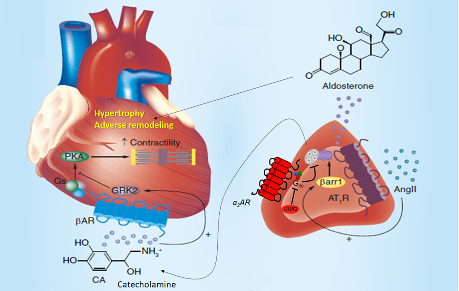
See main text for details and for all molecular acronym descriptions.
The role of locally released NE in the failing myocardium, as well as the perturbations in cardiac sympathetic innervation and NE synthesis, storage, and reuptake in chronic HF are well-established and extensively documented [41–46]. What had not been investigated at all, until about 15 years ago, was the role (if any) the adrenal CA secretion component of the SNS plays in chronic HF. As it turned out, the role of the adrenal source of CAs is equally important as that of cardiac sympathetic neuron-derived NE and, in the following sections, we review all these studies that helped elucidate this, with a focus on the two best studied adrenal GPCR types, α2ARs in the adrenal medulla and AT1Rs in the adrenal cortex, both of which fine-tune cardiac function via modulation of CA and aldosterone secretions, respectively.
Adrenal ARs and the failing heart
A salient pathophysiological feature of chronic HF is SNS hyperactivity, reflected by increased levels of circulating Epi and NE [47–49]. Adrenal CA secretion is stimulated by nicotinic cholinergic receptors in the membranes of chromaffin cells and is fine-tuned by presynaptic inhibitory α2ARs [49–52] (Figure 1). Of note, the human adrenal medulla also expresses all three βAR subtypes but these receptors operate as facilitatory autoreceptors, i.e., they augment CA release upon CA binding and activation [53]. In fact, α2AR is the only one or one of the only two adrenal GPCR types (the possible other one being adenosine receptors) identified to date that inhibits CA secretion [52]. α2ARs, similarly to β1- and β2ARs (but not β3ARs), undergo GRK-dependent desensitization [54–57]. Out of the seven human GRK isoforms (GRK1–7), GRK2 is the most abundant GRK in the heart and adrenals and it is significantly upregulated in the adrenal gland during chronic HF as it is in the heart [58]. As we and others have documented over the past 15 years, this GRK2 upregulation in the adrenal medulla is responsible for severe adrenal α2AR dysfunction in chronic HF, leading to a loss of the sympatho-inhibitory function by these receptors; thus, CA secretion is chronically elevated [58–62] (Figure 1). Importantly, GRK2 inhibition in the adrenal chromaffin cells, without any simultaneous intervention to cardiac GRK2, results in improved cardiac function and βAR number and signaling, as well as lower GRK2 levels in the failing myocardium [58]. In other words, just by lowering circulating Epi and NE levels, adrenal GRK2 blockade restores or at least ameliorates cardiac inotropic and adrenergic reserves [58]. Therefore, a crucial crosstalk at the level of entire organs seems to exist in chronic HF, via which adrenal GRK2 tightly regulates circulating CA levels, SNS activity, and cardiac function, as well as levels of its own activity in the heart, and vice versa. Another important ramification of this cardio-adrenal GRK2 axis is that, since circulating CA levels are kept at bay thanks to preservation of adrenal α2AR function, GRK2 inhibition in both of these organs (heart and adrenals) would be projected to be a safe positive inotropic therapy for HFrEF. In other words, cardiac contractility would be raised without concomitant SNS hyperactivity, which carries significant cardio-toxic risks such as arrhythmias, adverse remodeling, increased oxygen/metabolic demand, etc. [63].
Another study demonstrating the advantages of therapeutic targeting of adrenal GRK2 used transgenic mice having GRK2 genetically deleted only in cells expressing PNMT. Thus, these animals lack GRK2 expression in adrenal chromaffin cells [59]. The absence of GRK2 in the adrenal medulla led to significantly reduced SNS activity during progression to chronic HF secondary to myocardial infarction (MI), as reflected by circulating CA levels measured at 4 weeks post-MI [59]. In addition, cardiac contractility, structure/morphology (dilatation), and β-adrenergic responses were all improved [59]. Notably, the absence (from birth) of GRK2 in adrenal chromaffin cells led to a significant upregulation of α2ARs in the adrenals of these transgenic mice, in terms of both functional receptor number and G protein signaling activity (i.e., diminished α2AR functional desensitization) [59]. Thus, GRK2 and α2ARs regulate each other reciprocally to fine-tune CA secretion from the adrenal medulla. This study clearly demonstrated that halting the sympathetic activation immediately after an MI thanks to adrenal GRK2 inhibition can help the heart work close to normal and limit MI-induced damage. Therefore, adrenal GRK2 blockade can achieve the exact same goal for which β-blocker treatment is initiated in MI survivors.
Notably, adrenal GRK2 regulates CA secretion also under normal conditions, as its inhibition via adrenal-specific delivery of βARKct lowers circulating CA levels in normal, healthy animals, while GRK2 overexpression increases CA secretion [60]. In addition, exercise training, beneficial for the cardiovascular system as it reduces HF-related SNS hyperactivation, can also normalize adrenal GRK2 expression and α2AR function in HF animal models [61,62]. It should also be emphasized that adrenal GRK2 is upregulated in chronic HF irrespective of its etiology: apart from post-MI HF, adrenal GRK2 is elevated in pressure overload-induced hypertrophic cardiomyopathy, as well [64]. In fact, the degree of cardiac hypertrophy was found to directly correlate with adrenal weight and function (as measured by CA production) in this study [64].
Finally, one important question emanating from the studies described above was whether adrenal GRK2 upregulation was indeed the cause of the elevated SNS activity that accompanies and aggravates chronic HFrEF or merely an epiphenomenon of HFrEF, i.e., a symptom lacking causal relationship with the disease. A series of in vitro experiments in chromaffin cells helped answer this question by showing that chronic exposure of α2ARs to CAs by itself can directly upregulate GRK2 (transcriptionally) in chromaffin cells, thereby instigating α2AR desensitization/dysfunction that leads to chronic hypersecretion and excessive production of CAs [65]. This study was done in vitro and in a cellular context completely unrelated to HF or any other disease [65]. Consequently, its findings provide good evidence in support of the notion that it is the elevated SNS activity, i.e., the chronic exposure to elevated CA levels, that drives adrenal GRK2 upregulation rather than the presence of a certain pathology or disease, including chronic HF.
Other adrenal GPCRs and the failing heart
In addition to ARs, the adrenal medulla expresses a plethora of various other GPCR types that regulate CA secretion, including, but not limited to, muscarinic cholinergic receptors (mAChRs) [66], histaminergic H1 receptors [67], neuropeptide (NP)Y receptors Y1, Y2, Y4, and Y5 [68–70], pituitary adenylyl cyclase-activating polypeptide (PACAP) receptor PAC-1 [71,72], and adrenomedullin receptors [73,74]. All of these typically facilitate (enhance) CA secretion in response to acetylcholine stimulation. In contrast, very few adrenal GPCRs, other than α2ARs, inhibit CA secretion, mainly μ-opioid receptors (MORs) and purinergic P2Y receptors for adenosine triphosphate (ATP) [75–79]. Nevertheless, apart from the ARs, none of these other adrenal GPCRs (and their effects on CA secretion) have been studied in relation to HF or regulation of cardiac function in general thus far.
Perhaps the most important, in terms of cardiac regulation, adrenal GPCR type apart from ARs is the AT1R of AngII. This receptor can also augment CA secretion from the adrenal medulla via mobilization of intracellular calcium that promotes exocytosis/secretion [76]. This AT1R-induced, Ca2+-dependent exocytosis in adrenal chromaffin cells can be mediated by β-arrestin1, a GPCR adapter protein and signal transducer. β-arrestin1 directly interacts with the Ca2+ channel TRPC3 (short transient receptor potential channel-3) at the plasma membrane eliciting CA secretion [80]. However, the main physiological role of the AT1R in the adrenal gland is mapped in the adrenal cortex, where it induces synthesis and secretion of aldosterone [81–84] (Figure 1). More specifically, aldosterone is a mineralocorticoid hormone produced and secreted from adrenocortical zona glomerulosa (AZG) cells in response to hyperkalemia or AngII activating its AT1 receptors [85,86]. Aldosterone is elevated in chronic human HF, especially post-MI, and exerts a variety of indirect (hypertension, hypervolemia, etc.) and direct deleterious effects on the failing heart [87,88].
AngII-activated AT1R is typically a Gq/11 protein-coupled receptor, which means it activates phospholipase C-β [81]. The Gq-dependent signaling pathway elicited by the AT1R in AZG cells and leading to aldosterone production is well documented [85,86]. Diacylglycerol (DAG) and inositol 1`, 4`, 5`-trisphosphate (IP3), produced by phospholipase C (PLC)-β directly activated by Gαq subunits, ultimately lead to aldosterone secretion, via classic intracellular Ca2+ release-triggered exocytosis, and to aldosterone biosynthesis, via extracellular signal-regulated kinase (ERK)/mitogen-activated protein kinase (MAPK) activation, which, in turn, stimulates the gene transcription of StAR (steroidogenic acute regulatory) protein [86]. The latter mediates the mitochondrial uptake of the adrenal steroid precursor cholesterol, which is the rate-limiting step in aldosterone biosynthesis inside AZG cells [86]. GRKs phosphorylate the active AT1R causing the binding of the β-arrestins to the receptor, which effectively terminates this Gq/11-dependent signaling [12]. However, as we uncovered several years ago, β-arrestin1 binding to the adrenal AT1R elicits a second, G protein-independent wave of signaling towards aldosterone synthesis and secretion in response to AngII in AZG cells [89–91] (Figure 1). This action of β-arrestin1 centers on sustained ERK activation and subsequent transcriptional StAR upregulation, which has the same end-result as Gq protein signaling in AZG cells, i.e., mitochondrial cholesterol uptake and initiation of aldosterone biosynthesis [89]. Importantly, adrenal βarrestin1-dependent AT1R signaling promotes aldosterone synthesis and secretion both in vitro and in vivo [90,91] and, in fact, it is essential for the post-MI elevation of the endogenous production of this mineralocorticoid, since transgenic mice genetically lacking βarrestin1 have normal circulating aldosterone levels during post-MI HF progression [91]. Furthermore, CAs stimulate βARs to boost AT1R-dependent aldosterone production via GRK2-mediated receptor crosstalk in AZG cells [92].
Since adrenal AT1R induces aldosterone production via both G proteins and β-arrestin1, the question arose of how effective traditional angiotensin receptor blockers (ARBs), i.e., AT1R antagonists/inverse agonists (sartans), are at silencing AngII-dependent aldosterone production/secretion via both pathways in AZG cells [93]. Indeed, the prototypic drug in the class, losartan, was found to be a much more potent inverse agonist toward G proteins than toward β-arrestins at the AT1R, which made it incapable of suppressing the β-arrestin-dependent component of AngII-elicited aldosterone production and thus, inefficient aldosterone suppressor [94–96]. Irbesartan proved to act in a similar manner as losartan, i.e., a weak β-arrestin-dependent aldosterone suppressor [94]. In contrast, candesartan and valsartan demonstrated strong inverse agonist activity towards both G proteins and β-arrestins at the AT1R and excellent potency at suppressing aldosterone in vitro and in vivo [94–96]. Importantly, the effects of these drugs on adrenal AngII-dependent signaling to aldosterone secretion directly translated into effects on the function of the failing post-MI heart, since candesartan and valsartan markedly improved ejection fraction and cardiac function, accompanied by lower circulating aldosterone levels, in post-MI rats [95]. Conversely, losartan and irbesartan failed to halt the cardiac functional decline of these animals, as they were progressing to HF post-MI, as well as to lower circulating aldosterone levels [90,95]. Taken together, these studies strongly suggest that the adrenal AT1R can tightly regulate cardiac function and structure/remodeling from afar, i.e., from the adrenal gland, just by fine-tuning the circulating aldosterone levels in the body that reach the failing myocardium to exert their deleterious effects.
Finally, from the drug design/medicinal chemistry standpoint, it appears that the presence of a substitution both bulky and anionic at the opposite end of the biphenyl-methyl backbone of the compound to the tetrazole ring is required for efficient inverse agonism of β-arrestin activation at the AT1R [97–107]. Conversely, for potent β-arrestin agonism/activation, the aromatic side-chain of AngII peptidès Phe8 or Tyr4 are only required for G protein (not for β-arrestin) activation, but the aliphatic size of AngII position 5 (isoleucine in the natural agonist AngII) affects the extent of β-arrestin activation [97–107].
Conclusions/ Future Perspectives
In the present review, we provide an overview of the studies that have paved the way for the elucidation of the physiological and pathological roles the adrenal production of CAs and aldosterone play in regulation of cardiac function and in its dysregulation in chronic HF. Admittedly, this field is still in its infancy and a lot of questions remain unanswered. For instance, what is the role of adrenal CA secretion, if any, in the imbalance between the high SNS and low parasympathetic activities, which is the actual culprit of the neurohormonal dysregulation in human chronic HF? Does perhaps regulation of adrenal CA secretion hold the key for unlocking the true therapeutic potential of the once failed sympatholytic drugs, like moxonidine, for chronic human HF treatment? Does the adrenal β-arrestin-mediated hyperlaldosteronism play a role in the other major type of human chronic HF, HF with preserved ejection fraction (HFpEF), known to be accompanied and complicated by elevated circulating aldosterone levels? On the other hand, could adrenal β-arrestin-mediated hyperlaldosteronism be a pitfall for the therapeutic utility in HF of AT1R ligands that are purportedly β-arrestin-“biased” agonists? Hailed as major innovative breakthroughs for HF treatment more than a decade ago, these drugs, unfortunately, failed in major clinical trials for human HF about 5 years ago. All these questions are very important and await delineation in future studies.
Nevertheless, despite several outstanding questions, it appears that targeting of adrenal α2AR or AT1R signaling has some potential for lowering the hormonal burden of the failing heart (catecholaminergic and aldosteronic, respectively), thereby ameliorating cardiac function or at least halting its decline during HF development in humans. Additionally, therapeutic targeting of adrenal GPCR signaling in general, via GRK2 and/or β-arrestin1 blockade in the adrenal glands, could also be of value in heart disease therapy in that it may help restore normal SNS and RAAS functions/activities throughout the systemic circulation. Given the fact that the currently available anti-adrenergic (β-blockers) and anti-RAAS (ACE inhibitors, ARBs) drugs exert most of their actions directly on the failing myocardium itself, which may increase risk for adverse effects, adrenal GPCR therapeutic targeting has the major potential advantage of ameliorating cardiac function in HF without the drug or treatment acting on the heart itself, i.e., acting from a distance. Thus, future investigations will help add new “tricks up the sleeves” of sympatholytic and/or anti-RAAS treatments in a hope to yield novel, safer, and thus, better medications for human chronic HF. As most cardiologists and cardiovascular pharmacologists know all too well, this devastating disease has not seen many new therapies succeeding in a very long time, so it desperately needs them.
Acknowledgments
A.L. is supported by a research grant from the NIH/NHLBI (R01 #HL155718-01).
Footnotes
Conflict of interest
The authors declare no conflict of financial or of any other type of interest.
References
- [1].Ahmad FB, Anderson RN. The Leading Causes of Death in the US for 2020. JAMA. 2021. May 11;325(18):1829–30. doi: 10.1001/jama.2021.5469. [DOI] [PMC free article] [PubMed] [Google Scholar]
- [2].Rahamim E, Nachman D, Yagel O, Yarkoni M, Elbaz-Greener G, Amir O, Asleh R. Contemporary Pillars of Heart Failure with Reduced Ejection Fraction Medical Therapy. J Clin Med. 2021. Sep 26;10(19):4409. doi: 10.3390/jcm10194409. [DOI] [PMC free article] [PubMed] [Google Scholar]
- [3].Masarone D, Martucci ML, Errigo V, Pacileo G. The Use of β-Blockers in Heart Failure with Reduced Ejection Fraction. J Cardiovasc Dev Dis. 2021;8(9):101. doi: 10.3390/jcdd8090101. [DOI] [PMC free article] [PubMed] [Google Scholar]
- [4].Lymperopoulos A; et al. Signaling and function of cardiac autonomic nervous system receptors: Insights from the GPCR signalling universe. FEBS J. 2021;288:2645–59. [DOI] [PubMed] [Google Scholar]
- [5].Cohn JN, Levine TB, Olivari MT, Garberg V, Lura D, Francis GS, Simon AB, Rector T. Plasma norepinephrine as a guide to prognosis in patients with chronic congestive heart failure. N Engl J Med. 1984;311(13):819–23. [DOI] [PubMed] [Google Scholar]
- [6].Lymperopoulos A, Rengo G, Koch WJ. Adrenergic nervous system in heart failure: Pathophysiology and therapy. Circ. Res 2013;113:739–53. [DOI] [PMC free article] [PubMed] [Google Scholar]
- [7].Desimine VL, McCrink KA, Parker BM, Wertz SL, Maning J, Lymperopoulos A. Biased Agonism/Antagonism of Cardiovascular GPCRs for Heart Failure Therapy. Int Rev Cell Mol Biol. 2018;339:41–61. [DOI] [PubMed] [Google Scholar]
- [8].Lymperopoulos A; Garcia D; Walklett K Pharmacogenetics of cardiac inotropy. Pharmacogenomics 2014;15:1807–21. [DOI] [PubMed] [Google Scholar]
- [9].Lymperopoulos A, Rengo G, Koch WJ. Adrenal adrenoceptors in heart failure: fine-tuning cardiac stimulation. Trends Mol Med. 2007;13(12):503–11. [DOI] [PubMed] [Google Scholar]
- [10].Capote LA, Mendez Perez R, Lymperopoulos A. GPCR signaling and cardiac function. Eur J Pharmacol. 2015;763:143–48. [DOI] [PubMed] [Google Scholar]
- [11].DeWire SM, Ahn S, Lefkowitz RJ, Shenoy SK. Beta-arrestins and cell signaling. Annu Rev Physiol. 2007;69:483–510. [DOI] [PubMed] [Google Scholar]
- [12].Gurevich EV, Gurevich VV. GRKs as Modulators of Neurotransmitter Receptors. Cells. 2020;10:52. [DOI] [PMC free article] [PubMed] [Google Scholar]
- [13].McCrink KA, Maning J, Vu A, Jafferjee M, Marrero C, Brill A, Bathgate-Siryk A, Dabul S, Koch WJ, Lymperopoulos A. β-Arrestin2 Improves Post-Myocardial Infarction Heart Failure via Sarco(endo)plasmic Reticulum Ca2+-ATPase-Dependent Positive Inotropy in Cardiomyocytes. Hypertension. 2017;70(5):972–81. [DOI] [PubMed] [Google Scholar]
- [14].Lefkowitz RJ, Shenoy SK. Transduction of receptor signals by beta-arrestins. Science. 2005;308:512–7. [DOI] [PubMed] [Google Scholar]
- [15].Lymperopoulos A Arrestins in the Cardiovascular System: An Update. Prog Mol Biol Transl Sci. 2018;159:27–57. [DOI] [PubMed] [Google Scholar]
- [16].Yoshikawa T, Port JD, Asano K, Chidiak P, Bouvier M, Dutcher D, Roden RL, Minobe W, Tremmel KD, Bristow MR. Cardiac adrenergic receptor effects of carvedilol. Eur Heart J. 1996;17 Suppl B:8–16. [DOI] [PubMed] [Google Scholar]
- [17].Salazar NC, Vallejos X, Siryk A, Rengo G, Cannavo A, Liccardo D, De Lucia C, Gao E, Leosco D, Koch WJ, Lymperopoulos A. GRK2 blockade with βARKct is essential for cardiac β2-adrenergic receptor signaling towards increased contractility. Cell Commun Signal. 2013;11:64. [DOI] [PMC free article] [PubMed] [Google Scholar]
- [18].Communal C, Singh K, Sawyer DB, Colucci WS. Opposing effects of beta(1)- and beta(2)-adrenergic receptors on cardiac myocyte apoptosis : role of a pertussis toxin-sensitive G protein. Circulation. 1999;100(22):2210–2. [DOI] [PubMed] [Google Scholar]
- [19].Chesley A, Lundberg MS, Asai T, Xiao RP, Ohtani S, Lakatta EG, Crow MT. The beta(2)-adrenergic receptor delivers an antiapoptotic signal to cardiac myocytes through G(i)-dependent coupling to phosphatidylinositol 3’-kinase. Circ Res. 2000;87(12):1172–9. [DOI] [PubMed] [Google Scholar]
- [20].Zhu WZ, Zheng M, Koch WJ, Lefkowitz RJ, Kobilka BK, Xiao RP. Dual modulation of cell survival and cell death by beta(2)-adrenergic signaling in adult mouse cardiac myocytes. Proc Natl Acad Sci U S A. 2001;98(4):1607–12. [DOI] [PMC free article] [PubMed] [Google Scholar]
- [21].Zhang X, Szeto C, Gao E, Tang M, Jin J, Fu Q, Makarewich C, Ai X, Li Y, Tang A, Wang J, Gao H, Wang F, Ge XJ, Kunapuli SP, Zhou L, Zeng C, Xiang KY, Chen X. Cardiotoxic and cardioprotective features of chronic β-adrenergic signaling. Circ Res. 2013;112(3):498–509. [DOI] [PMC free article] [PubMed] [Google Scholar]
- [22].Eisenhofer G, Friberg P, Rundqvist B, Quyyumi AA, Lambert G, Kaye DM, Kopin IJ, Goldstein DS, Esler MD. Cardiac sympathetic nerve function in congestive heart failure. Circulation. 1996;93(9):1667–76. [DOI] [PubMed] [Google Scholar]
- [23].Xu X, Kaindl J, Clark MJ, Hübner H, Hirata K, Sunahara RK, Gmeiner P, Kobilka BK, Liu X. Binding pathway determines norepinephrine selectivity for the human β1AR over β2AR. Cell Res. 2021;31(5):569–79. [DOI] [PMC free article] [PubMed] [Google Scholar]
- [24].Wong DL. Epinephrine biosynthesis: hormonal and neural control during stress. Cell Mol Neurobiol. 2006;26(4–6):891–900. [DOI] [PMC free article] [PubMed] [Google Scholar]
- [25].Wong DL, Tank AW. Stress-induced catecholaminergic function: transcriptional and post-transcriptional control. Stress. 2007;10(2):121–30. [DOI] [PubMed] [Google Scholar]
- [26].Bristow MR. Mechanism of action of beta-blocking agents in heart failure. Am J Cardiol. 1997;80:26L–40L. [DOI] [PubMed] [Google Scholar]
- [27].Bristow MR, Hershberger RE, Port JD, Gilbert EM, Sandoval A, Rasmussen R, Cates AE, Feldman AM. Beta-adrenergic pathways in nonfailing and failing human ventricular myocardium. Circulation. 1990;82(2 Suppl):I12–25. [PubMed] [Google Scholar]
- [28].Port JD, Bristow MR. Altered beta-adrenergic receptor gene regulation and signaling in chronic heart failure. J Mol Cell Cardiol. 2001;33(5):887–905. [DOI] [PubMed] [Google Scholar]
- [29].Roghi A, Pedrotti P, Milazzo A, Bonacina E, Bucciarelli-Ducci C. Adrenergic myocarditis in pheochromocytoma. J Cardiovasc Magn Reson. 2011;13(1):4. [DOI] [PMC free article] [PubMed] [Google Scholar]
- [30].Rengo G, Lymperopoulos A, Zincarelli C, Donniacuo M, Soltys S, Rabinowitz JE, Koch WJ. Myocardial adeno-associated virus serotype 6-betaARKct gene therapy improves cardiac function and normalizes the neurohormonal axis in chronic heart failure. Circulation. 2009;119(1):89–98. [DOI] [PMC free article] [PubMed] [Google Scholar]
- [31].Prejbisz A, Lenders JW, Eisenhofer G, Januszewicz A. Cardiovascular manifestations of phaeochromocytoma. J Hypertens. 2011;29(11):2049–60. [DOI] [PubMed] [Google Scholar]
- [32].Iaccarino G, Tomhave ED, Lefkowitz RJ, Koch WJ. Reciprocal in vivo regulation of myocardial G protein-coupled receptor kinase expression by beta-adrenergic receptor stimulation and blockade. Circulation. 1998;98(17):1783–9. [DOI] [PubMed] [Google Scholar]
- [33].Lefkowitz RJ, Rockman HA, Koch WJ. Catecholamines, cardiac beta-adrenergic receptors, and heart failure. Circulation. 2000;101(14):1634–7. [DOI] [PubMed] [Google Scholar]
- [34].Rengo G, Lymperopoulos A, Koch WJ. Future g protein-coupled receptor targets for treatment of heart failure. Curr Treat Options Cardiovasc Med. 2009;11(4):328–38. [DOI] [PubMed] [Google Scholar]
- [35].Ungerer M, Böhm M, Elce JS, Erdmann E, Lohse MJ. Altered expression of beta-adrenergic receptor kinase and beta 1-adrenergic receptors in the failing human heart. Circulation. 1993;87(2):454–63. [DOI] [PubMed] [Google Scholar]
- [36].Koch WJ, Rockman HA, Samama P, Hamilton RA, Bond RA, Milano CA, Lefkowitz RJ. Cardiac function in mice overexpressing the beta-adrenergic receptor kinase or a beta ARK inhibitor. Science. 1995;268(5215):1350–3. [DOI] [PubMed] [Google Scholar]
- [37].Chen M, Sato PY, Chuprun JK, Peroutka RJ, Otis NJ, Ibetti J, Pan S, Sheu SS, Gao E, Koch WJ. Prodeath signaling of G protein-coupled receptor kinase 2 in cardiac myocytes after ischemic stress occurs via extracellular signal-regulated kinase-dependent heat shock protein 90-mediated mitochondrial targeting. Circ Res. 2013;112(8):1121–34. [DOI] [PMC free article] [PubMed] [Google Scholar]
- [38].Fusco A, Santulli G, Sorriento D, Cipolletta E, Garbi C, Dorn GW 2nd, Trimarco B, Feliciello A, Iaccarino G. Mitochondrial localization unveils a novel role for GRK2 in organelle biogenesis. Cell Signal. 2012;24(2):468–75. [DOI] [PMC free article] [PubMed] [Google Scholar]
- [39].Johnson JA, Liggett SB. Cardiovascular pharmacogenomics of adrenergic receptor signaling: clinical implications and future directions. Clin Pharmacol Ther 2011;89(3):366–78. [DOI] [PMC free article] [PubMed] [Google Scholar]
- [40].Dorn GW 2nd. Adrenergic signaling polymorphisms and their impact on cardiovascular disease. Physiol Rev 2010;90(3):1013–62. [DOI] [PubMed] [Google Scholar]
- [41].Small KM, Wagoner LE, Levin AM, Kardia SL, Liggett SB. Synergistic polymorphisms of beta1- and alpha2C-adrenergic receptors and the risk of congestive heart failure. N Engl J Med. 2002;347:1135–42. [DOI] [PubMed] [Google Scholar]
- [42].Port JD, Gilbert EM, Larrabee P, Mealey P, Volkman K, Ginsburg R, Hershberger RE, Murray J, Bristow MR. Neurotransmitter depletion compromises the ability of indirect-acting amines to provide inotropic support in the failing human heart. Circulation. 1990;81(3):929–38. [DOI] [PubMed] [Google Scholar]
- [43].Chen X, Kudo T, Lapa C, Buck A, Higuchi T. Recent advances in radiotracers targeting norepinephrine transporter: structural development and radiolabeling improvements. J Neural Transm (Vienna). 2020;127(6):851–73. [DOI] [PMC free article] [PubMed] [Google Scholar]
- [44].Schroeder C, Jordan J. Norepinephrine transporter function and human cardiovascular disease. Am J Physiol Heart Circ Physiol. 2012;303(11):H1273–82. [DOI] [PubMed] [Google Scholar]
- [45].Grassi G, D’Arrigo G, Pisano A, Bolignano D, Mallamaci F, Dell’Oro R, Quarti-Trevano F, Seravalle G, Mancia G, Zoccali C. Sympathetic neural overdrive in congestive heart failure and its correlates: systematic reviews and meta-analysis. J Hypertens. 2019;37(9):1746–56. [DOI] [PubMed] [Google Scholar]
- [46].Gupta S, Amanullah A. Radionuclide imaging of cardiac sympathetic innervation in heart failure: unlocking untapped potential. Heart Fail Rev. 2015;20(2):215–26. [DOI] [PubMed] [Google Scholar]
- [47].Mann DL, Bristow MR. Mechanisms and models in heart failure: the biomechanical model and beyond. Circulation 2005;111:2837–49. [DOI] [PubMed] [Google Scholar]
- [48].Mudd JO, Kass DA. Tackling heart failure in the twenty first century. Nature 2008;451:919–28. [DOI] [PubMed] [Google Scholar]
- [49].Esler MD, Jennings G, Schlaich M, Lambert G, Thompson J, Lambert E, Guo L, Alvarenga M, Esler D, Eikelis N, Kaye D. The adrenal medulla in cardiovascular medicine: an untold story. J Hypertens. 2021;39(5):819–29. [DOI] [PubMed] [Google Scholar]
- [50].Eaton MJ, Duplan H. Useful cell lines derived from the adrenal medulla. Mol Cell Endocrinol 2004;228:39–52. [DOI] [PubMed] [Google Scholar]
- [51].Moura E, Afonso J, Hein L, Vieira-Coelho MA. Alpha2-adrenoceptor subtypes involved in the regulation of catecholamine release from the adrenal medulla of mice. Br J Pharmacol 2006;149:1049–58. [DOI] [PMC free article] [PubMed] [Google Scholar]
- [52].Lymperopoulos A, Brill A, McCrink KA. GPCRs of adrenal chromaffin cells & catecholamines: The plot thickens. Int J Biochem Cell Biol. 2016;77(Pt B):213–9. [DOI] [PubMed] [Google Scholar]
- [53].Cortez V, Santana M, Marques AP, Mota A, Rosmaninho-Salgado J, Cavadas C. Regulation of catecholamine release in human adrenal chromaffin cells by β-adrenoceptors. Neurochem Int. 2012;60(4):387–93. [DOI] [PubMed] [Google Scholar]
- [54].Kurose H, Lefkowitz RJ. Differential desensitization and phosphorylation of three cloned and transfected alpha 2-adrenergic receptor subtypes. J Biol Chem. 1994;269(13):10093–9. [PubMed] [Google Scholar]
- [55].DeGraff JL, Gagnon AW, Benovic JL, Orsini MJ. Role of arrestins in endocytosis and signaling of alpha2-adrenergic receptor subtypes. J Biol Chem. 1999;274(16):11253–9. [DOI] [PubMed] [Google Scholar]
- [56].Nguyen K, Kassimatis T, Lymperopoulos A. Impaired desensitization of a human polymorphic α2B-adrenergic receptor variant enhances its sympatho-inhibitory activity in chromaffin cells. Cell Commun Signal. 2011;9(1):5. [DOI] [PMC free article] [PubMed] [Google Scholar]
- [57].Benovic JL. Historical Perspective of the G Protein-Coupled Receptor Kinase Family. Cells. 2021;10(3):555. [DOI] [PMC free article] [PubMed] [Google Scholar]
- [58].Lymperopoulos A, Rengo G, Funakoshi H, Eckhart AD, Koch WJ. Adrenal GRK2 upregulation mediates sympathetic overdrive in heart failure. Nat Med. 2007;13:315–23. [DOI] [PubMed] [Google Scholar]
- [59].Lymperopoulos A, Rengo G, Gao E, Ebert SN, Dorn GW II, Koch WJ. Reduction of sympathetic activity via adrenal-targeted GRK2 gene deletion attenuates heart failure progression and improves cardiac function after myocardial infarction. J Biol Chem 2010;285:16378–86. [DOI] [PMC free article] [PubMed] [Google Scholar]
- [60].Lymperopoulos A, Rengo G, Zincarelli C, Soltys S, Koch WJ. Modulation of adrenal catecholamine secretion by in vivo gene transfer and manipulation of G protein-coupled receptor kinase-2 activity. Mol Ther 2008;16:302–7. [DOI] [PubMed] [Google Scholar]
- [61].Rengo G, Leosco D, Zincarelli C, Marchese M, Corbi G, Liccardo D, Filippelli A, Ferrara N, Lisanti MP, Koch WJ, Lymperopoulos A. Adrenal GRK2 lowering is an underlying mechanism for the beneficial sympathetic effects of exercise training in heart failure. Am J Physiol Heart Circ Physiol 2010;298:H2032–8. [DOI] [PubMed] [Google Scholar]
- [62].Rengo G, Lymperopoulos A, Zincarelli C, Femminella G, Liccardo D, Pagano G, de Lucia C, Cannavo A, Gargiulo P, Ferrara N, Perrone Filardi P, Koch W, Leosco D. Blockade of β-adrenoceptors restores the GRK2-mediated adrenal α(2) -adrenoceptor-catecholamine production axis in heart failure. Br J Pharmacol. 2012;166(8):2430–40. [DOI] [PMC free article] [PubMed] [Google Scholar]
- [63].Packer M How should physicians view heart failure? The philosophical and physiological evolution of three conceptual models of the disease. Am J Cardiol. 1993;71(9):3C–11C. [DOI] [PubMed] [Google Scholar]
- [64].Schneider J, Lother A, Hein L, Gilsbach R. Chronic cardiac pressure overload induces adrenal medulla hypertrophy and increased catecholamine synthesis. Basic Res Cardiol. 2011;106(4):591–602. [DOI] [PubMed] [Google Scholar]
- [65].Jafferjee M, Reyes Valero T, Marrero C, McCrink KA, Brill A, Lymperopoulos A. GRK2 Up-Regulation Creates a Positive Feedback Loop for Catecholamine Production in Chromaffin Cells. Mol Endocrinol. 2016;30(3):372–81. [DOI] [PMC free article] [PubMed] [Google Scholar]
- [66].Zaika OL, Pochynyuk OM, Kostyuk PG, Yavorskaya EN, Lukyanetz EA. Acetylcholine-induced calcium signalling in adrenaline- and noradrenaline-containing adrenal chromaffin cells. Arch Biochem Biophys. 2004;424(1):23–32. [DOI] [PubMed] [Google Scholar]
- [67].Wallace DJ, Chen C, Marley PD. Histamine promotes excitability in bovine adrenal chromaffin cells by inhibiting an M-current. J Physiol. 2002;540(Pt 3):921–39. [DOI] [PMC free article] [PubMed] [Google Scholar]
- [68].Körner M, Waser B, Reubi JC. High expression of neuropeptide Y receptors in tumors of the human adrenal gland and extra-adrenal paraganglia. Clin Cancer Res. 2004;10(24):8426–33. [DOI] [PubMed] [Google Scholar]
- [69].Spinazzi R, Andreis PG, Nussdorfer GG. Neuropeptide-Y and Y-receptors in the autocrine-paracrine regulation of adrenal gland under physiological and pathophysiological conditions (Review). Int J Mol Med. 2005;15(1):3–13. [PubMed] [Google Scholar]
- [70].Cavadas C, Céfai D, Rosmaninho-Salgado J, Vieira-Coelho MA, Moura E, Busso N, Pedrazzini T, Grand D, Rotman S, Waeber B, Aubert JF, Grouzmann E. Deletion of the neuropeptide Y (NPY) Y1 receptor gene reveals a regulatory role of NPY on catecholamine synthesis and secretion. Proc Natl Acad Sci U S A. 2006;103(27):10497–502. [DOI] [PMC free article] [PubMed] [Google Scholar]
- [71].Payet MD, Bilodeau L, Breault L, Fournier A, Yon L, Vaudry H, Gallo-Payet N. PAC1 receptor activation by PACAP-38 mediates Ca2+ release from a cAMP-dependent pool in human fetal adrenal gland chromaffin cells. J Biol Chem. 2003;278(3):1663–70. [DOI] [PubMed] [Google Scholar]
- [72].Isobe K, Kaneko M, Kaneko S, Nissato S, Nanmoku T, Takekoshi K, Okuda Y, Kawakami Y. Expression of mRNAs for PACAP and its receptor in human neuroblastomas and their relationship to catecholamine synthesis. Regul Pept. 2004;123:29–32. [DOI] [PubMed] [Google Scholar]
- [73].Mazzocchi G, Albertin G, Andreis PG, Neri G, Malendowicz LK, Champion HC, Bahçelioglu M, Kadowitz PJ, Nussdorfer GG. Distribution, functional role, and signaling mechanism of adrenomedullin receptors in the rat adrenal gland. Peptides. 1999;20(12):1479–87. [DOI] [PubMed] [Google Scholar]
- [74].Kobayashi H, Yanagita T, Yokoo H, Wada A. Pathophysiological function of adrenomedullin and proadrenomedullin N-terminal peptides in adrenal chromaffin cells. Hypertens Res. 2003;26 Suppl:S71–8. [DOI] [PubMed] [Google Scholar]
- [75].Albillos A, Gandía L, Michelena P, Gilabert JA, del Valle M, Carbone E, García AG. The mechanism of calcium channel facilitation in bovine chromaffin cells. J Physiol. 1996;494:687–95. [DOI] [PMC free article] [PubMed] [Google Scholar]
- [76].Currie KP. Inhibition of Ca2+ channels and adrenal catecholamine release by G protein coupled receptors. Cell Mol Neurobiol. 2010;30(8):1201–8. [DOI] [PMC free article] [PubMed] [Google Scholar]
- [77].Harkins AB, Fox AP. Activation of purinergic receptors by ATP inhibits secretion in bovine adrenal chromaffin cells. Brain Res. 2000;885(2):231–9. [DOI] [PubMed] [Google Scholar]
- [78].Powell AD, Teschemacher AG, Seward EP. P2Y purinoceptors inhibit exocytosis in adrenal chromaffin cells via modulation of voltage-operated calcium channels. J Neurosci. 2000;20(2):606–16. [DOI] [PMC free article] [PubMed] [Google Scholar]
- [79].Ulate G, Scott SR, González J, Gilabert JA, Artalejo AR. Extracellular ATP regulates exocytosis in inhibiting multiple Ca(2+) channel types in bovine chromaffin cells. Pflugers Arch. 2000;439(3):304–14. [DOI] [PubMed] [Google Scholar]
- [80].Liu CH, Gong Z, Liang ZL, Liu ZX, Yang F, Sun YJ, Ma ML, Wang YJ, Ji CR, Wang MJ, Lin A, Zheng WS, He DF, Qu CX, Liu CY, Cahill TJ, Kahsai AW, Yi F, Xiao KH, Xue T, Zhou Z, Yu X, Sun JP. Arrestin-biased GPCR agonism induces acute catecholamine secretion through TRPC3 coupling. Nat Commun 2017;8:14335. [DOI] [PMC free article] [PubMed] [Google Scholar]
- [81].de Gasparo M, Catt KJ, Inagami T, Wright JW, Unger T. International union of pharmacology. XXIII. The angiotensin II receptors. Pharmacol Rev. 2000;52:415–72. [PubMed] [Google Scholar]
- [82].Lymperopoulos A, Aukszi B. Angiotensin receptor blocker drugs and inhibition of adrenal beta-arrestin-1-dependent aldosterone production: Implications for heart failure therapy. World J Cardiol. 2017;9:200–6. [DOI] [PMC free article] [PubMed] [Google Scholar]
- [83].Solesio ME, Mitaishvili E, Lymperopoulos A. Adrenal βarrestin1 targeting for tobacco-associated cardiac dysfunction treatment: Aldosterone production as the mechanistic link. Pharmacol Res Perspect. 2019;7:e00497. [DOI] [PMC free article] [PubMed] [Google Scholar]
- [84].Guitart-Mampel M, Urquiza P, Borges JI, Lymperopoulos A, Solesio ME. Impact of Aldosterone on the Failing Myocardium: Insights from Mitochondria and Adrenergic Receptors Signaling and Function. Cells. 2021;10:1552. [DOI] [PMC free article] [PubMed] [Google Scholar]
- [85].Ganguly A, Davis JS. Role of calcium and other mediators in aldosterone secretion from the adrenal glomerulosa cells. Pharmacol Rev 1994;46:417–47. [PubMed] [Google Scholar]
- [86].Rainey WE, Saner K, Schimmer BP. Adrenocortical cell lines. Mol Cell Endocrinol 2004;228:23–38. [DOI] [PubMed] [Google Scholar]
- [87].Brown NJ. Contribution of aldosterone to cardiovascular and renal inflammation and fibrosis. Nat Rev Nephrol. 2013;9(8):459–69. [DOI] [PMC free article] [PubMed] [Google Scholar]
- [88].Weber KT. Aldosterone in congestive heart failure. N Engl J Med. 2001;345:1689–97. [DOI] [PubMed] [Google Scholar]
- [89].Lymperopoulos A, Rengo G, Zincarelli C, Kim J, Soltys S, Koch WJ. An adrenal beta-arrestin 1-mediated signaling pathway underlies angiotensin II-induced aldosterone production in vitro and in vivo. Proc Natl Acad Sci USA 2009;106:5825–30. [DOI] [PMC free article] [PubMed] [Google Scholar]
- [90].Lymperopoulos A, Rengo G, Zincarelli C, Kim J, Koch WJ. Adrenal beta-arrestin 1 inhibition in vivo attenuates post-myocardial infarction progression to heart failure and adverse remodeling via reduction of circulating aldosterone levels. J Am Coll Cardiol 2011;57:356–65. [DOI] [PMC free article] [PubMed] [Google Scholar]
- [91].Bathgate-Siryk A, Dabul S, Pandya K, Walklett K, Rengo G, Cannavo A, De Lucia C, Liccardo D, Gao E, Leosco D, Koch WJ, Lymperopoulos A. Negative impact of β-arrestin-1 on post-myocardial infarction heart failure via cardiac and adrenal-dependent neurohormonal mechanisms. Hypertension. 2014;63:404–12. [DOI] [PMC free article] [PubMed] [Google Scholar]
- [92].Pollard CM, Ghandour J, Cora N, Perez A, Parker BM, Desimine VL, Wertz SL, Pereyra JM, Ferraino KE, Patel JJ, Lymperopoulos A. GRK2-Mediated Crosstalk Between β-Adrenergic and Angiotensin II Receptors Enhances Adrenocortical Aldosterone Production In Vitro and In Vivo. Int J Mol Sci. 2020;21:574. [DOI] [PMC free article] [PubMed] [Google Scholar]
- [93].Markan U, Pasupuleti S, Pollard CM, Perez A, Aukszi B, Lymperopoulos A. The place of ARBs in heart failure therapy: is aldosterone suppression the key? Ther Adv Cardiovasc Dis. 2019;13:1753944719868134. [DOI] [PMC free article] [PubMed] [Google Scholar]
- [94].Dabul S, Bathgate-Siryk A, Valero TR, Jafferjee M, Sturchler E, McDonald P, Koch WJ, Lymperopoulos A. Suppression of adrenal βarrestin1-dependent aldosterone production by ARBs: head-to-head comparison. Sci Rep. 2015;5:8116. [DOI] [PMC free article] [PubMed] [Google Scholar]
- [95].Lymperopoulos A, Sturchler E, Bathgate-Siryk A, Dabul S, Garcia D, Walklett K, Rengo G, McDonald P, Koch WJ. Different potencies of angiotensin receptor blockers at suppressing adrenal β-Arrestin1-dependent post-myocardial infarction hyperaldosteronism. J Am Coll Cardiol. 2014;64:2805–6. [DOI] [PubMed] [Google Scholar]
- [96].Maning J, Negussie S, Clark MA, Lymperopoulos A. Biased agonism/antagonism at the AngII-AT1 receptor: Implications for adrenal aldosterone production and cardiovascular therapy. Pharmacol Res. 2017;125:14–20. [DOI] [PubMed] [Google Scholar]
- [97].Imaizumi S, Miura S, Yahiro E, Uehara Y, Komuro I, Saku K. Class- and molecule-specific differential effects of angiotensin II type 1 receptor blockers. Curr Pharm Des. 2013;19:3002–8. [DOI] [PubMed] [Google Scholar]
- [98].Van Liefde I, Vauquelin G. Sartan-AT1 receptor interactions: in vitro evidence for insurmountable antagonism and inverse agonism. Mol Cell Endocrinol. 2009;302:237–43. [DOI] [PubMed] [Google Scholar]
- [99].Miura S, Fujino M, Hanzawa H, Kiya Y, Imaizumi S, Matsuo Y, Tomita S, Uehara Y, Karnik SS, Yanagisawa H, Koike H, Komuro I, Saku K. Molecular mechanism underlying inverse agonist of angiotensin II type 1 receptor. J Biol Chem. 2006;281:19288–95. [DOI] [PubMed] [Google Scholar]
- [100].Miura S, Kiya Y, Kanazawa T, Imaizumi S, Fujino M, Matsuo Y, Karnik SS, Saku K. Differential bonding interactions of inverse agonists of angiotensin II type 1 receptor in stabilizing the inactive state. Mol Endocrinol. 2008;22:139–46. [DOI] [PMC free article] [PubMed] [Google Scholar]
- [101].Takezako T, Unal H, Karnik SS, Node K. Current topics in angiotensin II type 1 receptor research: Focus on inverse agonism, receptor dimerization and biased agonism. Pharmacol Res. 2017;123:40–50. [DOI] [PMC free article] [PubMed] [Google Scholar]
- [102].Akazawa H, Yasuda N, Miura S, Komuro I. Assessment of inverse agonism for the angiotensin II type 1 receptor. Methods Enzymol. 2010;485:25–35. [DOI] [PubMed] [Google Scholar]
- [103].Balakumar P, Jagadeesh G. Structural determinants for binding, activation, and functional selectivity of the angiotensin AT1 receptor. J Mol Endocrinol. 2014;53:R71–R92. [DOI] [PubMed] [Google Scholar]
- [104].Valero TR, Sturchler E, Jafferjee M, Rengo G, Magafa V, Cordopatis P, McDonald P, Koch WJ, Lymperopoulos A. Structure-activity relationship study of angiotensin II analogs in terms of β-arrestin-dependent signaling to aldosterone production. Pharmacol Res Perspect. 2016;4:e00226. [DOI] [PMC free article] [PubMed] [Google Scholar]
- [105].Miura S, Kiya Y, Hanzawa H, Nakao N, Fujino M, Imaizumi S, Matsuo Y, Yanagisawa H, Koike H, Komuro I, Karnik SS, Saku K. Small molecules with similar structures exhibit agonist, neutral antagonist or inverse agonist activity toward angiotensin II type 1 receptor. PLoS One. 2012;7(6):e37974. [DOI] [PMC free article] [PubMed] [Google Scholar]
- [106].Fujino M, Miura S, Kiya Y, Tominaga Y, Matsuo Y, Karnik SS, Saku K. A small difference in the molecular structure of angiotensin II receptor blockers induces AT₁ receptor-dependent and -independent beneficial effects. Hypertens Res. 2010;33:1044–52. [DOI] [PMC free article] [PubMed] [Google Scholar]
- [107].Takezako T, Unal H, Karnik SS, Node K. Structure-Function Basis of Attenuated Inverse Agonism of Angiotensin II Type 1 Receptor Blockers for Active-State Angiotensin II Type 1 Receptor. Mol Pharmacol. 2015;88:488–501. [DOI] [PMC free article] [PubMed] [Google Scholar]