

Abstract
Extrapulmonary small cell carcinoma (EPSCC) is a rare cancer with a poor prognosis. It can arise from almost any site and is usually associated with extensive metastasis at the time of diagnosis. Due to the rarity of this cancer, very limited data is available in the literature and most of the recommendations for its evaluation and treatment are based on retrospective studies and expert opinion. This case report shares one such presentation of EPSCC. A 78-year-old male was admitted to the hospital with presenting symptoms of abdominal pain and discoloration of the eyes and urine for 2 months. Initial laboratory investigation revealed obstructive jaundice and leukocytosis. His infectious workup was negative. An ultrasound abdomen was performed, showing multiple liver deposits. He received a computed tomography chest, abdomen, and pelvis with contrast also showing multiple liver deposits highly indicative of metastatic disease. No other obvious abnormality or mass in other visceral organs was reported. He underwent endoscopy and endoscopic retrograde cholangiopancreatography, showing normal pancreatic-biliary ducts. A plastic stent was deployed to help with biliary drainage. A liver biopsy was performed and showed poorly differentiated small cell carcinoma of extrapulmonary origin. His abdominal pain improved after stent placement; however, liver tests continued to worsen. During his hospital stay, he was seen by oncology and given metastatic disease; he was offered palliative chemotherapy. Understanding his poor prognosis, the patient himself opted for comfort care and decided to go home with hospice care. Within days, he became lethargic, likely secondary to hepatic encephalopathy, and expired in the span of the next few days.
Keywords: Extrapulmonary small cell carcinoma, Obstructive jaundice, Metastatic neuroendocrine tumor
Introduction
Extrapulmonary small cell carcinoma (EPSCC) indicates small cell carcinoma that arises outside the lungs. It is a rare subtype of neuroendocrine cancer. Its incidence rate was around 0.1–0.4 per 100,000 during 2000–2004 and it is predominant in males. The age at diagnosis is usually around 70 years old. The origin could be from any organ, but the most common sites reported are in the gastrointestinal tract. Other sites reported include the genitourinary tract, prostate, head and neck, and breast [1, 2, 3]. It usually presents as a large aggressive tumor that is metastatic at the time of diagnosis. No discrete staging has been defined for EPSCC. However, most commonly, it is referred to as a limited versus extensive disease. Its staging is done with the help of a computed tomography (CT) chest/abdomen/pelvis and a positive emission tomography scan. Magnetic resonance imaging (MRI) brain is not a part of workup unless concerning symptoms for brain metastases are present [4]. It generally has a poor prognosis. Prognosis also depends on the site of origin and spread at the time of diagnosis. EPSCC with breast as primary is known to have the best prognosis, and EPSCC with gastrointestinal as primary has the worst prognosis [2].
Overall survival from diagnosis to death varies from 1 week to 22 months [5]. Multimodal therapy is currently used for its management. Surgery, chemotherapy, chemoradiation, and adjuvant chemotherapy are all utilized for limited disease. For extensive disease, palliative chemotherapy is offered. Recurrence is common, and survival remains limited despite treatment.
Case Report/Case Presentation
A 78-year-old Caucasian male with no significant past medical history and who has not seen a doctor in decades came to the hospital with abdominal pain for 2 months. He did not consume alcohol over the past 25 years. He was a current smoker and had a fifty-pack-year smoking history. He had no family history of malignancy. His abdominal pain was progressive and persistent, mainly located in the right upper quadrant and epigastric area. He also noticed yellowish discoloration in his eyes, and his urine color was dark. He denied any blood in stool, vomiting, fever, or loss of appetite. However, a review of the system was positive for weight loss of sixty pounds over the past 2 months. His worsening abdominal pain prompted him to come to the emergency department. His initial blood work was significant for leukocytosis: white count 20.1 × 10,000/uL (normal range 3.6–10.6 × 10,000/uL), total bilirubin 8.4 mg/dL (normal range 0.2–1.0 mg/dL), AST 67 U/L (normal range 15–37 U/L), ALT 40 U/L (normal range 15–61 U/L), alkaline phosphatase 256 U/L (normal range 45–115 U/L), INR 3 (normal range less than 1). His lipase level was normal. Urinalysis showed large bilirubin and urobilinogen. CT chest, abdomen, and pelvis with contrast showed liver cirrhosis with multiple hypodensities in the liver concerning metastatic disease. Other findings included lymphadenopathy in the lesser sac and hilum of the liver and in the retroperitoneal area. Ultrasound of the liver showed liver cirrhosis, multiple liver lesions, thickened gall bladder wall, and normal caliber of biliary and pancreatic ducts. MRI abdomen with and without contrast demonstrated diffuse T2 hyperintensive enhancing lesions throughout the liver, highly suspicious for metastatic disease (shown in Fig. 1), moderate ascites was noted, small focuses of T1 hyperintensity without enhancement were noted throughout the spine and left iliac bone. Pancreas and biliary and pancreatic ducts were reported to be normal. Gastroenterology was consulted, and further workup was ordered. His carcinoembryonic antigen level was 33.9. The hepatitis panel was nonreactive. For further evaluation of obstructive jaundice, he underwent endoscopic retrograde cholangiopancreatography (ERCP).
Fig. 1.
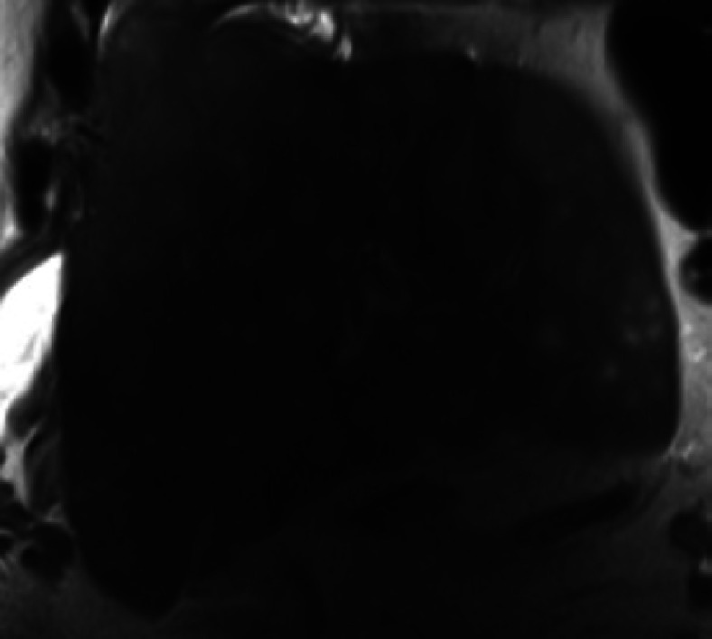
Diffuse T2 hyperintensive enhancing lesions throughout the liver.
He received vitamin K for elevated INR before ERCP. ERCP showed mild esophagitis, mild gastritis, and biliary sludge. No discrete mass was imaged. A biliary sphincterotomy was done, and a plastic stent was placed in the common bile duct. He then underwent a radiology assisted ultrasound-guided liver biopsy. Liver biopsy results showed a poorly differentiated small cell neuroendocrine tumor in the liver with a primary gastrointestinal versus pancreatic origin. Immune stains showed malignant cells diffusely positive for CAM5.2, CD56, and CDX-2 and negative for TTF-1 (shown in Fig. 2a-e). The further hospital course was complicated by acute kidney injury. His abdominal pain improved, but he continued to have worsening liver tests despite ERCP and stenting. Oncology was consulted and offered chemotherapy with carboplatin and etoposide versus comfort care. He ultimately decided on comfort care. He became lethargic in the next few days, likely secondary to hepatic encephalopathy. As he was in comfort care, blood work was not done for him. He was managed with analgesics and anxiolytics to keep him comfortable. While arrangements for home with home hospice were under process he expired before hospital discharge. His entire hospital course spanned 2 weeks.
Fig. 2.

a Liver biopsy showing poorly differentiated small cell neuroendocrine tumor. Hematoxylin and eosin stains of the core biopsy demonstrate malignant cells (left white arrow) infiltrating liver parenchyma (right black arrow). The malignant cells have enlarged nuclei with coarse chromatin, nuclear molding, and frequent mitoses (left white arrow), and scant cytoplasm. b Immune stains showing malignant cells diffusely positive for CAM5.2. c Immune stains showing malignant cells diffusely positive for CD56. d Immune stains showing malignant cells diffusely positive CDX-2. e Immune stains showing malignant cells diffusely positive for Synaptophysin.
Discussion/Conclusion
Poorly differentiated EPSCC is an extremely rare form of neuroendocrine carcinoma. It constitutes up to 5% of all small cell cancers and 1% of all gastrointestinal cancers [6]. The associated risk factor is cigarette smoking [7, 8]. It commonly arises from the gastrointestinal tract with the esophagus being the most common site. Other possible sites include the rectum, ileum, jejunum, pancreas, and gall bladder. Outside of the gastrointestinal system, there are reports that include the breast, prostate, cervix, bladder, head, and neck. Breast EPSCC has the best 3-year survival (30%), and gastrointestinal EPSCC has the worst (7%). The primary site cannot be identified in up to 30% of EPSCC patients. Symptoms associated with EPSCC are very variable and depend on the primary site location. Nausea, abdominal pain, and anorexia are common presentations for EPSCC with a gastrointestinal origin, whereas it can present as vaginal bleeding or hematuria if the primary is genitourinary. Being neuroendocrine in nature, in rare cases, it can present with paraneoplastic symptoms such as syndrome of inappropriate antidiuretic hormone and Lambert Eaton syndrome [2, 9]. Histopathologically, EPSCC is thought to arise from a multipotent stem cell that acquires neuroendocrine features. Diagnosis of EPSCC requires pathological identification of small cell carcinoma in biopsy cells and ruling out small cell lung cancers (SCLC). Immunohistochemistry stains can be helpful to identify the primary in some cases. Evaluation usually involves a CT chest, abdomen, pelvis, and positive emission tomography scan. MRI brain is not a part of routine investigation, unlike SCLC, as the incidence of brain metastasis is rare in EPSCC as compared to SCLC [4, 10].
EPSCC is staged as limited or extensive based on the radiation therapy field. Limited stage is defined as tumor burden that can be incorporated into one radiation treatment field, and extension is when it is beyond one radiation treatment field. Another opinion is to stage it the same way as its primary origin is historically staged [4].
Treatment depends on the stage at the time of presentation. Up to 50% have metastasized at the time of diagnosis. Common sites of metastases include liver, lymph nodes, bones, and bone marrow [11]. Multimodal therapy is the standard treatment protocol for limited disease and involves surgery, chemotherapy, or chemoradiation. Adjuvant therapy can also be added in some instances. Surgery alone is usually not curative. Extended survival can be achieved for limited disease with multimodal therapy. Multimodal therapy improves 2-year survival from 42% to 83%. For extensive disease, palliative chemotherapy can be considered but does not carry impressive outcomes. Survival, however, also depends on the histological site. For instance, breast EPSCC has the best survival whereas gastrointestinal has the worst survival of 6–12 months even after treatment [12, 13, 14]. First-line chemotherapy used is combination chemotherapy of cisplatin and etoposide for four cycles. Other alternative agents include carboplatin and irinotecan [13, 14, 15, 16].
This case presents an elderly male with abdominal pain and obstructive jaundice. His imaging was concerning for liver metastases with unknown primary. Further investigations including endoscopy, ERCP, ultrasound liver, CT chest abdomen pelvis, and MRI abdomen were deployed. ERCP did help in symptomatic relief; however, alkaline phosphatase and bilirubin continued to increase. Liver biopsy with immune stains narrowed the diagnosis of EPSCC to pancreatic versus gastrointestinal malignancy as primary. He was diffusely positive for CAM5.2, CD56, CDX-2, and Synaptophysin markers. CAM5.2 marker is seen in colorectal or pancreatic cancers, CD56 is seen in multiple myeloma, small cell lung cancer, neuroendocrine tumors, pancreatic cancers, lymphomas, and leukemias, CDX-2 is a very specific and sensitive marker for malignancies with intestinal origin [17]. Synaptophysin is sensitive and specific for neuroendocrine tumors and negative TTF-1 helped exclude SCLC [18]. Given no evidence or significant pathological abnormality noted in the above investigation, possible gastrointestinal sites concerning his primary site include colorectal and other gastrointestinal sites, except pancreas and esophagus. Further colonoscopy could be helpful in the diagnosis of primary site, but he refused further workup given the known poor prognosis and survival with his diagnosis. Unfortunately, he declined rapidly and passed away within 2 weeks of hospital admission.
Being a rare form of cancer, limited literature is available for EPSCC. Not many case reports are published. As per PubMed data using keywords extrapulmonary small cell carcinoma, case reports, a total of seven case reports were documented. Mostly referred to head and neck as primary and others mentioned, colon, rectum, and bladder as primary. Neither of the cases presented with obstructive jaundice. All the current recommendations for its management are based primarily on retrospective studies and institution-based expert opinions. More research, data, and prospective trials are needed to better understand EPSCC diagnosis, management, and treatment.
Statement of Ethics
Ethical approval is not required for this study in accordance with local or national guidelines. Written informed consent was obtained from the patient's next of kin for publication of the details of their medical case and any accompanying images.
Conflict of Interest Statement
The authors have no conflicts of interest to declare.
Funding Sources
No funding required.
Author Contributions
Ejaz Shah directly involved in patient care, reviewed the literature, and wrote the main body of the manuscript and discussion. Calvin Abro did the final review and editing of the case.
Data Availability Statement
All data generated or analyzed during this study are included in this article.
References
- 1.Remick SC, Hafez GR, Carbone PP. Extrapulmonary small-cell carcinoma. A review of the literature with emphasis on therapy and outcome. Medicine. 1987;66:457–71. [PubMed] [Google Scholar]
- 2.Wong YN, Jack RH, Mak V, Henrik M, Davies EA. The epidemiology and survival of extrapulmonary small cell carcinoma in South East England, 1970–2004. BMC Cancer. 2009;9:209. doi: 10.1186/1471-2407-9-209. [DOI] [PMC free article] [PubMed] [Google Scholar]
- 3.Haider K, Shahid RK, Finch D, Sami A, Ahmad I, Yadav S, et al. Extrapulmonary small cell cancer: a Canadian province's experience. Cancer. 2006 Nov 1;107((9)):2262–9. doi: 10.1002/cncr.22235. [DOI] [PubMed] [Google Scholar]
- 4.Berniker AV, Abdulrahman AA, Teytelboym OM, Galindo LM, Mackey JE. Extrapulmonary small cell carcinoma: imaging features with radiologic-pathologic correlation. Radiographics. 2015 Jan–Feb;35((1)):152–63. doi: 10.1148/rg.351140050. [DOI] [PubMed] [Google Scholar]
- 5.Theodros D, Goodwin CR, Crane GM, Liauw J, Kleinberg L, Lim M. Metastatic extrapulmonary small cell carcinoma to the cerebellopontine angle: a case report and review of the literature. Case Rep Oncol Med. 2015;2015:847058. doi: 10.1155/2015/847058. [DOI] [PMC free article] [PubMed] [Google Scholar]
- 6.Howard S, O'Regan K, Jagannathan J, Krajewski K, Giardino A, Ramaiya N. Extrapulmonary small cell carcinoma: a pictorial review. AJR Am J Roentgenol. 2011;197((3)):W392–8. doi: 10.2214/AJR.10.5757. [DOI] [PubMed] [Google Scholar]
- 7.Pervez N, El-Gehani F, Joseph K, Dechaphunkul A, Kamal M, Pertschy D, et al. Genitourinary small-cell carcinoma: a single-institution experience. Curr Oncol. 2013 Oct;20((5)):258–64. doi: 10.3747/co.20.1338. [DOI] [PMC free article] [PubMed] [Google Scholar]
- 8.Renner G. Small cell carcinoma of the head and neck: a review. Semin Oncol. 2007;34((1)):3–14. doi: 10.1053/j.seminoncol.2006.10.024. [DOI] [PubMed] [Google Scholar]
- 9.Walenkamp AM, Sonke GS, Sleijfer DT. Clinical and therapeutic aspects of extrapulmonary small cell carcinoma. Cancer Treat Rev. 2009;35((3)):228–36. doi: 10.1016/j.ctrv.2008.10.007. [DOI] [PubMed] [Google Scholar]
- 10.Strosberg JR, Coppola D, Klimstra DS, Phan AT, Kulke MH, Wiseman GA, et al. The NANETS consensus guidelines for the diagnosis and management of poorly differentiated (high-grade) extrapulmonary neuroendocrine carcinomas. Pancreas. 2010;39:799–800. doi: 10.1097/MPA.0b013e3181ebb56f. [DOI] [PMC free article] [PubMed] [Google Scholar]
- 11.Brenner B, Tang LH, Klimstra DS, Kelsen DP. Small-cell carcinomas of the gastrointestinal tract: a review. J Clin Oncol. 2004;22((13)):2730–9. doi: 10.1200/JCO.2004.09.075. [DOI] [PubMed] [Google Scholar]
- 12.Pignon JP, Arriagada R, Ihde DC, Johnson DH, Perry MC, Souhami RL, et al. A meta-analysis of thoracic radiotherapy for small-cell lung cancer. N Engl J Med. 1992;327:1618–24. doi: 10.1056/NEJM199212033272302. [DOI] [PubMed] [Google Scholar]
- 13.Hanna N, Bunn PA, Langer C, Einhorn L, Guthrie T, Beck T, et al. Randomized phase III trial comparing irinotecan/cisplatin with etoposide/cisplatin in patients with previously untreated extensive-stage disease small-cell lung cancer. J Clin Oncol. 2006 May 1;24((13)):2038–43. doi: 10.1200/JCO.2005.04.8595. [DOI] [PubMed] [Google Scholar]
- 14.Eckardt JR, von Pawel J, Papai Z, Tomova A, Tzekova V, Crofts TE, et al. Open-label, multicenter, randomized, phase III study comparing oral topotecan/cisplatin versus etoposide/cisplatin as treatment for chemotherapy-naive patients with extensive-disease small-cell lung cancer. J Clin Oncol. 2006 May 1;24((13)):2044–51. doi: 10.1200/JCO.2005.03.3332. [DOI] [PubMed] [Google Scholar]
- 15.Mitry E, Baudin E, Ducreux M, Sabourin JC, Rufié P, Aparicio T, et al. Treatment of poorly differentiated neuroendocrine tumours with etoposide and cisplatin. Br J Cancer. 1999 Dec;81((8)):1351–5. doi: 10.1038/sj.bjc.6690325. [DOI] [PMC free article] [PubMed] [Google Scholar]
- 16.Lassen U, Kristjansen PE, Osterlind K, Bergman B, Sigsgaard TC, Hirsch FR, et al. Superiority of cisplatin or carboplatin in combination with teniposide and vincristine in the induction chemotherapy of small-cell lung cancer. A randomized trial with 5 years follow up. Ann Oncol. 1996 Apr;7((4)):365–71. doi: 10.1093/oxfordjournals.annonc.a010603. [DOI] [PubMed] [Google Scholar]
- 17.Werling RW, Yaziji H, Bacchi CE, Gown AM. CDX2, a highly sensitive and specific marker of adenocarcinomas of intestinal origin: an immunohistochemical survey of 476 primary and metastatic carcinomas. Am J Surg Pathol. 2003 Mar;27((3)):303–10. doi: 10.1097/00000478-200303000-00003. [DOI] [PubMed] [Google Scholar]
- 18.Moldvay J, Jackel M, Bogos K, Soltész I, Agócs L, Kovács G, et al. The role of TTF-1 in differentiating primary and metastatic lung adenocarcinomas. Pathol Oncol Res. 2004;10((2)):85–8. doi: 10.1007/BF02893461. [DOI] [PubMed] [Google Scholar]
Associated Data
This section collects any data citations, data availability statements, or supplementary materials included in this article.
Data Availability Statement
All data generated or analyzed during this study are included in this article.