Abstract
A 16-year-old boy with elevated hyperopia presented to the office with a 24-h history of bilateral blurred vision, mainly of the left eye, and bilateral central serous chorioretinopathy. He showed a clinically recognizable bacillary layer detachment in one eye and excellent multimodal diagnostic image correlation, with the best-corrected visual acuity as 20/400. He had bilateral serous retinal detachment, as confirmed by optical coherence tomography. Laser photocoagulation was performed with good results, and reestablishment of the foveal anatomical structure was documented 16 days after treatment.
Keywords: Central serous chorioretinopathy, Bacillary layer detachment, High hyperopia, Optical coherence tomography
Introduction
Central serous chorioretinopathy (CSCR) is the fourth most common macular pathology, occurring typically in males between the second and fifth decades of life. Its most widely accepted pathophysiological explanation involves an increased choroidal hyperpermeability, secondary to congestion, ischemia, or local inflammation, and a secondary dysfunction of the retinal pigmentary epithelium. Its well-known risk factors include type A personality, elevated plasma levels of catecholamines and cortisol, alcohol consumption, antibiotics, pregnancy, Helicobacter pylori infection, sleep apnea, and arterial hypertension. At this time, we report a case of atypical CSCR with no known risk factors, associated with high hyperopia, with an acute, bilateral presentation and bacillary layer detachment in one eye.
Case Report
The patient was a 16-year-old male who complained of bilateral blurred vision, left eye greater than right, noticed within the previous 24 h. He had no significant personal or past medical history.
His ophthalmological examination revealed a best-corrected visual acuity of 20/20 OD (+6.00 D) and 20/400 OS (+6.50 D). Slit-lamp biomicroscopy was normal, and intraocular pressure was 14 mm Hg OU.
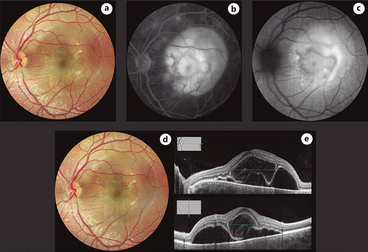
Fundus examination revealed a circular serous extrafoveal, superotemporal retinal detachment, of 1 disc diameter (DD), and a serous detachment centered around the fovea of 2 DD, in the right eye. The left eye revealed a 4-DD circular serous retinal detachment centered around the fovea, with well-defined margins, and inside of it, a second perifoveal border, limited by an ivory line with an irregular circular perimeter, interrupted by a wedge-shaped extension in its temporal margin (size: horizontal 3.390 μm; vertical 2.209 μm centered around the fovea) which created a clinically visible double-margin image, with the serous detachment border (Fig. 1). The axial length, anterior chamber depth, lens thickness, vitreous chamber length, and scleral thickness were measured using optical coherence tomography (OCT), enhanced depth imaging-OCT, and ultrabiomicroscopy (Table 1). OCT of the right eye showed a serous retinal detachment in the right eye, with central involvement and an extrafoveal, superotemporal serous detachment, with an adjacent focal retinal pigment epithelium detachment. The left eye showed a dome-shaped serous retinal detachment with a cystoid appearance (size: horizontal 3.391 μm; vertical 2.290 μm centered around the fovea), hyporeflective interiorly with abundant hypereflective granular images suspended inside, as well as adhered to its internal margin and to both sides of its external margin. At the extremes of the serous detachment, one could appreciate the division of the external retina and notice continuity between the external limiting membrane and ellipsoid zone which constitutes the internal and external limits of the cystoid area, respectively (Fig. 1).
Fig. 1.
a) Clinical picture of the posterior pole, b) corresponding fluorescein angiogram and c) autofluorescence image showing serous and bacillary layer detachment. Measurements of the bacillary layer detachment estimated in the fundus image: 3.39 × 2.29 mm (d) and OCT -horizontal (3,391 μm) and vertical (2,209 μm) diameters- (e), centered around the fovea.
Table 1.
Eye structure dimensions
| OD | OS | |
|---|---|---|
| Anterior chamber depth | 2.99 mm | 2.96 mm |
| Lens thickness | 4.16 mm | 4.14 mm |
| Axial length | 21.35 mm | 21.25 mm |
| Subfoveal choroidal thickness | 556 µm | >668 µm |
| Vitreous chamber length | 14.2 mm | 14.15 mm |
| Scleral thickness 2 mm from the scleral spur | 0.90 mm | 1.06 mm |
| Scleral thickness 6 mm from the scleral spur | 0.83 mm | 0.60 mm |
| Spherical equivalent | +6.00 D | +6.50 D |
Fluorescein angiography revealed in the early stages focal points of hyperfluorescence, nasal and superotemporal to the fovea in the right eye, and nasal to the fovea in the left eye, with leakage and pooling in late stages, that correspond to an area of greater intensity within the internal perimeter of the clinical image and an intermediate intensity corresponding to the external perimeter of the serous detachment (Fig. 1). The enhanced depth imaging-OCT showed congestion in the choriocapillaris registering subfoveal choroidal thickening of 556 μm in the right eye and 668 μm in the left eye.
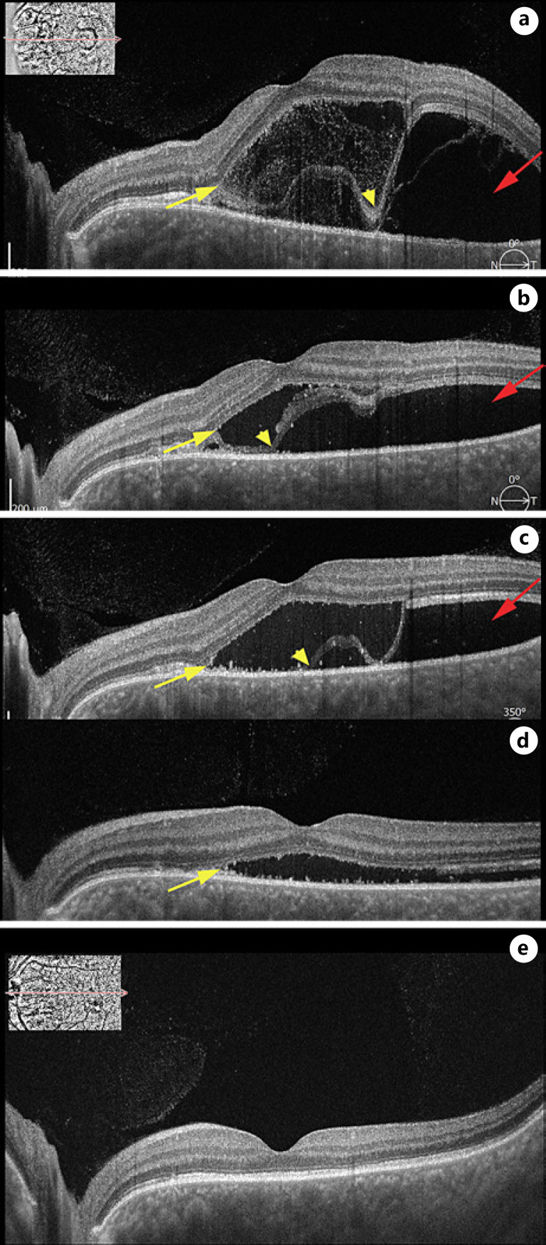
The patient was diagnosed with CSCR secondary to high hyperopia, and after informed consent was obtained, focal 532-nm green laser treatment was applied to the leakage points identified by fluorescein angiography, covering their surface, with a 250-μm spot size, 50 mW energy, and 200 ms duration. Reabsorption of subretinal fluid in both eyes was observed, at 48 h, and it was complete by day 12. In the left eye, reattachment preceded the resolution of the bacillary layer detachment, which was verified on day 16. Best-corrected visual acuity was 20/20 in both eyes, without any structural changes noted at 1-month follow-up (Fig. 2).
Fig. 2.
a–e Chronological images showing resolution of the serous retinal detachment (red arrows); dividing site in the photoreceptor layer (yellow arrows) and sequential images showing its gradual reunification (yellow arrowheads).
Discussion
Currently, neither the type or degree of refractive error nor the axial length is a recognized risk factor for CSCR. A review of the literature revealed CSCR to be more predominant among emmetropes, less common in hyperopes, and unusual in myopes [1]. Other studies however reflect a much higher proportion of CSCR in hyperopes [2]. Our patient would be considered a high hyperope, based on refractive error, although he had no clinical evidence or biometric measurements to suggest nanophthalmos or shortening restricted to the posterior segment as in posterior microphthalmos. A hyperopic eye however is not only associated with shorter axial length but also a smaller volume, greater compaction of scleral fibers, and scleral thickening, thus resulting in greater scleral rigidity [3]. A thickened and rigid sclera may result in a diminished return through the vortex veins, causing congestion and an elevation of hydrostatic pressure at the choriocapillaris, reducing protein transscleral flux, which in turn increases leakage, generating accumulation of liquid in the suprachoroidal space, secondary RPE dysfunction, and an exudative retinal detachment. Kishi et al. [4], support this theory by demonstrating an asymmetric venous return with a dominant vortex vein in 100% of patients with CSCR and pachychoroid pigment epitheliopathy, as well as areas of sluggish choroidal filling secondary to congestion of the dominant vortex that corresponds to those areas in patients with acute CSCR. Scleral thickness in our patient did not exceed normal values, and thus, the physical factor responsible for a diminished return of the vortex veins would be scleral rigidity alone. Determining the relationship between these two variables and establishing the level of vascular resistance generated in the venous system would contribute to clarify the pathophysiology of this and other diseases related to pachysclera.
Biometric characteristics of CSCR prove a direct relationship between a shorter axial length and increased thickness of the subfoveal choroid, as well as a shorter axial length in bilateral cases, compared with unilateral ones [5]. This decreased axial length may accentuate the choroidal dysfunction, given the shorter distance between the macula and the vortex vein ampullae, causing a steeper inclination and a shorter vascular path [5] since it has been shown that for each millimeter of axial length increase, there is a corresponding reduction of 0.6 mm Hg of pressure at the level of the retinochoroidal arterial tree and a consequent reduction of hydrostatic pressure in the choroid vascular bed [1].
Spontaneous resolution is always a possible outcome of CSCR; however, it usually involves a period of several weeks or even months [2]. On the other hand, the clinical course of this particular case marked by the extension of the serous detachment and the compromise of visual acuity contributed to treatment decision. CSCR rapid and progressive improvement, both anatomical and functional, started 48 h after treatment, suggesting a correlation of this response with the applied therapeutic effect.
The fundus image of the left eye in our patient showed an ivory, perifoveal margin located within the perimeter of the serous detachment that constituted the margin of the bacillary layer detachment, clearly delineated by the pooling of dye in the late phases of the angiogram and correlating in size and morphology with the structural OCT image.
Funduscopic round images with yellowish margins underlying an acute exudative detachment in patients with VKH and the corresponding accumulation of a multilobular pattern in the late phases of the FA were the original descriptions of these intraretinal fluid spaces that Ishihara et al. [6] suggested to be a separation between the inner and outer segments of photoreceptors. Similarly, Ouyang et al. [7], analyzing a patient with toxoplasmosis using a spectral-domain OCT, described a large cystoid space at the level of the external retina which contained hyperreflective material and was limited externally by a continuity hyperreflective line connecting with the ellipsoid zone [8]. This tomographic characteristic, also noted by Liakopoulos et al. [9] in a case of exudative age-related macular degeneration as possible delamination of photoreceptors and more recently labeled as a bacillary layer detachment by Mehta et al. [8], in a case of toxoplasmosis and pachychoroid is with increasing certainty understood as a division in the photoreceptor layer within the internal segment myoid, recognized histologically as the zone with the highest structural fragility and potentially divisible. In our patient, the layer division became evident since the external limit of the cystoid space was not in apposition with the RPE, allowing us to individualize its contour and establish continuity with the ellipsoid zone. Similarly, follow-up during the reattachment phase allowed us to observe the progressive apposition of its surface over the RPE, correlating to the reabsorption of the serous detachment, and final structural resolution, signed by the reunification of the photoreceptor layer.
Detachment of the bacillary layer has been reported recently, associated with different pathologies resulting in a hyperacute choroidal exudation, including posterior scleritis, systemic lupus erythematosus, toxicity secondary to anti-BRAF/anti-MEK therapy in melanoma, neovascularization secondary to choroidal osteoma, pulmonary and breast choroidal metastases [10], trauma [11], and peripapillary pachychoroid syndrome [12]. To our knowledge, this is the first report of a bacillary layer detachment in a case of CSCR in the absence of inflammatory disease, neoplasm, or associated trauma that was attributed to significant choroidal thickening and a sequence of events secondary to high hyperopia, capable of producing by themselves, a hyperacute accumulation of subretinal fluid, big enough to induce this unusual delaminating effect in the retina under the influence of hydrodynamic forces.
Conclusion
Elevated hyperopia, even in the absence of nanophthalmos or posterior microphthalmos, should be considered a risk factor for CSCR. Measuring certain biometric parameters in these eyes, including thickness and scleral rigidity, should be part of these patients' workup.
Bacillary layer detachment is a clinically recognizable entity, which shows excellent multimodal diagnostic image correlation and can be present not only as a result of hyperacute choroidal exudation associated with inflammatory events but also due to congestion and choroidal hyperpermeability linked to a circulatory abnormality.
Statement of Ethics
Written informed consent was obtained from the patient's parent for publication of the details of their medical case and any accompanying images. This study was reviewed and approved by our institutional Ethics Committee − Comité de ética Clínica del Ojo. Internal code: 2021CR12 MP.
Conflict of Interest Statement
No conflicting relationship exists for any author.
Funding Sources
No funding was provided for this study.
Author Contributions
Sergio A. Murillo: design of the work, writing process, and analysis and interpretation of results.
Silvia P. Medina: data and image acquisition, figures elaboration, and analysis of results.
Rosa Maria Romero: writing process and interpretation of results.
Fernando H. Murillo: writing process, translation, and interpretation of results.
Data Availability Statement
All data generated or analyzed during this study are included in this article. Further inquiries can be directed to the corresponding author.
References
- 1.Manayath GJ, Arora S, Parikh H, Shah PK, Tiwari S, Narendran V. Is myopia a protective factor against central serous chorioretinopathy? Int J Ophthalmol. 2016;9((2)):266–70. doi: 10.18240/ijo.2016.02.16. [DOI] [PMC free article] [PubMed] [Google Scholar]
- 2.Dalai R, Rout R, Pradhan M, Nanda P. Clinical study of central serous chorioretinopathy presenting in a tertiary care centre. Int J Res Med Sci. 2019 Nov;7((11)):4200–5. [Google Scholar]
- 3.Roberts CJ, Reinstein DZ, Archer TJ, Mahmoud AM, Gobbe M, Lee L. Comparison of ocular biomechanical response parameters in myopic and hyperopic eyes using dynamic bidirectional applanation analysis. J Cataract Refract Surg. 2014;40:929–36. doi: 10.1016/j.jcrs.2014.04.011. [DOI] [PubMed] [Google Scholar]
- 4.Kishi S, Matsumoto H, Sonoda S, Hiroe T, Sakamoto T, Akiyama H. Geographic filling delay of the choriocapillaris in the region of dilated asymmetric vortex veins in central serous chorioretinopathy. PLoS One. 2018;13((11)):e0206646. doi: 10.1371/journal.pone.0206646. [DOI] [PMC free article] [PubMed] [Google Scholar]
- 5.Terao N, Koizumi H, Kojima K, Kusada N, Nagata K, Yamagishi T, et al. Short axial length and hyperopic refractive error are risk factors of central serous chorioretinopathy. Br J Ophthalmol. 2020;104((9)):1260–5. doi: 10.1136/bjophthalmol-2019-315236. [DOI] [PubMed] [Google Scholar]
- 6.Ishihara K, Hangai M, Kita M, Yoshimura N. Acute Vogt-Koyanagi-Harada disease in enhanced spectral-domain optical coherence tomography. Ophthalmology. 2019;Sep((116 9)):1799–1807. doi: 10.1016/j.ophtha.2009.04.002. [DOI] [PubMed] [Google Scholar]
- 7.Ouyang Y, Pleyer U, Shao Q, Keane PA, Stübiger N, Joussen AM, et al. Evaluation of cystoid change phenotypes in ocular toxoplasmosis using optical coherence tomography. PLoS One. 2014;Feb((92)) doi: 10.1371/journal.pone.0086626. [DOI] [PMC free article] [PubMed] [Google Scholar]
- 8.Mehta N, Chong J, Tsui E, Duncan JL, Curcio CA, Freund KB, et al. Presumed foveal bacillary layer detachment in a patient with toxoplasmosis chorioretinitis and pachychoroid disease. Retin Cases Brief Rep. 2018;10:1097. doi: 10.1097/ICB.0000000000000817. [DOI] [PubMed] [Google Scholar]
- 9.Liakopoulos S, Keane PA, Ristau T, Kirchhof B, Walsh AC, Sadda SR. Atypical outer retinal fluid accumulation in choroidal neovascularization: a novel OCT finding. Ophthalmic Surg Lasers Imaging Retina. 2013;44((6 Suppl)):S11–8. doi: 10.3928/23258160-20131101-03. [DOI] [PubMed] [Google Scholar]
- 10.Cicinelli MV, Giuffré C, Marchese A, Jampol LM, Introini U, Miserocchi E, et al. The bacillary detachment in posterior segment ocular diseases. Ophthalmol Retina. 2020;4((4)):454–6. doi: 10.1016/j.oret.2019.12.003. [DOI] [PubMed] [Google Scholar]
- 11.Tekin K, Teke MY. Bacillary layer detachment: a novel optical coherence tomography finding as part of blunt eye trauma. Clin Exp Optom. 2019;102((3)):343–4. doi: 10.1111/cxo.12876. [DOI] [PubMed] [Google Scholar]
- 12.Ramtohul P, Comet A, Denis D. Bacillary layer detachment in peripapillary pachychoroid syndrome. Ophthalmol Retina. 2020;4((6)):587. doi: 10.1016/j.oret.2020.01.017. [DOI] [PubMed] [Google Scholar]
Associated Data
This section collects any data citations, data availability statements, or supplementary materials included in this article.
Data Availability Statement
All data generated or analyzed during this study are included in this article. Further inquiries can be directed to the corresponding author.


