Abstract
Soft tissue lipoma is one of the most common benign tumors rarely forming in hand. In this study, 11 cases of symptomatic hand lipoma were investigated. The chief complaint was a palpable mass in all 11 patients, out of whom 6 (55%) cases presented with neurological symptoms, including paresthesia and numbness in the median, ulnar, and superficial radial nerve pathways. One patient had an intramuscularly painful forearm with a large mass presentation. In the finger, the clinical manifestations were radial anesthesia of the finger. The mass sizes were above 5 cm3 and less than 5 cm3 in seven and four patients, respectively. The mean follow-up period was 25 months. No patient demonstrated a recurrence during the follow-up period. Although lipoma is a benign tumor and often presents itself as a palpable mass in the hand, it can cause neurological symptoms and decreased function. Regardless of the size of the tumor, mass removal can prevent symptoms
Key Words: Carpal tunnel syndrome, Hand, Lipoma, Microsurgery
Introduction
Soft tissue lipoma is one of the most common benign tumors rarely forming in the hand, accounting for about 1%-3.8% of benign tumors of the hand (1,2). It is most common in the thenar and hypothenar regions but can rarely occur in Parona’s space. Lipoma originates from preadipocyte mesenchymal cells. This benign tumor can have a progressive enlargement nature; moreover, due to the density of neurovascular elements in the hand, it can result in symptoms and functional disorders. It is considered a giant lipoma with a size greater than 50 mm, and removal is recommended (1,2). In addition, malignant sarcomatous transformation is possible but infrequent (1,2). The present study reported symptomatic hand lipomas.
Materials and Methods
This descriptive study was conducted on patients with a possible diagnosis of lipoma in 15 Khoradad and Imam Khomeini hospitals affiliated with Shahid Beheshti University of Medical Sciences and Urmia University of Medical Sciences, respectively, from 2015 to 2020. All patients underwent magnetic resonance imaging (MRI) before surgery. Demographic findings, anatomical location of the tumor, tumor size, duration of formation, clinical manifestations, and recurrence were investigated. The confirmation of the diagnosis in all cases was based on histopathological findings, such as the presence of stained mature fat cells (hematoxylin and eosin; magnification, x200), atypia, mitosis, or necrosis. All patients were followed up for at least 12 months. This study was conducted under the supervision of the Ethics Committee of Shahid Beheshti University of Medical Sciences.
Results
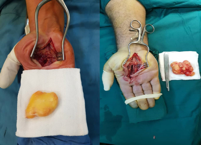
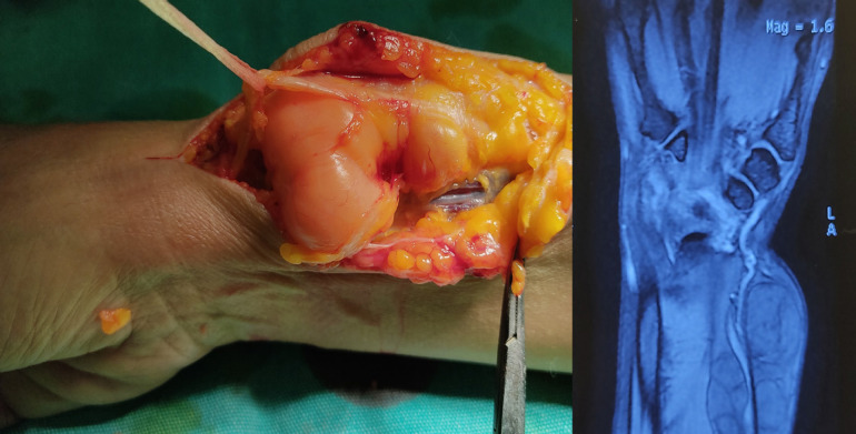
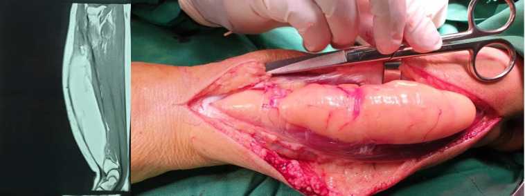
A total of 11 patients were enrolled, including 8 (72.7%) men and 3 (27.3%) women. The mean age of the patients was 46±13 years. In terms of anatomical location, the mass was in the following locations: 4 (36.3%) in thenar [Figure 1], 2 (18.1%) in hypothenar [Figure 2], 1 (9.09%) in volar index finger surface [Figure 3], 3 (27.2%) in the forearm with symptoms of paresthesia or decreased hand function, and 1 (9.09%) in the wrist. The chief complaint was a palpable mass in all patients, out of whom six (55%) cases presented with some degree of neurological involvement due to the effect of mass on the adjacent nerve [Figure 4]. One patient presented with a painful and large mass intramuscularly in the forearm [Figure 5]. The clinical symptom in the finger was radial side numbness. Other findings are reported separately in Table 1. Moreover, the mass sizes were above 5 cm3 and less than 5 cm3 in seven and four patients, respectively. The mean follow-up period was 25 months. No patient demonstrated a recurrence during the follow-up period, and in all patients, the neurological symptoms resolved after the mass excision. The diagnosis of lipoma was confirmed in all patients based on the histopathological findings. There was no cellular atypia and necrosis.
Figure 1.
Intraoperative thenar lipoma in two patients presenting with carpal tunnel syndrome
Figure 2.
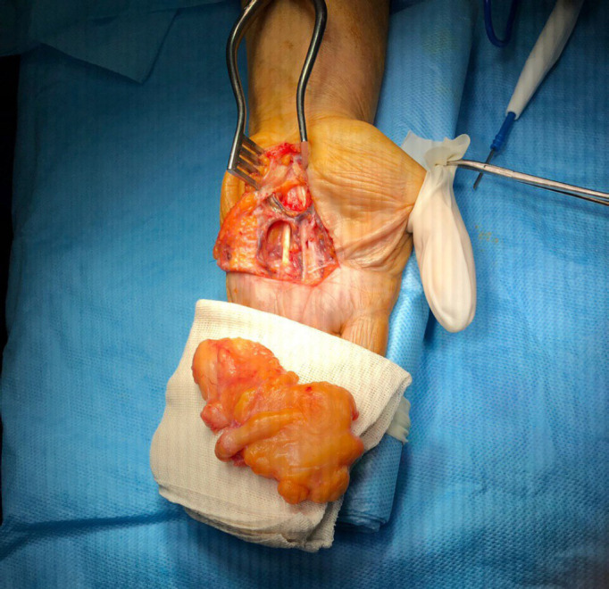
Intraoperative hypothenar lipoma presenting with an ulnar nerve compression neuropathy
Figure 3.
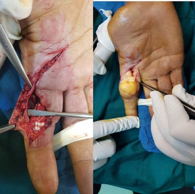
Lipoma of the index finger with digital nerve compression symptom
Figure 4.
Superficial radial nerve compression due to lipoma
Figure 5.
Intermuscular lipoma of the forearm with swelling and achiness
Table 1.
Patients characteristics with symptomatic lipoma
| Patient No | Age | Sex | Location | Presenting symptoms | Tumor size | Time of involvement | Follow up | Recurrence |
|---|---|---|---|---|---|---|---|---|
| 1 | 45 | M | Thenar, near to carpal tunnel | Palpable mass/Numbness and paresthesia in median nerve | 4.5×2×2cm3 | 2years | 24 | No |
| 2 | 48 | M | Radial side of forearm with compression of superficial radial nerve | Palpable mass/ Numbness and paresthesia in superficial radial nerve | 10×3.5×5cm3 | 3years | 12 | No |
| 3 | 31 | F | Volar of wrist in radial side | Palpable mass only | 3×2×2cm3 | 1.5year | 24 | No |
| 4 | 83 | M | Large mass in dorsal intermuscular extensor of forearm | Swelling, aching with flexion and extension of fingers | 5×4.5×3cm3 | 3years | 36 | No |
| 5 | 53 | M | Thenar with fixed to common digital nerve of thumb, index and long finger | Palpable mass/ Numbness and paresthesia in median nerve | 4.5×3×1.5cm3 | 2years | 24 | No |
| 6 | 62 | M | Thenar near to carpal tunnel and above the retinaculum flexor | Painless mass | 2×1.5×1.5cm3 | 2years | 12 | No |
| 7 | 43 | F | Not fixed in hypothenar | Painless mass | 2 ×2×1.5cm3 | 1.5year | 24 | No |
| 8 | 46 | F | Volar of forearm in intermuscular of flexor | Swelling, aching in flexion | 3.5×2×2cm3 | 2year | 48 | No |
| 9 | 45 | M | Thenar near to proximal crease | Palpable mass/ Numbness and paresthesia in median nerve | 4.5×3×2cm3 | a year | 36 | No |
| 10 | 54 | M | Hypothenar near to ulnar nerve at bifurcation of motor and sensory branches |
Palpable mass/ Numbness and paresthesia in superficial branch of ulnar nerve | 3×1.5×1 cm3 | a year | 36 | No |
| 11 | 25 | M | Proximal of index phalanx in volar side with fixed to radial digital nerve and artery of index finger |
Palpable mass/ Numbness and tenderness in digital nerve | 1.5×1×1cm3 | 2 years | 24 | No |
Discussion
Lipoma is one of the benign tumors originating from the mesenchymal soft tissue. It is usually encapsulated and infiltrating (1, 3). According to Bocchiotti et al., the cases of giant lipoma larger than 50 mm were more likely to be symptomatic (1). Nonetheless, in the present study, neurological symptoms were noted even with small lipomas depending on the location and depth of the tumor. The difference between lipomas of the hand and other anatomical areas is that they can involve multiple compartments in the hand and are multi-compartmental; therefore, they can cause a wide range of symptoms (1). Hand lipomas grow slowly and, in most cases, are asymptomatic and present with a palpable and mobile mass. The primary etiology and pathogenesis of its occurrence are not fully understood. Genetic factors, trauma, and metabolic factors have been suggested in lipoma. Unlike other body areas, it is recommended to remove this tumor in the hand to prevent such complications as sensory damage and hand function due to decreased grip strength (1-4). In 5% of cases, depending on the location, it can cause neurological symptoms in the hand and forearm and induce compression neuropathy symptoms. Vascular compression and subsequent distal ischemia have not been reported (5). Neurological symptoms are not related to the size of the tumor, and the position of the tumor relative to the nerve plays a vital role in the development of neurological symptoms. According to Chen et al., out of 779 patients with carpal tunnel syndrome (CTS), three cases were due to lipoma (6).
In the present study, there were neurological symptoms in 6 out of 11 (55%) patients with symptomatic lipoma of the hand and forearm. There was also superficial radial nerve involvement, in addition to previous reports of median and ulnar neurologic symptoms. Brand and Gelberman have identified deep palmar lipoma as one of the factors leading to CTS (7). Lipoma is much rarer in the finger. In four cases, it was reported in the index, thumb, middle, and third fingers, and there was no difference between the fingers in the likelihood of its occurrence (8-11). Another study presented a case of thumb lipoma with a size of more than 50 mm in a 5-year-old boy who presented with mass and discomfort (9). In the fingers, the clinical symptoms can vary from a palpable mass to motion restriction. In the report of Ramirez-Montaño et al., on the volar side, the third finger decreased range of motion in the interphalangeal joint was noted with a mass larger than 50 mm (11). As illustrated in Table 2, reports of cases marked in hand lipoma can be observed based on keyword search. The majority of clinical presentations have been reported as a CTS.
Table 2.
Reports of symptomatic lipoma in the hand
| Report by | Location and size | Clinical Manifestation | Time of follow up- Recurrence |
|---|---|---|---|
| Bocchiotti et al.(1) | Dorsal left hand 9×10×8.5mm |
a prominent swelling, discomfort | - |
| Kim et al.(2) | the left hypothenar area 8×5×2cm |
tingling sensation and paresthesia | 6 month- No |
| Sbai et al.(5) | Volar side of wrist in carpal tunnel 2.5 x 1.5 x 1 cm |
Median nerve compression neuropathy( CTS) | 2 month- |
| Hu et al.(8) | middle finger 35×15mm |
painless swelling and mass with distal interphalangeal joint was limited range of motion | - |
| Kamra et al.(9) | Right thumb 5 × 4 × 2.5 cm |
diffuse swelling, mass and discomfort | - |
| Chronopoulos et al.(10) | Right index finger 2 × 1 × 1 cm |
Difficulty in manual movements | 24 months- No |
| Ramirez-Montaño et al.(11) | The third finger 50 × 20 mm |
limited interphalangeal joint movement | 12 months- No |
| Pagonis et al.(12) | Hand and deep palmar 8.0 × 4.0 × 3.75 cm |
Median nerve compression neuropathy( CTS) | 3 month- |
| Papakostas et al.(13) | Lipoma of the Thenar 4.9 ×3.7×2.9 cm |
Median nerve compression neuropathy( CTS) | 12 months- No |
| Chatterton et al.(14) | Lipoma of the Thenar and thumb 8×6×3 cm |
an extremely large inconvenient swelling of the palm | No |
| Ribeiro et al.(15) | the palmar side of the left hand 6.5×6×6cm |
paresthesia and pain in the first three fingers of the left hand and CTS | 5 month-No |
| Fnini et al.(16) | the hand and palm 10×8cm |
Median nerve compression neuropathy( CTS) | - |
| Sonoda et al.(17) | The volar of wrist 4.8×2.5 |
Median nerve compression neuropathy( CTS) | 2 month-No |
| Berlund et al. (18) | The volar of wrist 1.5×1×1cm |
Median nerve compression neuropathy( CTS) | No |
| Imai et al.(19) | Intrasynovial lipoma of wrist | trigger wrist and carpal tunnel syndrome(CTS) | No |
In a study by Nada et al. conducted on 13 cases of hand lipoma, the most common clinical manifestations were mass, swelling, pain, weakness, and decreased performance (20). There was no recurrence after the removal of the tumor, and symptoms resolved after the removal. In the study by Leffert et al., out of 141 cases, upper extremity lipoma was asymptomatic in 109 cases and symptomatic in 32 cases, out of whom 26 cases presented with pain and tenderness, while neurological symptoms, including paresthesia and nerve dysfunction, were observed in 6 cases (21).
In 1853, Paget first introduced a type of intermuscular lipoma in the trapezius muscle (22), and Regan et al. then introduced the term infiltrating lipoma (23). Greenberg et al. stated that this type of lipoma could be intermuscular or intramuscular (24). In general, lipomas can be superficially located or deep-seated. The type of superficial location is more common and involves the subcutaneous fat, whereas lipomas, which are called deep-seated lipomas, may be localized deep under the enclosing fascia. The deep lesions are mature fat lesions that may occur in intermuscular (affecting the intermuscular connective tissue) or intramuscular locations (intramuscular lipomas are deep-seated lipomas that originate within the muscle).
The intermuscular and some of the intramuscular lipomas will grow by expansion and enclosure of other structures rather than infiltration (24-26). Intermuscular or intramuscular type of lipoma is often observed in the limbs. In terms of anatomical distribution, most reports were in the thigh and forearm (26, 27). In one of our patients, lipoma was detected in the forearm intramuscularly, which caused a painful mass. In a report by Liu et al., intramuscular lipoma has been more common in middle-aged men, with the most common presentation in the thigh as a painless mass. Nevertheless, it can be associated with clinical symptoms in the forearm and cause pain or dysfunction (26). Tumor removal can be helpful to prevent subsequent complications in these patients (26,27). It is essential to use appropriate preoperative imaging techniques to determine the extent of the involvement. The MRI is an assisted imaging modality with high detection and differentiation, as well as high signal intensity on T1 and T2 weighted images (22,27).
Hand lipoma can present with neurological symptoms, and regardless of the size of the tumor, mass removal can relieve symptoms.
Conflicts of Interest:
The authors declare that they have no conflict of interest to be reported.
References
- 1.Bocchiotti MA, Lovati AB, Pegoli L, Pivato G, Pozzi A. A case report of multi-compartmental lipoma of the hand. Case Reports Plast Surg Hand Surg. 2018;5(1):35–38. doi: 10.1080/23320885.2018.1469988. [DOI] [PMC free article] [PubMed] [Google Scholar]
- 2.Kim KS, Lee H, Lim DS, Hwang JH, Lee SY. Giant lipoma in the hand: A case report. Medicine (Baltimore). 2019;98(52):e18434. doi: 10.1097/MD.0000000000018434. [DOI] [PMC free article] [PubMed] [Google Scholar]
- 3.Ramirez-Montaño L, Lopez RP, Ortiz NS. Giant lipoma of the third finger of the hand. Springerplus. 2013;2(1):164. doi: 10.1186/2193-1801-2-164. [DOI] [PMC free article] [PubMed] [Google Scholar]
- 4.Chatterton BD, Moores TS, Datta P, Smith KD. An exceptionally large giant lipoma of the hand. BMJ Case Rep. 2013:2013:bcr2013200206. doi: 10.1136/bcr-2013-200206. [DOI] [PMC free article] [PubMed] [Google Scholar]
- 5.Sbai MA, Benzarti S, Msek H, Boussen M, Khorbi A. Carpal tunnel syndrome caused by lipoma: a case report. Pan Afr Med J. 2015;22:51. doi: 10.11604/pamj.2015.22.51.7650. [DOI] [PMC free article] [PubMed] [Google Scholar]
- 6.Chen CH, Wu T, Sun JS, Lin WH, Chen CY. Unusual causes of carpal tunnel syndrome: space occupying lesions. The Journal of hand surgery. 2012;37:14–9. doi: 10.1177/1753193411414352. [DOI] [PubMed] [Google Scholar]
- 7.Brand MG, Gelberman RH. Lipoma of the flexor digitorum superficialis causing triggering at the carpal canal and median nerve compression. J Hand Surg Am. 1988;13(3):342–4. doi: 10.1016/s0363-5023(88)80005-4. [DOI] [PubMed] [Google Scholar]
- 8.Hu Z, Yue Z, Tang Y, Zhu Y. Lipoma of the middle finger: A case report and review of literature. Medicine (Baltimore). 2017;96(42):e8309. doi: 10.1097/MD.0000000000008309. [DOI] [PMC free article] [PubMed] [Google Scholar]
- 9.Kamra HT, Munde SL. Lipoma on palmar aspect of thumb: a rare case report. J Clin Diagn Res. 2013;7(8):1706–7. doi: 10.7860/JCDR/2013/5683.3261. [DOI] [PMC free article] [PubMed] [Google Scholar]
- 10.Chronopoulos E, Nikolaos P, Karanikas C, Kalliakmanis A, Plessas S, Neofytou I, et al. Patient presenting with lipoma of the index finger: a case report. Cases J. 2010;3:20. doi: 10.1186/1757-1626-3-20. [DOI] [PMC free article] [PubMed] [Google Scholar]
- 11.Ramirez-Montaño L, Lopez RP, Ortiz NS. Giant lipoma of the third finger of the hand. Springerplus. 2013;2(1):164. doi: 10.1186/2193-1801-2-164. [DOI] [PMC free article] [PubMed] [Google Scholar]
- 12.Pagonis T, Givissis P, Christodoulou A. Complications arising from a misdiagnosed giant lipoma of the hand and palm: a case report. J Med Case Rep. 2011;5:552. doi: 10.1186/1752-1947-5-552. [DOI] [PMC free article] [PubMed] [Google Scholar]
- 13.Papakostas T, Tsovilis AE, Pakos EE. Intramuscular Lipoma of the Thenar: A Rare Case. Arch Bone Jt Surg. 2016;4(1):80–2. [PMC free article] [PubMed] [Google Scholar]
- 14.Chatterton BD, Moores TS, Datta P, Smith KD. An exceptionally large giant lipoma of the hand. BMJ Case Rep. 2013;2013:bcr2013200206. doi: 10.1136/bcr-2013-200206. [DOI] [PMC free article] [PubMed] [Google Scholar]
- 15.Ribeiro G, Salgueiro M, Andrade M, Fernandes VS. Giant palmar lipoma - an unusual cause of carpal tunnel syndrome. Rev Bras Ortop. 2017;52(5):612–615. doi: 10.1016/j.rboe.2017.08.001. [DOI] [PMC free article] [PubMed] [Google Scholar]
- 16.Fnini S, Hassoune J, Garche A, Rahmi M, Largab A. Giant lipoma of the hand: case report and literature review. Chirurgie de la Main. 2009;29(1):44–7. doi: 10.1016/j.main.2009.11.006. [DOI] [PubMed] [Google Scholar]
- 17.Sonoda H, Takasita M, Taira H, Higashi T, Tsumura H. Carpal tunnel syndrome and trigger wrist caused by a lipoma arising from flexor tenosynovium: a case report. J Hand Surg Am. 2002;27(6):1056–8. doi: 10.1053/jhsu.2002.36522. [DOI] [PubMed] [Google Scholar]
- 18.Berlund P, Kalamaras M. A case report of trigger wrist associated with carpal tunnel syndrome caused by an intramuscular lipoma. Hand Surg. 2014;19(2):237–9. doi: 10.1142/S0218810414720174. [DOI] [PubMed] [Google Scholar]
- 19.Imai S, Kodama N, Matsusue Y. Intrasynovial lipoma causing trigger wrist and carpal tunnel syndrome. Scand J Plast Reconstr Surg Hand Surg. 2008;42(6):328–30. doi: 10.1080/02844310701282104. [DOI] [PubMed] [Google Scholar]
- 20.Nadar MM, Bartoli CR, Kasdan ML. Lipomas of the hand: a review and 13 patient case series. Eplasty. 2010;10:e66. [PMC free article] [PubMed] [Google Scholar]
- 21.Leffert RD. Lipomas of the upper extremity. J Bone Joint Surg Am. 1972;54(6):1262–6. [PubMed] [Google Scholar]
- 22.McTighe S, Chernev I. Intramuscular lipoma: a review of the literature. Orthop Rev (Pavia) 2014;6(4):5618. doi: 10.4081/or.2014.5618. [DOI] [PMC free article] [PubMed] [Google Scholar]
- 23.Regan JM, Bickel WH, Broders AC. Infiltrating benign lipomas of the extremities. West J Surg. 1946;54:87–93. [PubMed] [Google Scholar]
- 24.Greenberg SD, Isensee C, Gonzalez-Angulo A, Wallace SA. Infiltrating lipomas of the thigh. Am J Clin Pathol. 1963;39:66–72. doi: 10.1093/ajcp/39.1.66. [DOI] [PubMed] [Google Scholar]
- 25.Lahrach K, el Kadi KI, Mezzani A, Marzouki A, Boutayeb F. An unusual case of an intramuscular lipoma of the biceps brachii. Pan Afr Med J. 2013;15:40. doi: 10.11604/pamj.2013.15.40.2654. [DOI] [PMC free article] [PubMed] [Google Scholar]
- 26.Balabram D, Cabral CCdSR, Filho OdPR, Barros CPd. Intramuscular lipoma of the subscapularis muscle. Sao Paulo Med J. 2014;132:65–7. doi: 10.1590/1516-3180.2014.1321537. [DOI] [PMC free article] [PubMed] [Google Scholar]
- 27.Liu DR, Li C, Chen L. Management of giant intermuscular lipoma of hips: A case report and review of literature. Mol Clin Oncol. 2013;1(2):369–372. doi: 10.3892/mco.2013.63. [DOI] [PMC free article] [PubMed] [Google Scholar]


