Abstract
Background:
This study describes a minimally invasive technique for the reconstruction of the medial collateral ligament (MCL) and posterior oblique ligament (POL) through minimal incisions on the tibial and femoral sides of the ligament using the modified Bosworth technique.
Methods:
This study included 19 consecutive patients who presented with chronic grade III injury; the mean age was 29.6 years (standard deviation ± 7.5 years, range 19–43 years), and five patients (26.3%) had no associated injuries. Ten patients (52.6%) had associated anterior cruciate ligament (ACL) injury and four patients (21.1%) had associated posterior cruciate ligament (PCL) injury. All patients were assessed 18 months postoperatively regarding functional outcome using the Lysholm score and medial joint space opening.
Results:
There was a statistically significant improvement in the patient functional outcome as the Lysholm score improved from 55.39 ± 6.9 to 89.42 ± 6.4 at 18 months postoperatively. (P< 0.001). At the end of the follow-up, 16 cases had grade 1 medial laxity, 3 cases with grade II laxity, and no patients with grade III medial laxity.
Conclusion:
Minimally invasive MCL reconstruction with modified Bosworth technique gives very good results regarding the functional outcome and residual medial laxity of the knee.
Key Words: nee, edial collateral ligament, inimally invasive techniques, osterior oblique ligament, econstruction
Introduction
The medial collateral ligament (MCL) injury is the most common injury to the knee accounting for about 40% of knee injuries (1). The incidence of MCL injuries is 0.24/1000, with a 2:1 male to female ratio (2). In athletes, the incidence increases to 7.3/1000 population (3). The main medial static stabilizers of the knee are the superficial MCL, deep MCL, and posterior oblique ligament (POL) (4).
The MCL lies in the second layer in the medial aspect of the knee, the femoral attachment lies 1-mm proximal and 37 mm posterior to the medial femoral epicondyle, the distal bony attachment is located just anterior to the posteromedial crest of the tibia 42–71 mm from the tibial joint line. The superficial MCL is the main medial static stabilizer of the knee all over the range of motion (4-6).
The deep MCL is a thickening of the joint capsule. It consists of two distinct components: the meniscofemoral ligament is distal and deep to the femoral attachment of the superficial MCL approximately 6-mm distal and 5-mm posterior to the medial epicondyle; the meniscotibial portion, which is shorter and thicker, attaches just distal to the edge of the articular surface of the medial tibial plateau (5,6).
The femoral attachment of the POL extends from the posterosuperior aspect of superficial attachment of MCL to the gastrocnemius tubercle and is divided into three components, superficial, capsular, and most importantly, the central arm (5-7). The central arm arises from the main tendon of the semimembranosus, directly attached to the posterior joint capsule and posterior meniscus, and blends its attachment on the tibia 5-mm below the tibial plateau (5,8). The POL is a valgus stabilizer when the knee is extended and has a role in maintaining rotational stability of the knee, especially in PCL-deficient knees (9).
The incidence of concomitant ligamentous injury with grade 3 MCL injury is about 80%, most of which are associated with anterior cruciate ligament (ACL) injury (10,11).
MCL is known for its good healing potential rather than intracapsular ligaments, so nonoperative treatment is the rule for treatment of MCL injuries in grades 1 and 2 and selected cases of grade 3 injuries (12, 13). Failed nonoperative treatment in grade 3 injuries leads to deterioration in knee function and subsequent osteoarthritis in 63% of cases after 10 years (14).
Several techniques have been described for MCL reconstruction however, no evidence supports one technique over the others (15).
This study aims to describe the minimally invasive reconstruction of the MCL of the knee and to evaluate the functional outcome and medial joint space opening 18 months postoperatively.
Materials and Methods
Twenty-one consecutive patients with chronic grade 3 MCL injury treated using minimally invasive MCL reconstruction from January 2016 to December 2018 have been enrolled in the case series. Two cases discontinued follow-up, which brings the total number of cases to 19. All patients consented to participate in the study and signed written informed consent. The study was approved by the local scientific committee before starting the first case.
The inclusion criteria included chronic (more than 6 weeks) grade 3 medial instability of the knee as an isolated injury or with a single ligament injury needing reconstruction in skeletally mature populations without vascular injury or significant arthritic changes in plain radiographs.
The exclusion criteria included an associated recent fracture in the lower extremity, skeletally immature patients, knee injury with multiple ligaments (3 or more), cases of knee malalignment, patients with chronic laxity requiring osteotomy, revision cases, and patients with knee osteoarthritis grade 2 or more.
Statistical analysis
Data were analyzed using Statistical Program for Social Science version 15.0. Quantitative data were expressed as mean ± SD after confirmation of its normal distribution. Qualitative data were expressed as frequency and percentage. P<0.05 was statistically significant.
Surgical technique
The indication for surgery is chronic grade 3 MCL injury according to the Hughston classification (17) after confirmation of injury using clinical, MRI, X-ray, and stress valgus views [Figure 1].
Figure 1.
A X-ray knee Anteroposterior view in resting position
B stress X-ray in full extension
C Stress X-ray at a 30-degree flexion shows a significant opening of the medial joint space.
The background of this technique follows Kim et al.’s modification of the Bosworth technique (18, 19).
Surgery is performed under general or regional anesthesia. The graft used for other ligament reconstructions (ACL and PCL) is contralateral hamstring tendon autograft in all cases.
In the supine position and with the use of a tourniquet, which is applied as high on the thigh as possible, examination under anesthesia has been performed for all cases, and stress valgus has been done at 0º and 30º.
Arthroscopy has been performed to confirm the diagnosis and to evaluate the state of the articular cartilage. All concomitant procedures have been performed, including meniscal repair or meniscectomy, ACL or PCL tunnel position, and femoral graft fixation; tibial fixation to be done after complete MCL reconstruction has been executed.
A 4–5-cm vertical skin incision has been done for the harvesting of the semitendinosus tendon [Figure 2]. In association with ACL/PCL reconstruction, the tibial tunnel was planned to be medial to the pes anserinus, and care is taken to avoid injury during reaming of the tibial tunnel.
Figure 2.
The skin incision with subcutaneous dissection
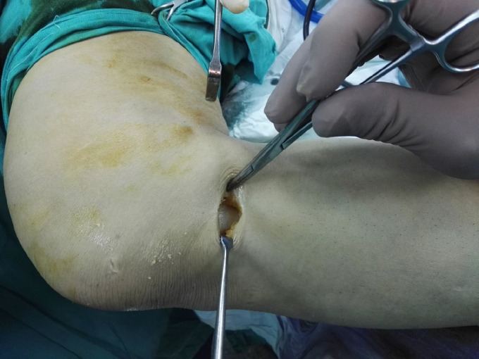
Care should be taken during harvesting of the semitendinosus tendon, as premature harvesting may abort the whole procedure. Careful dissection of the tendon with sectioning of the connection bands, especially the band connecting to the medial head of the gastrocnemius [Figure 3]. After harvesting the tendon proximally using the tendon stripper, the proximal edge of the tendon was cleared off of muscle tissue and stitched using baseball sutures.
Figure 3.
Identification of the semitendinosus tendon and the connection with the medial head of the gastrocnemius
The tendon was passed deep into the gracilis tendon to ensure that the location of the graft is as close as possible to its anatomical location [Figure 4].
Figure 4.
The graft passes underneath the gracilis tendon to ensure the location of the graft in layer 3
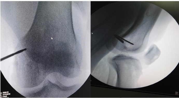
A similar incision centrally located on the medial femoral epicondyle has been made. A deep dissection that reached the medial femoral epicondyle was made. The femoral tunnel is made under fluoroscopic control as the entry point is performed posterosuperior to the medial femoral epicondyle and confirmed by anteroposterior and lateral fluoroscopic images, as described by Wijdicks CA et al. (20) [Figure 5].
Figure 5.
Fluoroscopic identification of the entry point in AP and lateral views
Care should be taken to avoid tunnel convergence, especially in combined MCL and PCL reconstruction, so the direction of the femoral tunnel should be far from the previously done tunnels, and to avoid this, the tunnel is directed superiorly.
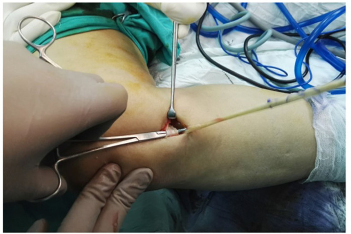
A curved hemostat is used for making the track in which the graft will pass from the tibia to the femur. The tip of the hemostat should be directly rested on the bone, sometimes due to fibrous tissue formation, then the graft is delivered to the femoral window [Figure 6]. Isometry of the graft is tested all over the range of motion by looping the tendon on the guidewire and moving the leg. After confirmation of isometry, the femoral tunnel (30–40 mm) is drilled using the appropriate femoral reamer, a suture loop is used to engage the middle of the graft in the femoral tunnel, and the tension of the graft is kept by the assistant. Once an extra length for the graft to reconstruct the POL has been reached, a biodegradable interference screw is inserted in the femoral tunnel, while the knee is flexed at 30ºwith axial loading and varus stress, and the assistant exerts tension on the suture loop [Figure 7].
Figure 6.
Delivery of the graft to the femoral window
Figure 7.
The position of the knee before fixation of the MCL with varus load and flexion of 30º while maintaining tension on the free edge of the graft
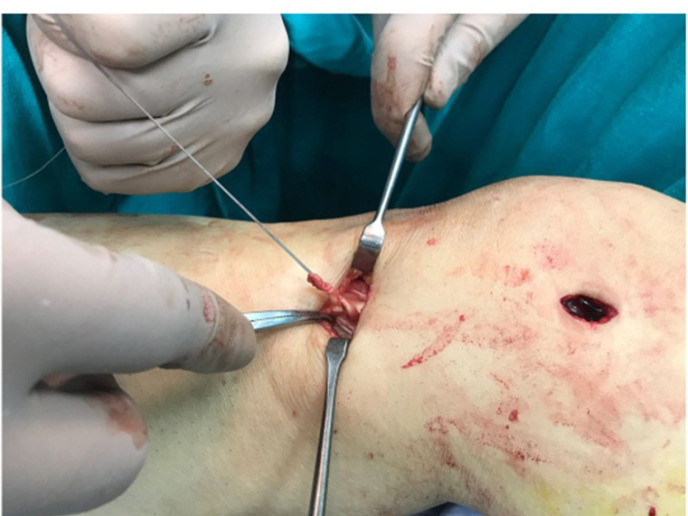
After confirmation of the stability of the MCL, the rest of the graft is pulled back through the tibial soft tissue window to be sutured to the direct arm of the semimembranosus in full extension using non-absorbable sutures. [Figure 8].
Figure 8.
Delivery of the graft to the tibial window before fixation in the direct arm of semimembranosus for POL reconstruction
The range of motion and medial stability is confirmed, and the procedure is finalized. The tourniquet is then released, and hemostasis and wound closure are performed. [Figure 9].
Figure 9.
The incision needed for the whole procedure
Postoperatively, a hinged knee brace is used, and the patient is allowed to touch the ground with two crutches. The patient is allowed to flex the knee at 30º in the first two weeks, and the range is increased gradually. The brace is removed at 6–8 weeks according to patient compliance, and patients were instructed to avoid pivoting for 16 weeks. The milestones of rehabilitation continue under the supervision of the rehabilitation team with monitoring of the surgeon in regular outpatient clinic visits. The rehabilitation is tailored according to the concomitant surgeries performed.
Results
All patients who enrolled in this case series were subjected to initial clinical and radiographic evaluation.
The mean age was 29.6 years (standard deviation (SD) ± 7.5 years, range 19–43 years), and there were 15 males and four females (3.75:1 ratio). All patients were presented with chronic (more than 6 weeks) grade 3 MCL injury. The mean duration of surgery since injury was 11.2 ± 3 weeks, and five patients (26.3%) had isolated injuries, 10 patients (52.6%) had associated ACL injury, and four patients (21.1%) had associated posterior cruciate ligament (PCL) injury [Table 1].
Table 1.
Demographic data
| CharacteRISTICS | Value |
|---|---|
| Age (years) | |
| Minimum | 19 |
| Maximum | 43 |
| Mean ( SD) | 29,63 (±7.5) |
| Gender | |
| Males | 15 |
| Females | 4 |
| Associated injuries | |
| Isolated injury | 5 (26.3%) |
| ACL+MCL | 10 (52,6%) |
| PCL+MCL | 4 (21,1%) |
| Duration (weeks) | |
| Minimum | 7 |
| Maximum | 17 |
| Mean ( SD) | 11,15 (2,98) |
Magnetic resonance imaging (MRI) evaluation of the MCL was in accordance with the classification proposed by Makhmalbaf and Shahpari (16). Type 2 (proximal) injury was detected in 8 patients, type 3 (midsubstance) injury in 10 patients, and type 4 (distal) injury was found in 1 case.
There were eight medial meniscal injuries (42.1%), four of them were associated with an ACL injury, two cases with PCL injuries, and two cases with isolated MCL injury [Table 1]. All of these were treated by all-inside meniscal repair.
Logistic regression analysis found no difference between cases with and without meniscal injuries.
The mean preoperative Lysholm score was 55.37 (SD ± 6.89, range 44–67) [Table 2].
Table 2.
preoperative and postoperative Lysholm scores
| Total | Minimum | Maximum | Mean | SD | P-value | |
|---|---|---|---|---|---|---|
| preoperative Lysholm | 19 | 44,00 | 67,00 | 55,3684 | 6,88969 | |
| postoperative Lysholm | 19 | 78,00 | 98,00 | 89,4211 | 6,40586 | <0.001 |
A regular evaluation was done for all patients who have been enrolled in this study during their outpatient clinic appointments.
There was a statistically significant improvement in patient function, assessed by the Lysholm score, as it improved from 55.39 ± 6.9 to 89.42 ± 6.4 24 months postoperatively. (P-value < 0.001) [Table 2].
At the final follow-up evaluation (18 months postoperatively), the mean final postoperative Lysholm score was 89.42 ((SD ± 6,4, range 78–98). The clinical examination shows that 16 patients show grade I laxity, three patients (15.8% ) had grade II laxity, and no patients in the grade III laxity according to Hughston classification (16). To minimize information bias, all patients were evaluated by two other surgeons aside from the operating surgeon [Table 3].
Table 3.
Post-operative medial laxity
| Frequency | Percent | ||
|---|---|---|---|
| Medial laxity | Grade I (0-5mm) |
16 | 84,2 |
| Grade II (5-10mm) |
3 | 15,8 | |
| Grade III (≥ 10 mm) |
0 | 0 | |
| Total | 19 | 100,0 | |
There were no major complications (deep or superficial infection, deep vein thrombosis), although three patients complained of tingling at the tibial surgical site, which improved during follow-up. Two patients had limited flexion (110º–120º) with a full extension (one had combined ACL/MCL reconstruction and the other with PCL/MCL reconstruction) without significant complaint. No patients had a failed procedure that required revision at the end of follow-up.
Discussion
MCL and POL reconstruction aims to restore valgus and rotational stability of the knee. The study shows a marked improvement outcome of the knee at 18 months postoperatively. The study included 19 patients, with isolated injury in 4 cases (21%), combined MCL and PCL injury in 5 cases (26%), and combined MCL and ACL injury in 10 cases (52.5%). This means that 74% of cases had an associated ligamentous injury.
In a systematic review by Varelas et al., the isolated MCL reconstruction cases were 48 over 275 cases (17.5%), taking into consideration that cases of multi-ligament injury were included (21). When cases with multi-ligament injury have been excluded, their incidence of isolated MCL reconstruction was 19.67%, which means that isolated reconstruction is not a common procedure, and the rule is combined injury.
As the healing probability of MCL is confirmed thanks to the presence of epiligament, as first described by Bray et al., and confirmed in the human knee by Georgiev GP et al., such epilegament is formed; the shorter the distance between both ends of the tear, the more stable and reliable the healing of the ligament will be (22-24).
In a biomechanical study comparing Bosworth with modified Bosworth with single reconstruction and modified Bosworth with double-bundle MCL reconstruction using computer-assisted navigation, modified Bosworth was found to be comparable to double-bundle reconstruction in the restoration of the valgus stability as well as external rotation stability of the knee at 0º and 30º; however, modified Bosworth technique is a more economic and less time-consuming procedure, especially when considering that most cases of medial instability of the knee require an additional surgical procedure (ligament or meniscal injury) (25).
The technique has the advantages of being economic (one interference screw is needed rather than four screws in anatomical reconstruction), not technically demanding, and leads to less scarring of the knee, which is important in chronic cases of osteoarthritis who may require arthroplasty.
However, there are limitations of this study: the small sample size as the injury is not commonly treated surgically and the confounding effect of associated ligament reconstruction. However, the isolated MCL injury requiring reconstruction is very rare in clinical scenarios and it has been minimized by excluding multiple injuries (three or more ligament injuries). Furthermore, the follow-up time did not enable us to determine the risk of osteoarthritis; thus, a longer follow-up is needed with a larger sample size and more focus on an isolated chronic MCL injury.
In conclusion, the minimally invasive MCL reconstruction with modified Bosworth technique gives very good results regarding the functional outcome and residual medial laxity of the knee with less knee scarring and could be a good option, especially when associated with another ligament reconstruction.
Competing interests:
The authors declare that they have no conflict of interest.
Funding:
The authors didn’t get a fund from a third party or institution or any organization or company.
References
- 1.Wijdicks CA, Griffith CJ, Johansen S, Engebretsen L, LaPrade RF. Injuries to the medial collateral ligament and associated medial structures of the knee. JBJS. 2010;92(5):1266–80. doi: 10.2106/JBJS.I.01229. [DOI] [PubMed] [Google Scholar]
- 2.Roach CJ, Haley CA, Cameron KL, Pallis M, Svoboda SJ, Owens BD. The epidemiology of medial collateral ligament sprains in young athletes. The American journal of sports medicine. 2014;42(5):1103–9. doi: 10.1177/0363546514524524. [DOI] [PubMed] [Google Scholar]
- 3.LaPrade RF, Engebretsen AH, Ly TV, Johansen S, Wentorf FA, Engebretsen L. The anatomy of the medial part of the knee. JBJS. 2007;89(9):2000–10. doi: 10.2106/JBJS.F.01176. [DOI] [PubMed] [Google Scholar]
- 4.Warren LF, Marshall JL, Girgis F. The prime static stabilizer of the medial side of the knee. JBJS. 1974;56(4):665–74. [PubMed] [Google Scholar]
- 5.Athwal KK, Willinger L, Shinohara S, Ball S, Williams A, Amis AA. The bone attachments of the medial collateral and posterior oblique ligaments are defined anatomically and radiographically. Knee surgery, sports traumatology, arthroscopy. 2020;28(12):3709–19. doi: 10.1007/s00167-020-06139-6. [DOI] [PMC free article] [PubMed] [Google Scholar]
- 6.Griffith CJ, LaPrade RF, Johansen S, Armitage B, Wijdicks C, Engebretsen L. Medial knee injury: part 1, static function of the individual components of the main medial knee structures. The American journal of sports medicine. 2009;37(9):1762–70. doi: 10.1177/0363546509333852. [DOI] [PubMed] [Google Scholar]
- 7.Hughston JC, Eilers AF. The role of the posterior oblique ligament in repairs of acute medial (collateral) ligament tears of the knee. JBJS. 1973;55(5):923–40. [PubMed] [Google Scholar]
- 8.Tibor LM, Marchant Jr MH, Taylor DC, Hardaker Jr WT, Garrett Jr WE, Sekiya JK. Management of medial-sided knee injuries, part 2: posteromedial corner. The American journal of sports medicine. 2011;39(6):1332–40. doi: 10.1177/0363546510387765. [DOI] [PubMed] [Google Scholar]
- 9.Petersen W, Loerch S, Schanz S, Raschke M, Zantop T. The role of the posterior oblique ligament in controlling posterior tibial translation in the posterior cruciate ligament-deficient knee. The American journal of sports medicine. 2008;36(3):495–501. doi: 10.1177/0363546507310077. [DOI] [PubMed] [Google Scholar]
- 10.Daniel KCM, Stone ML, Hirshman P. The incidence of knee ligament injuries in the general population. American journal of knee surgery. 1991;4:3–8. [Google Scholar]
- 11.Fetto JF, Marshall JL. Medial collateral ligament injuries of the knee: a rationale for treatment. Clinical orthopaedics and related research. . 1978;132:206–18. [PubMed] [Google Scholar]
- 12.Zhang J, Pan T, Im HJ, Fu FH, Wang JH. Differential properties of human ACL and MCL stem cells may be responsible for their differential healing capacity. BMC medicine. 2011;9(1):68. doi: 10.1186/1741-7015-9-68. [DOI] [PMC free article] [PubMed] [Google Scholar]
- 13.Chen L, Kim PD, Ahmad CS, Levine WN. Medial collateral ligament injuries of the knee: current treatment concepts. Current reviews in musculoskeletal medicine. 2008;1(2):108–13. doi: 10.1007/s12178-007-9016-x. [DOI] [PMC free article] [PubMed] [Google Scholar]
- 14.Kannus PE. Long-term results of conservatively treated medial collateral ligament injuries of the knee joint. Clinical orthopaedics and related research. . 1988;(226):103–12. [PubMed] [Google Scholar]
- 15.Bonasia DE, Bruzzone M, Dettoni F, Marmotti A, Blonna D, Castoldi F, et al. Treatment of medial and posteromedial knee instability: indications, techniques, and review of the results. The Iowa orthopaedic journal. 2012;32:173. [PMC free article] [PubMed] [Google Scholar]
- 16.Makhmalbaf H, Shahpari O. Medial collateral ligament injury; a new classification based on MRI and clinical findings A guide for patient selection and early surgical intervention. Archives of bone and joint surgery. 2018;6(1) [PMC free article] [PubMed] [Google Scholar]
- 17.Hughston JC. The importance of the posterior oblique ligament in repairs of acute tears of the medial ligaments in knees with and without an associated rupture of the anterior cruciate ligament Results of long-term follow-up. The Journal of bone and joint surgery. American volume. 1994;76(9):1328–44. doi: 10.2106/00004623-199409000-00008. [DOI] [PubMed] [Google Scholar]
- 18.Kim SJ, Lee DH, Kim TE, Choi NH. Concomitant reconstruction of the medial collateral and posterior oblique ligaments for medial instability of the knee. The Journal of bone and joint surgery. British volume. 2008;90(10):1323–7. doi: 10.1302/0301-620X.90B10.20781. [DOI] [PubMed] [Google Scholar]
- 19.Bosworth DM. Transplantation of the semitendinosus for repair of laceration of medial collateral ligament of the knee. JBJS. 1952;34(1):196–202. [PubMed] [Google Scholar]
- 20.Wijdicks CA, Griffith CJ, LaPrade RF, Johansen S, Sunderland A, Arendt EA, et al. Radiographic identification of the primary medial knee structures. JBJS. 2009;91(3):521–9. doi: 10.2106/JBJS.H.00909. [DOI] [PubMed] [Google Scholar]
- 21.Varelas AN, Erickson BJ, Cvetanovich GL, Bach Jr BR. Medial collateral ligament reconstruction in patients with medial knee instability: a systematic review. Orthopaedic journal of sports medicine. 2017;5(5):2325967117703920. doi: 10.1177/2325967117703920. [DOI] [PMC free article] [PubMed] [Google Scholar]
- 22.Bray RC, Fisher AW, Frank CB. Fine vascular anatomy of adult rabbit knee ligaments. Journal of anatomy. 1990;172:69–79. [PMC free article] [PubMed] [Google Scholar]
- 23.Georgiev GP, Iliev A, Kotov G, Kinov P, Slavchev S, Landzhov B. Light and electron microscopic study of the medial collateral ligament epiligament tissue in human knees. World journal of orthopedics. 2017;8(5):372–8. doi: 10.5312/wjo.v8.i5.372. [DOI] [PMC free article] [PubMed] [Google Scholar]
- 24.Loitz-Ramage BJ, Frank CB, Shrive NG. Injury size affects the long-term strength of the rabbit medial collateral ligament. Clinical orthopaedics and related research®. 1997;337:272–80. doi: 10.1097/00003086-199704000-00031. [DOI] [PubMed] [Google Scholar]
- 25.Feeley BT, Muller MS, Allen AA, Granchi CC, Pearle AD. Biomechanical comparison of medial collateral ligament reconstructions using computer-assisted navigation. The American journal of sports medicine. 2009;37(6):1123–30. doi: 10.1177/0363546508331134. [DOI] [PubMed] [Google Scholar]


