Abstract
Purpose
This study aimed to compare the treatment outcomes of radiofrequency ablation (RFA) and stereotactic body radiation therapy (SBRT) for colorectal cancer liver metastases (CRLM) and to determine the favorable treatment modality according to tumor characteristics.
Materials and Methods
We retrospectively analyzed the records of 222 colorectal cancer patients with 330 CRLM who underwent RFA (268 tumors in 178 patients) or SBRT (62 tumors in 44 patients) between 2007 and 2014. Kaplan-Meier method and Cox models were used by adjusting with inverse probability of treatment weighting (IPTW).
Results
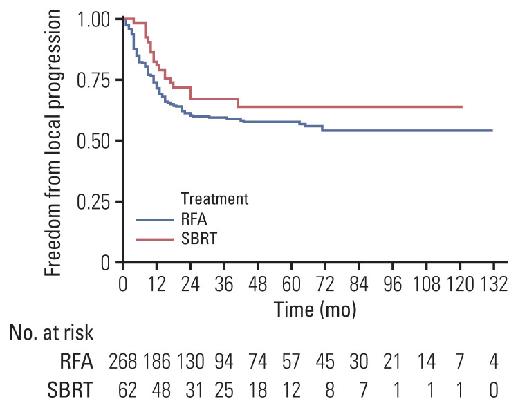
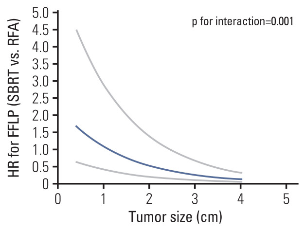
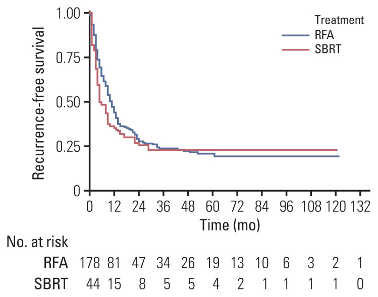
The median follow-up duration was 30.5 months. The median tumor size was significantly smaller in the RFA group than in the SBRT group (1.5 cm vs 2.3 cm, p < 0.001). In IPTW-adjusted analysis, difference in treatment modality was not associated with significant differences in 1-year and 3-year recurrence-free survival (35% vs. 43%, 22% vs. 23%; p=0.198), overall survival (96% vs. 91%, 58% vs. 56%; p=0.508), and freedom from local progression (FFLP; 90% vs. 72%, 78% vs. 60%; p=0.106). Significant interaction effect between the treatment modality and tumor size was observed for FFLP (p=0.001). In IPTW-adjusted subgroup analysis of patients with tumor size > 2 cm, the SBRT group had a higher FFLP compared with the RFA group (hazard ratio, 0.153; p < 0.001).
Conclusion
SBRT and RFA showed similar local control in the treatment of patients with CRLM. Tumor size was an independent prognostic factor for local control and SBRT may be preferred for larger tumors.
Keywords: Radiofrequency ablation, Stereotactic body radiation therapy, Colorectal cancer liver metastases, Prognosis
Introduction
Colorectal cancer is the third most common type of malignancy worldwide, and liver metastases develop in as much as 50% of the patients with colorectal cancer during the disease course [1,2]. Surgery is the standard treatment in patients with resectable colorectal liver metastases (CRLM) and has been shown to result in 5-year survival rates of up to 60% [3]. Unfortunately, only 20% to 30% of patients with CRLM are amenable to surgery [3,4]. As such, different types of nonsurgical approaches such as radiofrequency ablation (RFA) and stereotactic body radiation therapy (SBRT) have been attempted for tumor ablation in such patients [3,5–8].
RFA is the most frequently used method and has been well-validated [9,10]. Two randomized controlled trials from the EORTC (European Organisation for Research and Treatment of Cancer) showed that RFA resulted in comparable local control rates to surgical resection in patients with CRLM [10,11]. However, larger tumor size and particular tumor locations such as subcapsular proximity to extrahepatic vital organs or perivascular area have been reported to be associated with higher rates of local recurrence after RFA [12–14]. Additionally, RFA is not suitable in tumors located in poorly visible areas on ultrasonography [15].
Recently, SBRT has become a valuable addition to the nonsurgical approaches for hepatic metastatic tumors including CRLM [5–7]. According to two prospective studies, SBRT provided 2-year local control rates of over 90% in patients with liver metastases from various primary malignancies [7,16]. Another recent retrospective comparative study showed that RFA and SBRT had similar local control rates for intrahepatic metastases that are < 2 cm in size, while SBRT showed superior results in lesions ≥ 2 cm [17]; however, this study did not exclusively analyze CRLM but included liver metastases from various primary cancer as well. Likewise, there is a paucity of studies that solely analyzed patients with CRLM for comparing the treatment outcomes between RFA and SBRT. In addition, given the differences between the two treatment modalities, we suppose that it would be meaningful to investigate the tumor- and patient-related characteristics that favor one treatment over the other.
Therefore, we aimed to compare the outcomes of RFA and SBRT for the treatment of CRLM and identify the important factors affecting the treatment outcomes.
Materials and Methods
1. Study population
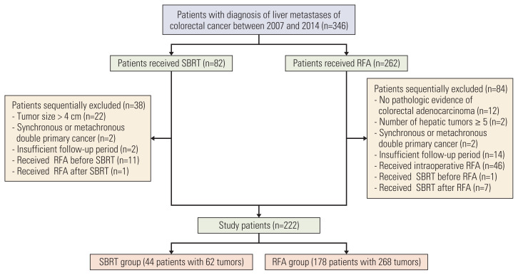
We included patients who received RFA or SBRT for CRLM between January 2007 and December 2014 at Asan Medical Center (Seoul, Korea) and met the following inclusion criteria: (1) confirmed with primary colorectal adenocarcinoma based on pathologic evidence; (2) Eastern Cooperative Oncology Group score 0–2; and (3) more than three months of follow-up after treatment. The exclusion criteria were as follows: (1) evidence of synchronous or metachronous double primary cancer other than colorectal cancer within five years of the initial diagnosis; (2) five or more treated liver lesions; (3) received intraoperative RFA; and (4) received both SBRT and RFA for CRLM (Fig. 1).
Fig. 1.
Flow diagram of patient selection. RFA, radiofrequency ablation; SBRT, stereotactic body radiation therapy.
2. RFA or SBRT treatments
All patients who underwent RFA or SBRT were considered to have unresectable CRLM because of insufficient liver remnant, proximity to critical structures, prohibitive comorbidity, or patient refusal. RFA was performed percutaneously under ultrasonographic guidance by one of the five radiologists at Asan Medical Center, all of whom had more than 8 years of experience in RFA. The tumors were ablated by using an internally cooled electrode system, which was either a single-type with a 3-cm active tip (Cool-tip RF System, Covidien, Mansfield, MA) or a cluster-type (Cluster, RF Medical Co. Ltd., Seoul, Korea) depending on the size and location of the tumor. In order to deliver maximum power with an automatic impedance control method, radiofrequency current was emitted for 12 to 15 minutes by a 200 W generator set. The end point was complete ablation of the visible tumor along with at least a 0.5–1.0 cm margin of the normal liver parenchyma surrounding the tumor. Immediately after the ablation, all patients underwent contrast-enhanced computed tomography (CT) for a final evaluation of the ablation and detection of potential complications. If residual tumors were observed on the immediate post-RFA imaging, repeat ablation was attempted on the same day or the following day.
Details on the SBRT procedure used at our institution have been described in our previous study [15]. Briefly, each patient was immobilized using a vacuum cushion and pillow in a supine position during the SBRT procedure. By using a four-dimensional respiratory gating system, an intravenous contrast-enhanced CT (GE LightSpeed RT 16, GE Healthcare, Waukesha, WI) scan was obtained with a thickness of 2.5-mm. The four-dimensional CT images were synchronized with the respiratory data sorted into 10 CT series in conformity with the respiratory phase by using the Real-time Position Management gating system (Varian Medical Systems, Palo Alto, CA).
Contouring for the gross tumor volume (GTV) was performed in end-expiratory phase CT images by referring to the liver dynamic CT, magnetic resonance imaging (MRI), and positron emission tomography-CT. The clinical target volume was set to be equal to the GTV, and the internal target volume (ITV) was expanded to reflect the movement of the tumor resulting from respiration. The planning target volume was set by a 5-mm margin on ITV.
SBRT planning was developed using a 3-dimensional radiotherapy planning system (Eclipse, Varian Medical Systems) that used the 4 to 8 coplanar/non-coplanar beams or volumetric-modulated arc therapy technique, which were produced by a linear accelerator (TrueBeam STx, Varian Medical Systems) with energies of 6 to 15 MV.
A total dose of 36 to 60 Gy was delivered in three to five fractions to the isodose line covering the planning target volume. In the planning of SBRT, the attending physician decided on the final prescribed dose in accordance with the normal tissue constraint dose. For image guidance, the cone-beam CT was carried out in each treatment and a linear accelerator was used to administer the radiation.
3. Follow-up and outcomes
All patients were followed up using hepatic imaging, which was initiated approximately 3 months after the treatment; generally, hepatic imaging was carried out in 3-month intervals using CT and/or MRI. The therapeutic outcomes were compared between the RFA group and the SBRT group by assessing the freedom from local progression (FFLP), recurrence-free survival (RFS), and overall survival (OS). FFLP was defined as the time from the date of RFA or SBRT initiation until the detection of local progression of treated tumor in follow-up CT or MRI according to the Response Evaluation Criteria in Solid Tumors criteria. RFS was defined as the time from the date of RFA or SBRT initiation until the date of intrahepatic or extrahepatic metastasis in follow-up CT or MRI. The OS was calculated from the date of RFA or SBRT initiation to the date of death. The treatment-related complication was graded using the Common Terminology Criteria for Adverse Events (CTCAE) ver. 5.0. Non-classic radiation-induced liver disease (nRILD) was defined as an elevation in liver transaminase or bilirubin within 3-months after SBRT, and was graded according to the CTCAE ver. 5.0. Difficult locations of tumors were defined as those within 1 cm of the hilum or main portal vein, liver surface, hepatic dome, or gallbladder.
4. Statistical analysis
In the comparison of patient characteristics between the RFA and SBRT groups, categorical variables were analyzed using the chi-square test and continuous variables were analyzed using the Mann-Whitney U-test. The RFA and SBRT groups were compared at the tumor level using FFLP and at the patient level using RFS or OS. By using the Cox proportional hazards regression model, univariate and multivariable analyses were performed to identify the potential factors affecting FFLP, RFS, and OS. To find the prognostic factors, we used a stepwise variable selection for multivariable analysis. The interaction of treatment modality and the tumor size was evaluated using the cox proportional hazard model with interaction term. We applied the inverse probability of treatment weighting (IPTW) method to the Cox models and Kaplan-Meier method to adjust for the imbalances in potential confounders in the treatment modalities. For IPTW adjustment, the weights for patients in the SBRT group were the inverse of propensity score (PS), and the weights for patients in the RFA group were the inverse of 1-PS. The PS was the probability of receiving SBRT, which was estimated by using multiple logistic regression analysis. To evaluate the estimated propensity score, the Hosmer-Lemeshow goodness of fit test and c-statistic were conducted as well as the balance check of each covariate after IPTW adjustment (S1 Table). The multivariable analysis included all covariates that were used for the calculation of propensity scores. All statistical analyses were performed using SAS ver. 9.4 (SAS Institute, Cary, NC). p-values < 0.05 were considered statistically significant.
Results
1. Patient characteristics
A total of 222 patients with 330 lesions were included in the study, whose age (mean±standard deviation) was 60.6±10.7 years and 161 (72.5%) were men (Table 1). Of the 222 patients, 178 (80.2%) patients with 268 (81.2%) metastases received RFA (RFA group) and 44 (19.8%) patients with 62 (18.8%) metastases were treated with SBRT (SBRT group). Out of the 86 patients with synchronous liver metastasis, 60 received pre-treatment systemic therapy followed by resection for colorectal cancer and the remaining patients underwent primary resection for colorectal cancer. The RFA group had a higher proportion of patients with metachronous liver metastasis than did the SBRT group (65.2% vs. 45.4%, p=0.016). The median size of liver tumors in the RFA group was significantly smaller than that in the SBRT group (1.5 vs. 2.3 cm, p < 0.001). The median SBRT dose was 48 Gy (range, 36 to 60 Gy) and the median biologically effective dose (BED) was 112.5 Gy10 (range, 68.4 to 180 Gy10). The details of the SBRT regimens used for each tumor are provided in S2 Table. After IPTW adjustment, the absolute standardized difference was less than 0.1 for all variables (S1 Table).
Table 1.
Patient characteristics
| Characteristic | RFA (n=178) | SBRT (n=44) | p-value |
|---|---|---|---|
| Age (yr) | |||
| ≤ 70 | 144 (80.9) | 36 (81.8) | 0.89 |
| > 70 | 34 (19.1) | 8 (18.2) | |
| Mean±SD | 60.8±10.9 | 60.3±9.9 | 0.80 |
| Male sex | 130 (73.0) | 31 (70.5) | 0.73 |
| ECOG score ≤ 1 | 177 (99.4) | 44 (100) | > 0.99 |
| Primary tumor location | |||
| Left colona) | 41 (23.0) | 7 (15.9) | 0.20 |
| Right colonb) | 29 (16.3) | 12 (27.3) | |
| Rectum | 108 (60.7) | 25 (56.8) | |
| Primary tumor differentiation | |||
| Well | 15 (8.8) | 4 (9.5) | 0.86 |
| Moderate | 145 (85.3) | 37 (88.1) | |
| Poor | 10 (5.9) | 1 (2.4) | |
| Timing of liver metastasis | |||
| Synchronous | 62 (34.8) | 24 (54.6) | 0.016 |
| Metachronous | 116 (65.2) | 20 (45.4) | |
| Pre-treatment systemic therapy (yes) | 139 (78.1) | 38 (86.4) | 0.22 |
| Post-treatment systemic therapy (yes) | 132 (74.2) | 30 (68.2) | 0.42 |
| CEA < 6 ng/mL | 137 (77.0) | 31 (70.5) | 0.37 |
| No. of liver metastases | |||
| 1 | 120 (67.4) | 30 (68.2) | 0.48 |
| 2 | 40 (22.5) | 10 (22.7) | |
| 3 | 10 (5.6) | 4 (9.1) | |
| ≥ 4 | 8 (4.5) | 0 | |
| Liver tumor size (per-lesion) (cm) | |||
| ≤ 2 | 211 (78.7) | 22 (35.5) | < 0.001 |
| > 2 | 57 (21.3) | 40 (64.5) | |
| Mean±SD | 1.54±0.74 | 2.31±0.79 | < 0.001 |
Values are presented as number (%) unless otherwise indicated. CEA, carcinoembryonic antigen; ECOG, Eastern Cooperative Oncology Group; RFA, radiofrequency ablation; SBRT, stereotactic body radiation therapy; SD, standard deviation.
From the cecum to the proximal half of the transverse colon,
From the distal half of the transverse colon to the sigmoid colon.
2. Local progression
The median follow-up duration of the RFA group and the SBRT group was 30.5 (range, 3.1 to 138.9) and 31.8 months (range, 3.2 to 122.9 months), respectively. During follow-up, local progression occurred in 103 of 330 lesions (31.2%); specifically, 86 of the 268 lesions (32.1%) in the RFA group and 16 of the 62 lesions (25.8%) in the SBRT group showed local progression.
Univariate and multivariate analyses for the predictors of FFLP are summarized in Table 2. Univariate analysis revealed that the risk for local progression did not differ significantly between the RFA and SBRT groups (p=0.426); conversely, multivariate analysis showed that treatment modality (hazard ratio [HR], 0.309; 95% confidence interval [CI], 0.152 to 0.631; p=0.001), pre-treatment systemic therapy (HR, 2.608; 95% CI, 1.259 to 5.405; p=0.009), and liver tumor size (HR, 4.714; 95% CI, 2.806 to 7.922; p < 0.001) were significant factors for FFLP. After IPTW adjustment, however, FFLP was not significantly different between the two groups (Table 3). The 1-, 3-, and 5-year rates of FFLPs were 90%, 78%, and 76% in the SBRT group and 72%, 60%, and 58% in the RFA group, respectively (Fig. 2).
Table 2.
Prognostic factor analysis for freedom from local progression
| Variable (reference) | Univariate analysis | Multivariate analysis | ||||
|---|---|---|---|---|---|---|
|
|
|
|||||
| HR | 95% CI | p-value | HR | 95% CI | p-value | |
| Treatment (RFA) | 0.764 | 0.395–1.481 | 0.43 | 0.309 | 0.152–0.631 | 0.001 |
|
| ||||||
| Male sex | 0.979 | 0.554–1.730 | 0.94 | |||
|
| ||||||
| Age (≤ 70 yr) | 0.881 | 0.448–1.733 | 0.71 | |||
|
| ||||||
| CEA (< 6 ng/mL) | 1.964 | 1.161–3.324 | 0.011 | |||
|
| ||||||
| Pre-treatment systemic therapy (no) | 2.493 | 1.200–5.182 | 0.014 | 2.608 | 1.259–5.405 | 0.009 |
|
| ||||||
| Post-treatment systemic therapy (no) | 0.887 | 0.500–1.574 | 0.68 | |||
|
| ||||||
| Primary cancer AJCC stage (≤ II) | 2.622 | 1.286–5.346 | 0.008 | |||
|
| ||||||
| Primary cancer location (colon)a) | 0.932 | 0.558–1.556 | 0.79 | |||
|
| ||||||
| Liver tumor size (≤ 2 cm) | 3.462 | 2.140–5.602 | < 0.001 | 4.714 | 2.806–7.922 | < 0.001 |
|
| ||||||
| Timing of metastasis (synchronous)b) | 0.860 | 0.524–1.411 | 0.55 | |||
|
| ||||||
| Difficult location c) | 2.180 | 1.268–3.745 | 0.004 | |||
AJCC, American Joint Committee on Cancer; CEA, carcinoembryonic antigen; CI, confidence interval; HR, hazard ratio; RFA, radiofrequency ablation.
Colon compared with rectum,
Synchronous liver metastasis compared with metachronous metastasis,
Locations including central, subcapsular area, hepatic dome, or near gallbladder.
Table 3.
Hazard ratios for oncological outcomes according to treatment modality
| Oncologic outcome | Method | HRa) | 95% CI | p-value |
|---|---|---|---|---|
| Freedom from local progression | Univariate | 0.764 | 0.395–1.481 | 0.43 |
| Multivariable-adjustedb) | 0.423 | 0.198–0.903 | 0.026 | |
| IPTW-adjusted | 0.590 | 0.311–1.120 | 0.11 | |
| Recurrence-free survival | Univariate | 1.422 | 0.988–2.046 | 0.06 |
| Multivariable-adjustedb) | 1.265 | 0.855–1.870 | 0.24 | |
| IPTW-adjusted | 1.152 | 0.929–1.428 | 0.20 | |
| Overall survival | Univariate | 1.249 | 0.836–1.867 | 0.28 |
| Multivariable-adjustedb) | 1.062 | 0.687–1.640 | 0.79 | |
| IPTW-adjusted | 1.082 | 0.857–1.365 | 0.51 |
CI, confidenceinterval; HR, hazard ratio; IPTW, inverse probability of treatment weighting.
Radiofrequency ablation compared with stereotactic body radiation therapy,
A multivariate analysis was performed using the variables used for calculating the propensity score.
Fig. 2.
Inverse probability of treatment weighting–adjusted freedom from local progression according to treatment modality. RFA, radiofrequency ablation; SBRT, stereotactic body radiation therapy.
To further characterize the relationship between treatment modality, liver tumor size, and local progression, we calculated the HR between SBRT and RFA according to tumor size. As a result, we found a significant interaction effect between the treatment modality and liver tumor size for local progression (p < 0.001) (Fig. 3). The HR for local progression between the two treatment groups increased as the tumor size increased (Fig. 3). In subgroup analysis, patients with tumor size ≤ 2 cm did not show significant differences according to treatment modality. In contrast, among patients with tumor size > 2 cm, SBRT was associated with a significantly better FFLP compared with RFA (HR, 0.153; 95% CI, 0.103 to 0.228; p < 0.001) in the IPTW-adjusted analysis (Table 4).
Fig. 3.
FFLP according to treatment modality stratified by tumor size. Y-axis is the ratio of the HR of RFA to SBRT. FFLP, freedom from local progression; HR, hazard ratio; RFA, radiofrequency ablation; SBRT, stereotactic body radiation therapy.
Table 4.
Hazard ratios for FFLP after RFA and SBRT stratified by tumor size (≤2 cm vs. > 2 cm)
| Tumor size | Method | HRa) | 95% CI | p-value |
|---|---|---|---|---|
| ≤ 2 cm | Univariate | 0.734 | 0.265–2.030 | 0.55 |
| Multivariable-adjustedb) | 0.478 | 0.164–1.393 | 0.18 | |
| IPTW-adjusted | 0.648 | 0.386–1.085 | 0.10 | |
| > 2 cm | Univariate | 0.330 | 0.171–0.638 | 0.001 |
| Multivariable-adjustedb) | 0.268 | 0.129–0.553 | < 0.001 | |
| IPTW-adjusted | 0.153 | 0.103–0.228 | < 0.001 |
CI, confidenceinterval; FFLP, freedom from local progression; HR, hazard ratio; IPTW, inverse probability of treatment weighting; RFA, radiofrequency ablation; SBRT, stereotactic body radiation therapy.
RFA compared with SBRT,
A multivariate analysis was performed using the variables used for calculating the propensity score.
We performed another subgroup analysis by stratifying the patients according to pre-treatment systemic therapy. Among the patients who underwent pre-treatment systemic therapy, the FFLP did not significantly differ between the RFA group and the SBRT group (HR, 0.645; 95% CI, 0.365–1.141; p=0.13). Consistently, among the patients who did not undergo pre-treatment systemic therapy, RFA and SBRT showed no significant differences in FFLP (HR, 2.429; 95% CI, 0.514 to 11.470; p=0.26).
3. RFS and OS
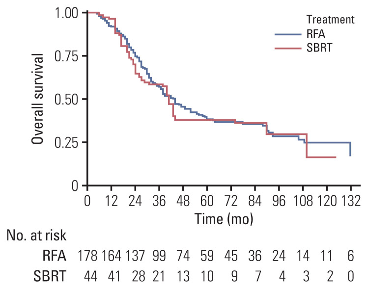
In the RFA group, 135 patients experienced tumor recurrence (intrahepatic recurrence, n=89; extrahepatic recurrence, n=101) and 119 died during follow-up; in the SBRT group, 37 patients experienced tumor recurrence (intrahepatic recurrence, n=29; extrahepatic recurrence, n=29) and 30 died during follow-up. For the treatment of patients with intrahepatic recurrence, additional local therapy (RFA or SBRT) or systemic chemotherapy were performed depending on the disease status and patient performance. On multivariate and IPTW-adjusted analyses, there were no significant differences in RFS or OS between the RFA and SBRT groups (Table 3). Univariate and multivariate analyses for the predictors of RFS are summarized in S3 and S4 Tables. The 1- and 3-year rates of RFS were 35% and 22% in the SBRT group and 43% and 23% in the RFA group, respectively (Fig. 4). The 1- and 3-year rates of OS were 96% and 58% in the SBRT group and 91% and 56% in the RFA group, respectively (Fig. 5).
Fig. 4.
Kaplan-Meier curves of recurrence-free survival after inverse probability of treatment weighting adjustment according to treatment modality. RFA, radiofrequency ablation; SBRT, stereotactic body radiation therapy.
Fig. 5.
Kaplan-Meier curves of overall survival after inverse probability of treatment weighting adjustment according to treatment modality. RFA, radiofrequency ablation; SBRT, stereotactic body radiation therapy.
4. Toxicity
Both RFA and SBRT were well-tolerated, with no patients showing treatment-related mortality or grade ≥ 3 treatment-related toxicity. RFA-related toxicity was all grade 1 and included peritoneal hemorrhage (n=6), biliary track injury (n=3), pneumothorax (n=2), and portal vein thrombus (n=1). In the SBRT group, 23 patients (52.2%) had nRILD, and elevation of liver enzyme levels of grade 1 in 21 patients (47.7%) and grade 2 in two patients (4.5%).
Discussion
To our knowledge, there are no randomized controlled trials that directly compared SBRT and RFA in patients with CRLM, and this is the first retrospective analysis on this subject that exclusively studied patients with CRLM. In this study, we analyzed a cohort of 222 CRLM patients with 330 tumors who were treated with SBRT or RFA. In IPTW-adjusted analysis, FFLP, RFS, and OS in the SBRT group were not significantly different from those in the RFA group. Our multivariate analysis revealed that tumor size was an independent predictor for FFLP. Therefore, we performed a stratified analysis based on tumor size and found that the difference in HR for FFLP according to treatment modality was increased with increasing tumor size. We also found significant interaction effects between the treatment modality and tumor size for FFLP (p=0.001). Lastly, subgroup analysis according to tumor size showed that among patients with tumor sizes larger than 2 cm, those receiving SBRT had a significantly higher FFLP than did those receiving RFA.
Our study results are consistent with previous studies that compared RFA with SBRT in other types of cancer. Wahl et al. [18] reported that SBRT was associated with a significantly better FFLP than RFA (HR, 3.35; 95% CI, 1.17 to 9.62; p=0.025) in patients with hepatocellular carcinomas (HCCs) that are 2 cm or larger. Similarly, Jackson et al. [17] recently reported that SBRT was associated with a better FFLP than RFA (HR, 3.54; 95% CI, 1.08 to 11.6; p < 0.01) in ≥ 2-cm-sized liver metastases from multiple primary origin cancer. These findings can be attributed to differences in the therapeutic efficacy of RFA and SBRT for large-sized tumors, in which SBRT appears to be less affected by the sizes of tumors. Most RF devices are capable of ablation with a diameter of 3 cm [9,19] and require a safety margin of at least 0.5 cm [20,21]; as such, 2 cm seems to be an important cutoff value for local control rate after RFA. Currently, there are limited data on the association between tumor size and local control rate after SBRT in CRLM patients, and the existing studies have shown conflicting results [6,15,22,23]. A pooled analysis of CRLM showed that maximum tumor size was associated with local control rates in patients who underwent SBRT (p=0.04) [6], while also reporting that high-dose (BED ≥ 79 Gy) SBRT can provide a 1-year local control rate of approximately 90%. While McPartlin et al. [22] and van der Pool et al. [23] suggested that tumor size was not a predictor of local control. We speculate that these results may be related to the dose of SBRT because several previous studies reported that high doses of SBRT may result in improved local control rates [6,7,15,22]. Scorsetti et al. [7] conducted a phase II study in patients with CRLM and reported that SBRT resulted in high 1- and 2-year local control rates of 95% (95% CI, 89 to 100) and 91% (95 % CI, 82 to 99), respectively; moreover, they showed high local control rates using 75 Gy in three fractions even when 45% of patients had tumors larger than 3 cm. Similarly, Joo et al. [15] found that patients in the high-dose group (132 to 180 Gy) had a good 1-year local control rate of 93% and that tumor size was not a prognostic factor for local control in patients treated with high doses of SBRT. We believe that higher radiation doses may provide improved FFLP, even in patients with larger CRLMs.
There is increasing evidence that SBRT dose is associated with the local control of liver metastases. Klement et al. [24] analyzed the tumor control probability (TCP) in 363 patients who underwent SBRT by using data from the German Society of Radiation Oncology and found that BED was an important predictive factor of TCP in the results of histology-adjusted analysis. From a histological point of view, CRLM is known to be more radioresistant than metastases from other primary cancers [24,25]. Accordingly, it has been suggested that CRLM needs a higher dose of 51 Gy in three fractions to achieve 90% TCP [25]. In our study, the median BED was 112.5 Gy10, which was lower than 51 Gy in three fractions (BED, 137.7 Gy10). Thus, we carefully assume that the SBRT group in our study has room for improvement in FFLP by dose escalation.
The benefits of dose escalation in our study were likely augmented by the high proportion of patients who received pre-treatment systemic therapy in the SBRT group. A previous report showed that in order to achieve 90% local control, patients who had received chemotherapy prior to SBRT required higher radiation doses compared with patients who had not received chemotherapy [24]; according to their predictive model, patients without prior chemotherapy required 48 Gy in three fractions or above to achieve 90% local control at 2 years, and patients with prior chemotherapy required 57 Gy in three fractions. In our study, most patients (86.4%) in the SBRT group had received chemotherapy prior to SBRT. Therefore, we believe that increasing the dose of SBRT may prolong FFLP, especially in patients who received chemotherapy before SBRT.
The relationship between improvement in FFLP and the translation to survival benefit is unclear. There were no significant differences in OS and RFS between the two groups in our study, which is in line with the results of previous comparative studies conducted on HCC and liver metastases [17,18]. We believe that further well-controlled randomized clinical trials are needed to draw definitive conclusions on this issue.
Because the treatment outcome of RFA is largely dependent on the technical skill of the radiologist, the level of proficiency of the performing radiologists may have influenced the study results. However, the local progression rate, 3-year FFLP, and 5-year FFLP in the RFA group were 32.1%, 60%, and 58%, respectively, which are within the ranges reported in previous studies (local progression rate, 9%–60%; 3-year FFLP, 53%–76%; 5-year FFLP, 43%–70%) [26]. Therefore, we assume that the technical factors of the radiologists who performed RFA did not significantly affect the outcomes of our study.
SBRT treatment outcomes can also vary from institution to institution. A recent multi-institutional study including 510 patients with HCC showed that SBRT dose was an independent prognostic factor of local control and that the prescription dose at an experienced center was significantly higher than that at a less experienced center [27]. Such differences in institutional treatment results may also occur in cases of liver metastases from colorectal cancer. Therefore, we believe that a wide generalization of our result requires external validation studies.
The present study has some limitations. First, the major limitation of our study was its retrospective nature, although we tried to mitigate the effects of the selection bias by applying IPTW adjustment. Our results may have been influenced by a selection bias stemming from the fact that RFA is considered a first-line treatment for CRLM patients who refuse surgery or are unsuitable for surgery, and SBRT is considered an alternative treatment option in the clinic. Furthermore, the various reasons why patients were considered to have unresectable CRLM might also have introduced a selection bias. Second, the overall sample size was rather small to achieve a strong statistical strength; especially, the number of patients who received SBRT was relatively small compared with those who received RFA. Third, there is a possibility that treatment-related toxicity was underestimated. However, previous prospective studies reported low rates of toxicity with both treatments [7,22,23]. Fourth, MRI was not performed in only a subset of patients (45% in the SBRT group and 40% in the RFA group) prior to SBRT and RFA. Considering that pre-treatment liver MRI can provide a more precise target delineation that offers the possibility for a better local control rate, further study is needed to determine whether pre-treatment MRI is indeed useful for improving local control. Lastly, a portion of the included patients had extrahepatic metastasis at the time of local therapy (RFA group: 5/178, SBRT group: 21/44), which may have affected the treatment outcome. Large-sized randomized controlled clinical trials are needed to directly compare the efficacy and safety of SBRT and RFA in patients with CRLM.
SBRT and RFA showed similar rates of local control in patients with CRLM. We found that liver tumor size was an independent prognostic factor for local control and that there was a significant interaction between treatment modality and tumor size for local control. For patients with tumor size >2 cm, SBRT was associated with a significantly better localcontrol compared with RFA. These results suggest that SBRTmay be preferred over RFA for larger tumors.
Acknowledgments
We thank Dr. Joon Seo Lim from the Scientific Publications Team at Asan Medical Center for his editorial assistance in preparing this manuscript.
Footnotes
Ethical Statement
This study was approved by the institutional review board of Asan Medical Center (no. 2018-1487) and written informed consent was waived because of the retrospective study.
Author Contributions
Conceived and designed the analysis: Park JH, Kim SY, Yoon SM, Jung J.
Collected the data: Yu J, Kim DH.
Contributed data or analysis tools: Shin YM, Yoon SM, Jung J, Kim JH, Kim JC, Yu CS, Lim SB, Park IJ, Kim TW, Hong YS, Kim SY, Kim JE, Park JH, Kim SY.
Performed the analysis: Lee J.
Wrote the paper: Yu J, Kim DH, Park JH, Kim SY.
Review the manuscript:: Park JH, Kim SY.
Conflicts of Interest
Conflict of interest relevant to this article was not reported.
Electronic Supplementary Material
Supplementary materials are available at Cancer Research and Treatment website (https://www.e-crt.org).
References
- 1.Ferlay J, Soerjomataram I, Dikshit R, Eser S, Mathers C, Rebelo M, et al. Cancer incidence and mortality worldwide: sources, methods and major patterns in GLOBOCAN 2012. Int J Cancer. 2015;136:E359–86. doi: 10.1002/ijc.29210. [DOI] [PubMed] [Google Scholar]
- 2.National Cancer Institute. Surveillance, Epidemiology, and End Results Program. Cancer stat facts: colorectal cancer [Internet] Bethesda, MD: National Cancer Institute; 2021. [cited 2021 Feb 2]. Available from: https://seer.cancer.gov/statfacts/html/colorect.html . [Google Scholar]
- 3.Hackl C, Neumann P, Gerken M, Loss M, Klinkhammer-Schalke M, Schlitt HJ. Treatment of colorectal liver metastases in Germany: a ten-year population-based analysis of 5772 cases of primary colorectal adenocarcinoma. BMC Cancer. 2014;14:810. doi: 10.1186/1471-2407-14-810. [DOI] [PMC free article] [PubMed] [Google Scholar]
- 4.Kopetz S, Chang GJ, Overman MJ, Eng C, Sargent DJ, Larson DW, et al. Improved survival in metastatic colorectal cancer is associated with adoption of hepatic resection and improved chemotherapy. J Clin Oncol. 2009;27:3677–83. doi: 10.1200/JCO.2008.20.5278. [DOI] [PMC free article] [PubMed] [Google Scholar]
- 5.Venkat SR, Mohan PP, Gandhi RT. Colorectal liver metastasis: overview of treatment paradigm highlighting the role of ablation. AJR Am J Roentgenol. 2018;210:883–90. doi: 10.2214/AJR.17.18574. [DOI] [PubMed] [Google Scholar]
- 6.Chang DT, Swaminath A, Kozak M, Weintraub J, Koong AC, Kim J, et al. Stereotactic body radiotherapy for colorectal liver metastases: a pooled analysis. Cancer. 2011;117:4060–9. doi: 10.1002/cncr.25997. [DOI] [PubMed] [Google Scholar]
- 7.Scorsetti M, Comito T, Tozzi A, Navarria P, Fogliata A, Clerici E, et al. Final results of a phase II trial for stereotactic body radiation therapy for patients with inoperable liver metastases from colorectal cancer. J Cancer Res Clin Oncol. 2015;141:543–53. doi: 10.1007/s00432-014-1833-x. [DOI] [PMC free article] [PubMed] [Google Scholar]
- 8.Hoyer M, Roed H, Traberg Hansen A, Ohlhuis L, Petersen J, Nellemann H, et al. Phase II study on stereotactic body radiotherapy of colorectal metastases. Acta Oncol. 2006;45:823–30. doi: 10.1080/02841860600904854. [DOI] [PubMed] [Google Scholar]
- 9.Wong SL, Mangu PB, Choti MA, Crocenzi TS, Dodd GD, 3rd, Dorfman GS, et al. American Society of Clinical Oncology 2009 clinical evidence review on radiofrequency ablation of hepatic metastases from colorectal cancer. J Clin Oncol. 2010;28:493–508. doi: 10.1200/JCO.2009.23.4450. [DOI] [PubMed] [Google Scholar]
- 10.Ruers T, Van Coevorden F, Punt CJ, Pierie JE, Borel-Rinkes I, Ledermann JA, et al. Local treatment of unresectable colorectal liver metastases: results of a randomized phase II trial. J Natl Cancer Inst. 2017;109:djx015. doi: 10.1093/jnci/djx015. [DOI] [PMC free article] [PubMed] [Google Scholar]
- 11.Tanis E, Nordlinger B, Mauer M, Sorbye H, van Coevorden F, Gruenberger T, et al. Local recurrence rates after radiofrequency ablation or resection of colorectal liver metastases: analysis of the European Organisation for Research and Treatment of Cancer #40004 and #40983. Eur J Cancer. 2014;50:912–9. doi: 10.1016/j.ejca.2013.12.008. [DOI] [PubMed] [Google Scholar]
- 12.Mulier S, Ni Y, Jamart J, Ruers T, Marchal G, Michel L. Local recurrence after hepatic radiofrequency coagulation: multivariate meta-analysis and review of contributing factors. Ann Surg. 2005;242:158–71. doi: 10.1097/01.sla.0000171032.99149.fe. [DOI] [PMC free article] [PubMed] [Google Scholar]
- 13.Teratani T, Yoshida H, Shiina S, Obi S, Sato S, Tateishi R, et al. Radiofrequency ablation for hepatocellular carcinoma in so-called high-risk locations. Hepatology. 2006;43:1101–8. doi: 10.1002/hep.21164. [DOI] [PubMed] [Google Scholar]
- 14.Komorizono Y, Oketani M, Sako K, Yamasaki N, Shibatou T, Maeda M, et al. Risk factors for local recurrence of small hepatocellular carcinoma tumors after a single session, single application of percutaneous radiofrequency ablation. Cancer. 2003;97:1253–62. doi: 10.1002/cncr.11168. [DOI] [PubMed] [Google Scholar]
- 15.Joo JH, Park JH, Kim JC, Yu CS, Lim SB, Park IJ, et al. Local control outcomes using stereotactic body radiation therapy for liver metastases from colorectal cancer. Int J Radiat Oncol Biol Phys. 2017;99:876–83. doi: 10.1016/j.ijrobp.2017.07.030. [DOI] [PubMed] [Google Scholar]
- 16.Rusthoven KE, Kavanagh BD, Cardenes H, Stieber VW, Burri SH, Feigenberg SJ, et al. Multi-institutional phase I/II trial of stereotactic body radiation therapy for liver metastases. J Clin Oncol. 2009;27:1572–8. doi: 10.1200/JCO.2008.19.6329. [DOI] [PubMed] [Google Scholar]
- 17.Jackson WC, Tao Y, Mendiratta-Lala M, Bazzi L, Wahl DR, Schipper MJ, et al. Comparison of stereotactic body radiation therapy and radiofrequency ablation in the treatment of intrahepatic metastases. Int J Radiat Oncol Biol Phys. 2018;100:950–8. doi: 10.1016/j.ijrobp.2017.12.014. [DOI] [PMC free article] [PubMed] [Google Scholar]
- 18.Wahl DR, Stenmark MH, Tao Y, Pollom EL, Caoili EM, Lawrence TS, et al. Outcomes after stereotactic body radiotherapy or radiofrequency ablation for hepatocellular carcinoma. J Clin Oncol. 2016;34:452–9. doi: 10.1200/JCO.2015.61.4925. [DOI] [PMC free article] [PubMed] [Google Scholar]
- 19.Crocetti L, de Baere T, Lencioni R. Quality improvement guidelines for radiofrequency ablation of liver tumours. Cardiovasc Intervent Radiol. 2010;33:11–7. doi: 10.1007/s00270-009-9736-y. [DOI] [PMC free article] [PubMed] [Google Scholar]
- 20.Kudo M. Radiofrequency ablation for hepatocellular carcinoma: updated review in 2010. Oncology. 2010;78(Suppl 1):113–24. doi: 10.1159/000315239. [DOI] [PubMed] [Google Scholar]
- 21.Lencioni R, Crocetti L. Radiofrequency ablation of liver cancer. Tech Vasc Interv Radiol. 2007;10:38–46. doi: 10.1053/j.tvir.2007.08.006. [DOI] [PubMed] [Google Scholar]
- 22.McPartlin A, Swaminath A, Wang R, Pintilie M, Brierley J, Kim J, et al. Long-term outcomes of phase 1 and 2 studies of SBRT for hepatic colorectal metastases. Int J Radiat Oncol Biol Phys. 2017;99:388–95. doi: 10.1016/j.ijrobp.2017.04.010. [DOI] [PubMed] [Google Scholar]
- 23.van der Pool AE, Mendez Romero A, Wunderink W, Heijmen BJ, Levendag PC, Verhoef C, et al. Stereotactic body radiation therapy for colorectal liver metastases. Br J Surg. 2010;97:377–82. doi: 10.1002/bjs.6895. [DOI] [PubMed] [Google Scholar]
- 24.Klement RJ, Guckenberger M, Alheid H, Allgauer M, Becker G, Blanck O, et al. Stereotactic body radiotherapy for oligometastatic liver disease: influence of pre-treatment chemotherapy and histology on local tumor control. Radiother Oncol. 2017;123:227–33. doi: 10.1016/j.radonc.2017.01.013. [DOI] [PubMed] [Google Scholar]
- 25.Klement RJ. Radiobiological parameters of liver and lung metastases derived from tumor control data of 3719 metastases. Radiother Oncol. 2017;123:218–26. doi: 10.1016/j.radonc.2017.03.014. [DOI] [PubMed] [Google Scholar]
- 26.Yang G, Wang G, Sun J, Xiong Y, Li W, Tang T, et al. The prognosis of radiofrequency ablation versus hepatic resection for patients with colorectal liver metastases: a systematic review and meta-analysis based on 22 studies. Int J Surg. 2021;87:105896. doi: 10.1016/j.ijsu.2021.105896. [DOI] [PubMed] [Google Scholar]
- 27.Kim N, Cheng J, Huang WY, Kimura T, Zeng ZC, Lee VH, et al. Dose-response relationship in stereotactic body radiation therapy for hepatocellular carcinoma: a pooled analysis of an Asian Liver Radiation Therapy Group Study. Int J Radiat Oncol Biol Phys. 2021;109:464–73. doi: 10.1016/j.ijrobp.2020.09.038. [DOI] [PubMed] [Google Scholar]
Associated Data
This section collects any data citations, data availability statements, or supplementary materials included in this article.


