Abstract
Background
Percutaneous endoscopic gastrostomy (PEG) was developed by Ponsky-Gauderer in the early 1980s. These tubes are placed through the abdominal wall mainly to administer fluids, drugs and/or enteral nutrition but can also be used for drainage or decompression. The tubes consist of an internal and external retention device. It is a generally safe technique but major or minor complications may arise during and after tube placement.
Method
A narrative review of the literature investigating minor complications after PEG placement.
Results
This review was written from a clinical viewpoint focusing on prevention and management of minor complications and documented with real cases from more than 21 years of clinical practice.
Conclusions
Depending on the literature the incidence of minor complications after gastrostomy placement can be high. To decrease associated morbidity, prevention, early recognition and popper management of these complications are important.
Keywords: gastrostomy, endoscopic gastrostomy, enteral nutrition
Key messages.
Minor PEG complications have been described in review articles and guidelines.
This narrative review comprehensively summerizes all existing evidence-based practice about prevention and management options.
Having access to one clear overview can optimize care and cure for patients with a PEG.
Introduction
The first and still most widely used ‘pull’ technique to introduce a percutaneous endoscopic gastrostomy (PEG) was developed by Ponsky-Gardener in the early 1880s.1 If patients require enteral access for more than 4 to 6 weeks, a PEG is recommended by international guidelines.2 A PEG-tube can serve as a vehicle for liquid feeding formulas, fluids and/or liquid medications into the stomach but can also be used for decompression, drainage or management of gastric volvulus.3 It is retained in position by an internal and external fixation device, fixator or bumper. The internal bumper holds the device securely inside the stomach. It may be in the form of a flange, dome, string, basket or balloon. The external bumper may be in the form of a triangle, circle or other shape; it can be soft or hard and secures the gastrostomy tube externally against the abdominal wall, limiting unnecessary tube movement and leakage of gastric content.4 PEG tube insertion is usually considered a safe procedure, however, complications can occur with a variable rate based on the study population. These complications can be classified as minor or major.5 Major postprocedural complications include buried bumper syndrome, bleeding, tube dislodgement, gastric erosions and ulcers, (pneumoperitoneum) peritonitis, necrotising fasciitis, colonic injury, liver injury and PEG tract tumour seeding. A comprehensive overview of major complications with preventive actions and management was recently published.6 Fortunately, most of complications are minor (13%–40%) but, nevertheless, can be linked to a high incidence of morbidity. Minor complications include peristomal site infection, overgranulation tissue, peristomal leakage and tube blockage.7 In this narrative review, existing evidence of minor postprocedural PEG complications is explored while focusing on prevention and management. Furthermore, the evidence is illustrated with real cases from more than 21 years of clinical practice.
Peristomal site infection
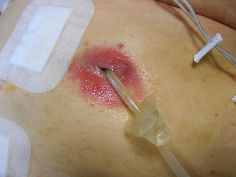
Peristomal site infection is characterised by increased erythema, tenderness, induration and a purulent discharge. It is the most common complication following PEG (percutaneous endoscopic jejunostomy) tube placement and its incidence ranges from 4% to 30%.8 PEG insertion sites are frequently colonised with multiple micro-organisms. A Dutch study found in 85 of a 100 patients Candida albicans (n=37; 44%), Staphylococcus aureus (n=28; 33%), Escherichia coli, Klebsiella, Enterobacter and enterococci (5%–20%) after culturing. Although, this did not result in any major discomfort besides some itching and local pain in approximately one fourth of patients.9 In a small study, fungi were isolated from the stomach in 13 (65%) of 20 patients. They found that the isolated species from the oral cavity, the stomach and later the gastrostomy tube were identical in most cases.10 In a retrospective review of 297 medical records of patients receiving prophylactic cefazolin before PEG placement, wound infection occurred in 36 patients (12.1%). Staphylococcus aureus resistant to methicillin was the most frequently isolated microorganism (33.3%), followed by Pseudomonas aeruginosa (30.6%).11 In a more recent, retrospective study over 16 years, 67 episodes of PEG site infection were diagnosed in patients with head and neck cancer, with an overall prevalence of 21.2%.12 Those undergoing PEG tube placement are often vulnerable to infection because of age, compromised nutritional intake, immunosuppression or underlying disease such as malignancy and diabetes mellitus.13–16 Additionally, patients who underwent chemotherapy or radiotherapy before PEG placement had a higher incidence of peristomal infections.17 18 Apart from patient-related factors, other variables can influence infectious outcomes, for example, placement technique, procedural differences, diameter of the tube, the presence of leakage and/or hypergranulation tissue and the differences in experience of stoma aftercare.18 19 Peristomal infection is mostly mild and generally well controlled by local therapy. Rarely, cases are severe or involve an abscess within the soft tissue surrounding the tube (figure 1). Even more rare, abscesses develop in the deeper tissue layers which are not easily visualised on inspection. Patients usually report excessive pain around the tube and may exhibit signs of systemic infection such as leukocytosis or fever. CT scan can be helpful in the diagnosis of these abscesses.20 The overall incidence of infections at PEG sites can be decreased by the use of periprocedural antibiotics (a single intravenous dose of a beta-lactam antibiotic or a suitable alternative in case of allergy).13 21 Alternatively, 20 mL of a co-trimoxazole solution deposited immediately through a newly inserted PEG catheter could be as effective as the intravenous administration.22 PEG insertion should be performed using a strict sterile/aseptic technique (skin disinfection, sterile surgical drapes, sterile gloves, sterile dressing, etc).21 A close professional relationship and good communication between the care givers (eg, nurses team, the nutrition support (nurse) specialist, wound ostomy nurse, endoscopist or radiologist) result in good periprocedural preparation and early identification and management of potential problems.19–23 Preventive actions and management options are summarised below.2–4 6 19 21 24–29
Figure 1.
Severe peristomal gastrostomy infection resulting in removal of the tube afterwards.
Prevention
Prior to the procedure (< 30 min before)
Use an oral antiseptic mouthwash (chlorhexidine or aqueous iodine) to reduce bacterial presence.
Decolonise the nasopharynx if diagnosed (but not yet eradicated) presence of methicillin-resistant Staphylococcus aureus.
If the body hair is abundant at the insertion site, use an electric shaver.
Stop a proton pump inhibitor 24 hours before the procedure.
Use a single intravenous dose of periprocedural antibiotics (a first-generation cephalosporin); unless in patients already receiving antibiotics covering skin-flora.
Apply standard measures for infection prevention including aseptic preparation of the surgical field and preoperative handwashing and/or disinfection.
Use a checklist that serves as a reminder of all necessary steps prior to and after tube placement.
Following the procedure
Alternatively, consider administering a 20 mL co-trimoxazole solution through the newly inserted PEG catheter just after placement, instead of the periprocedural intravenous dose.
Clean the stoma and peristomal skin with a sterile solution (normal saline or local disinfection) daily for the first week and consider applying a skin protecting film or cream.
Alternatively, use a glycerin hydrogel or glycogel dressing instead of classical aseptic wound care during the first week.
Apply a (split) gauze dressing (not too thick) to remove any discharge, above or under the external bumper (with a free distance of 0.5–1 cm).
Protect the skin with a nonocclusive dressing.
Avoid excessive pressure between the skin and the external bumper.
Assess the stoma and peristomal skin daily for signs and symptoms of infection such as loss of skin integrity, maceration, erythema, purulent and/or malodorous exudate, fever and pain.
Reduce (after stoma healing) dressings to once or twice a week. The entry site can be cleansed using an additive-free pH 5.5 soap and water of drinking quality.
Alternatively, dressings can be omitted and the site can be left open.
Management options
Apply, in case of mild infection, a thin layer of an antiseptic ointment (eg, iodine paste) to the entry site of the tube and the surrounding tissue (in combination with a skin protecting film or cream).
Ask for a wound consultant in case of severe infection or failure of first-line treatment.
Administer a short course of a broad-spectrum antibiotics, either orally or enterally if infections occur early after PEG placement (within 3–5 days).
Intravenous antibiotics are only indicated in more severe infections.
Treat accordingly if infection and granulation tissue occur simultaneously (also see paragraph ‘overgranulation tissue’).
Review effectiveness of any treatment at regular intervals.
Remove the tube if the infection cannot be resolved (not responding to antibiotics or deterioration) or if the tube is affected by a fungus (also see paragraph ‘Tube replacement’)
Consider urgent surgical intervention if a patient has signs of peritonitis, abscess or necrotising fasciitis.
Overgranulation tissue
Over time, a spongy, friable, deep red coloured tissue above the gastrostomy site may develop (figure 2). Overgranulation or hypergranulation is an aberrant response with overgrowth of fibroblasts and endothelial cells with a structure similar to normal granulation tissue. It is vascular, so it bleeds easily and can sometimes be painful. The presence of this excess tissue usually leads to excess moisture with increased site drainage. It hinders keratinocytes progress on the wound bed surface to achieve complete re-epithelialisation, hereby compromising an adequate seal of healthy tissue around the tube. Risk factors for granulation tissue development are friction movement at the wound interface (eg, due to poor or incorrect positioning of the external fixator); and critical colonisation or true infection. Preventive actions and management options are summarised below.2 4 8 19 25 30–32
Figure 2.
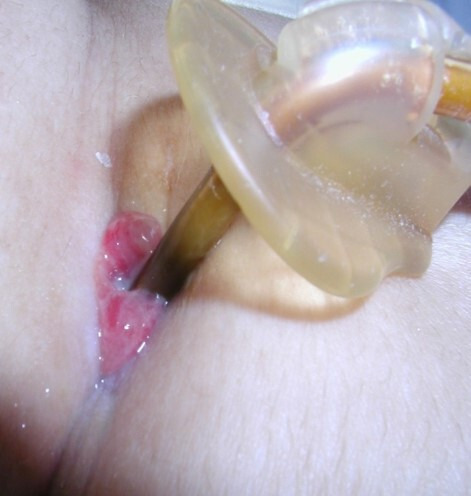
Overgranulation tissue.
Prevention
Keep the gastrostomy site as dry as possible.
Secure the tube properly and minimise friction/movement.
Apply preventive actions against peristomal infection after the procedure (also see previous paragraph ‘Peristomal site infection’).
Check if a low-profile device is in situ, if the device comfortably fits in the tract and has minimal movement.
Management options
Cauterise excess tissue with topical silver nitrate (should be performed by an experienced person) and be aware that the healthy tissue around the granulation tissue might also be harmed if not performed properly.
Apply a topical corticosteroid cream or ointment on the overgranulation tissue once or twice a day for a maximum of 7–10 days.
Use salt (sprinkle about one-third of a 5 mL teaspoon of table salt over the tissue once a day until the overgranulation is flattened). This is an inexpensive and safe approach and is very feasible in a home environment if required.
Apply, if overgranulation tissue is extensive, a foam dressing in addition to hydrocortisone cream (depending on the level of exudate, foams can be left in place for up to 7 days).
In case of inflammation or signs of infection, consider a foam impregnated with an antimicrobial agent such as silver, honey or cadexomer iodine under the fixation device.
Change to an alternative brand or type of gastrostomy tube (low-profile or skin-level device (button)).
Apply argon plasma coagulation on the overgranulation tissue.
Peristomal leakage
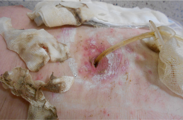
Peristomal leakage of gastric contents due to enlarging diameter of the PEG tract is a common complication and reported in some studies as high as 10%.25 Several factors contributing to the risk of peristomal leakage have been identified, including excessive cleansing with hydroperoxide, infection, gastric hypersecretion and excessive side torsion along the PEG tube. The latter causes ulceration at the skin and enlargement of the gastrostomy (figure 3). Also patient-related factors (eg, malnutrition, immunodeficiency, diabetes, increased workload of breathing/chronic cough and constipation) can compromise wound healing. Preventive actions and management options are summarised below.4 7 8 19 25
Figure 3.
Peristomal leakage with skin damage and enlarged gastrostomy tract.
Prevention
Avoid side torsion on the tract wall.
Evaluate regularly if the tube is not fixed too loosely or too tightly to the skin and check for a potential buried bumper syndrome.
Check balloon inflation volume at weekly intervals (if the tube is a balloon retained gastrostomy tube) and inspect the water for evidence of stomach contents indicating balloon rupture.
Observe the ostomy site closely for infection or overgranulation tissue.
Check gastric residual volume if any signs of gastrointestinal intolerance are present (eg, nausea, vomiting, abdominal distention, constipation).
Management options
Assess first whether leakage is caused by a problem of the tube itself (eg, poorly fitting tube or connected administration set, inside blockage, kinking, degeneration or cracking).
Specify the nature of the leakage (eg, feed, fluids, gastric contents). The pH can be tested using a pH indicator strip.
Stop tube feeding and investigate if leakage is seen within the first 72 hours and if it is associated with pain following initial gastrostomy tube insertion.
Protect the surrounding skin using a barrier film or cream.
Place a foam dressing or a super absorbent gauze dressing under the fixation device to absorb exudate and protect the exit site from further irritation/maceration.
Ask for a wound care consultant.
Uncap the tube and connect it to a drainage bag; or use a stoma bag (with adequate skin protection) placed over the ostomy site to collect excess leakage.
Review medication and consider starting antisecretory therapy (proton pump inhibitor).
Do not replace the initial tube by a larger diameter tube as this may cause enlargement of the tract, resulting in exacerbation of the leakage.
Remove and place a new PEG at a different site allowing the original site to close and heal.
Convert the PEG to a PEG-J (postpyloric feeding line through the PEG tube) for jejunal feeding, potentially combined with gastric drainage.
Tube blockage
Occlusion, clogging or blockage is a common complication of enteral tube feeding. The incidence of clogged feeding tubes in PEG is reported to be as high as 23%–35%. There are several risk factors for tube blockage: increased tube length, smaller tube calibre, medication administration and/or dissolving (eg, crushed (mixed) tablets), inadequate flushing, viscous solution (eg, high-fibre, caloriedense or blended foods), slow flow rates of the feed, contact of enteral formula with acidic gastric secretions and regular aspiration to measure residual volumes. Prevention is the key factor but once tube blockage occurs, several management options can be tried before resorting to removal and/or replacement.3 7 8 19 23 33–37
Prevention
Replace the tube feeding set every 24 hours.
Flush the tube using 30 mL of pure water every 4 hours during continuous tube feedings, before and after intermittent feedings and after checking gastric residuals.
Flush with ±15 mL of water after and between each medication through the tube.
Consider adapting flushing protocols in people with restricted fluid intake, for example, 10 mL every 6 hours with continuous infusions; and 5 mL before and 10 mL after administering drugs; or interrupting or starting enteral nutrition.
Pay particular attention to avoid obstruction with jejunal tubes because they tend to have smaller calibres than gastric tubes.
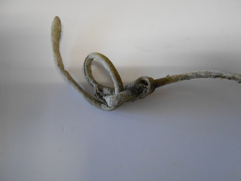
Never rotate a PEG with a jejunal extension (PEG-J) (figure 4).
Critically evaluate the medication: which drugs are really necessary, which medication has an alternative form (eg, liquid, effervescent tablet, syrup).
Crush, dissolve and administrate drugs separate from each other to prevent incompatibility.
Use sterile water in immunocompromised or critically ill patients if there are concerns about the safety of pure water.
Figure 4.
A percutaneous endoscopic gastrostomy with a knotted jejunal extension.
Management options
First assess if the tube is not kinked or compressed in any way.
Try to unclog using a (lukewarm) water-filled syringe (20 mL) using a back-and-forth motion for about 5 min.
Try to unclog using a small (lukewarm) water-filled syringe (5–10 mL) to apply more pressure (but avoid excessive force).
Acidic carbonated soft drinks (eg, Coca-Cola) can be tried (low pH) but its effect is not superior to water.
Do not use cranberry juice or sodas.
Irrigate with an 8.4% NaHCO3 solution and close the tube for 5–10 min.
Irrigate with pancreatic enzymes diluted in water plus NaHCO3 and close the tube for 5–10 min.
Consider the use of commercial unclogging devices, for example, preloaded enzyme cocktails, a brush, a machine-operated unclogger or corrugated plastic rod.
Replace the tube if occlusion is caused by fungal infection or if all previous strategies have failed (see next paragraph ‘tube replacement’).
Tube replacement
Most transoral bumper-type tubes can be maintained for 1 or 2 years (or sometimes longer), but eventually replacement will be required because of breakage, occlusion, dislodgement or degradation.25 The replacement can be performed in several ways: endoscopically, radiologically, surgically and bedside replacement (depending on the type of gastrostomy tube being replaced).4 23 38 39
Prevention
See preventive measures in the paragraph ‘tube blockage’.
Management actions
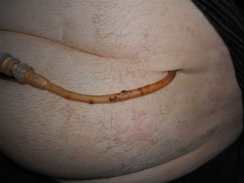
Replace the tube in a non-urgent but timely manner if it is diagnosed with signs of fungal colonisation, with material deterioration or compromised structural integrity. Especially silicon tubes are at risk for colonisation (figure 5).
Consider a balloon-type tube that can be inserted ‘blindly’ (without endoscopy) in a matured tract.
For a bumper-type tube, cut the tube just above the skin and push the internal bumper into the stomach (‘cut and push’ method). Migration is usually uneventful, even with large-calibre tubes.
Endoscopic retrieval of the bumper is advocated in case of previous bowel surgery and in patients at risk of strictures, which could hinder spontaneous migration and elimination of the sectioned bumper.
Figure 5.
Invasion of a percutaneous feeding tube with a fungus.
The most relevant discussed preventive measures are summarised in an overview in table 1.
Table 1.
Overview of minor postprocedural percutaneous endoscopic gastrostomy complications and their prevention
| Complication | Prevention |
| Peristomal site infection | Prior to the procedure (<30 min before)
Following the procedure
|
| Overgranulation tissue |
|
| Peristomal leakage |
|
| Tube blockage and replacement |
|
PEG, percutaneous endoscopic gastrostomy.
Conclusion
After gastrostomy placement several minor complications can occur, resulting in associated morbidity, affecting quality of life, increasing healthcare costs (eg, hospital (re) admissions, length of stay) and potentially interrupting nutritional treatment. Systematic long-term nutrition team follow-up of patients after PEG is therefore recommended. A nutrition support team with a nutrition nurse specialist can play a very important role in preventing, reducing and managing these complications.40–42 This review was written from a clinical viewpoint and focuses on relevant existing literature and evidence-based recommendations.
Footnotes
Contributors: KB and ID contributed to the design of the study, KB drafted the manuscript and KB, ID and WV critically revised the manuscript for important intellectual content and final approval.
Funding: The authors have not declared a specific grant for this research from any funding agency in the public, commercial or not-for-profit sectors.
Competing interests: None declared.
Provenance and peer review: Not commissioned; internally peer reviewed.
Data availability statement
No data are available.
Ethics statements
Patient consent for publication
Not applicable.
References
- 1.Gauderer MW, Ponsky JL, Izant RJ. Gastrostomy without laparotomy: a percutaneous endoscopic technique. J Pediatr Surg 1980;15:872–5. 10.1016/S0022-3468(80)80296-X [DOI] [PubMed] [Google Scholar]
- 2.Bischoff SC, Austin P, Boeykens K, et al. ESPEN guideline on home enteral nutrition. Clin Nutr 2020;39:5–22. 10.1016/j.clnu.2019.04.022 [DOI] [PubMed] [Google Scholar]
- 3.Lord LM. Enteral access devices: types, function, care, and challenges. Nutr Clin Pract 2018;33:16–38. 10.1002/ncp.10019 [DOI] [PubMed] [Google Scholar]
- 4.National Nurses Nutrition Group (NNNG) . Good practice guideline – care of gastrostomy tubes and exit site management in adults and children UK, 2020. [Google Scholar]
- 5.Rahnemai-Azar AA, Rahnemaiazar AA, Naghshizadian R, et al. Percutaneous endoscopic gastrostomy: indications, technique, complications and management. World J Gastroenterol 2014;20:7739–51. 10.3748/wjg.v20.i24.7739 [DOI] [PMC free article] [PubMed] [Google Scholar]
- 6.Boeykens K, Duysburgh I. Prevention and management of major complications in percutaneous endoscopic gastrostomy. BMJ Open Gastroenterol 2021;8:e000628. 10.1136/bmjgast-2021-000628 [DOI] [PMC free article] [PubMed] [Google Scholar]
- 7.Blumenstein I, Shastri YM, Stein J. Gastroenteric tube feeding: techniques, problems and solutions. World J Gastroenterol 2014;20:8505–24. 10.3748/wjg.v20.i26.8505 [DOI] [PMC free article] [PubMed] [Google Scholar]
- 8.Roveron G, Antonini M, Barbierato M, et al. Clinical practice guidelines for the nursing management of percutaneous endoscopic gastrostomy and jejunostomy (PEG/PEJ) in adult patients: an executive summary. J Wound Ostomy Continence Nurs 2018;45:326–34. 10.1097/WON.0000000000000442 [DOI] [PubMed] [Google Scholar]
- 9.de Vries T, de Ruiter A, Westendorp A, et al. Microorganisms and complaints in outpatients with a percutaneous endoscopic gastrostomy catheter. Am J Infect Control 2015;43:802–4. 10.1016/j.ajic.2015.04.002 [DOI] [PubMed] [Google Scholar]
- 10.Gottlieb K, Iber FL, Livak A, et al. Oral Candida colonizes the stomach and gastrostomy feeding tubes. JPEN J Parenter Enteral Nutr 1994;18:264–7. 10.1177/0148607194018003264 [DOI] [PubMed] [Google Scholar]
- 11.Duarte H, Santos C, Capelas ML, et al. Peristomal infection after percutaneous endoscopic gastrostomy: a 7-year surveillance of 297 patients. Arq Gastroenterol 2012;49:255–8. 10.1590/S0004-28032012000400005 [DOI] [PubMed] [Google Scholar]
- 12.Oh J, Park SY, Lee JS, et al. Clinical characteristics and pathogens in percutaneous endoscopic gastrostomy site infection in patients with head and neck cancer: a 16-year retrospective study. Laryngoscope Investig Otolaryngol 2021;6:1325–31. 10.1002/lio2.666 [DOI] [PMC free article] [PubMed] [Google Scholar]
- 13.Lipp A, Lusardi G. Systemic antimicrobial prophylaxis for percutaneous endoscopic gastrostomy. Cochrane Database Syst Rev 2013:CD005571. 10.1002/14651858.CD005571.pub3 [DOI] [PMC free article] [PubMed] [Google Scholar]
- 14.Lee JH, Kim JJ, Kim YH, et al. Increased risk of peristomal wound infection after percutaneous endoscopic gastrostomy in patients with diabetes mellitus. Dig Liver Dis 2002;34:857–61. 10.1016/S1590-8658(02)80256-0 [DOI] [PubMed] [Google Scholar]
- 15.Shangab MOM, Shaikh NA. Prediction of risk of adverse events related to percutaneous endoscopic gastrostomy: a retrospective study. Ann Gastroenterol 2019;32:469–75. 10.20524/aog.2019.0409 [DOI] [PMC free article] [PubMed] [Google Scholar]
- 16.Dormann AJ, Huchzermeyer H, Lippert H. The relevance of systemic complications and the different outcomes of subgroups after percutaneous endoscopic gastrostomy (PEG). Am J Gastroenterol 2001;96:1951–2. 10.1111/j.1572-0241.2001.03915.x [DOI] [PubMed] [Google Scholar]
- 17.Vizhi K, Rao HB, Venu RP. Percutaneous endoscopic gastrostomy site infections-Incidence and risk factors. Indian J Gastroenterol 2018;37:103–7. 10.1007/s12664-018-0822-4 [DOI] [PubMed] [Google Scholar]
- 18.Zopf Y, Konturek P, Nuernberger A, et al. Local infection after placement of percutaneous endoscopic gastrostomy tubes: a prospective study evaluating risk factors. Can J Gastroenterol 2008;22:987–91. 10.1155/2008/530109 [DOI] [PMC free article] [PubMed] [Google Scholar]
- 19.Pars H, Çavuşoğlu H. A literature review of percutaneous endoscopic gastrostomy: dealing with complications. Gastroenterol Nurs 2019;42:351–9. 10.1097/SGA.0000000000000320 [DOI] [PubMed] [Google Scholar]
- 20.Miller KR, McClave SA, Kiraly LN, et al. A tutorial on enteral access in adult patients in the hospitalized setting. JPEN J Parenter Enteral Nutr 2014;38:282–95. 10.1177/0148607114522487 [DOI] [PubMed] [Google Scholar]
- 21.Gkolfakis P, Arvanitakis M, Despott EJ, et al. Endoscopic management of enteral tubes in adult patients - Part 2: Peri- and post-procedural management. European Society of Gastrointestinal Endoscopy (ESGE) Guideline. Endoscopy 2021;53:178–95. 10.1055/a-1331-8080 [DOI] [PubMed] [Google Scholar]
- 22.Blomberg J, Lagergren P, Martin L, et al. Novel approach to antibiotic prophylaxis in percutaneous endoscopic gastrostomy (PEG): randomised controlled trial. BMJ 2010;341:c3115. 10.1136/bmj.c3115 [DOI] [PMC free article] [PubMed] [Google Scholar]
- 23.McClave SA, DiBaise JK, Mullin GE, et al. Acg clinical guideline: nutrition therapy in the adult hospitalized patient. Am J Gastroenterol 2016;111:315–34. 10.1038/ajg.2016.28 [DOI] [PubMed] [Google Scholar]
- 24.Horiuchi A, Nakayama Y, Kajiyama M, et al. Nasopharyngeal decolonization of methicillin-resistant Staphylococcus aureus can reduce PEG peristomal wound infection. Am J Gastroenterol 2006;101:274–7. 10.1111/j.1572-0241.2006.00366.x [DOI] [PubMed] [Google Scholar]
- 25.Toussaint E, Van Gossum A, Ballarin A, et al. Enteral access in adults. Clin Nutr 2015;34:350–8. 10.1016/j.clnu.2014.10.009 [DOI] [PubMed] [Google Scholar]
- 26.Fernandez R, Griffiths R. Water for wound cleansing. Cochrane Database Syst Rev 2012:CD003861. 10.1002/14651858.CD003861.pub3 [DOI] [PubMed] [Google Scholar]
- 27.Alsunaid S, Holden VK, Kohli A, et al. Wound care management: tracheostomy and gastrostomy. J Thorac Dis 2021;13:5297–313. 10.21037/jtd-2019-ipicu-13 [DOI] [PMC free article] [PubMed] [Google Scholar]
- 28.Wounds UK . Best practice statement: the use of topical antiseptic/ antimicrobial agents in wound management, 2011. Available: www.wounds-uk.com/pdf/content_9969.pdf [Accessed Feb 2022].
- 29.Im JP, Cha JM, Kim JW, et al. Proton pump inhibitor use before percutaneous endoscopic gastrostomy is associated with adverse outcomes. Gut Liver 2014;8:248–53. 10.5009/gnl.2014.8.3.248 [DOI] [PMC free article] [PubMed] [Google Scholar]
- 30.León AH, Hebal F, Stake C, et al. Prevention of hypergranulation tissue after gastrostomy tube placement: a randomised controlled trial of hydrocolloid dressings. Int Wound J 2019;16:41–6. 10.1111/iwj.12978 [DOI] [PMC free article] [PubMed] [Google Scholar]
- 31.Tanaka H, Arai K, Fujino A, et al. Treatment for hypergranulation at gastrostomy sites with sprinkling salt in paediatric patients. J Wound Care 2013;22:17–20. 20. 10.12968/jowc.2013.22.1.17 [DOI] [PubMed] [Google Scholar]
- 32.Townley A, Wincentak J, Krog K, et al. Paediatric gastrostomy stoma complications and treatments: a rapid scoping review. J Clin Nurs 2018;27:1369–80. 10.1111/jocn.14233 [DOI] [PubMed] [Google Scholar]
- 33.Guenter P, Boullata J. Nursing2013 survey results: drug administration by enteral feeding tube. Nursing 2013;43:26–33. quiz 34-5. 10.1097/01.NURSE.0000437469.13218.7b [DOI] [PubMed] [Google Scholar]
- 34.Phillips NM, Nay R. A systematic review of nursing administration of medication via enteral tubes in adults. J Clin Nurs 2008;17:2257–65. 10.1111/j.1365-2702.2008.02407.x [DOI] [PubMed] [Google Scholar]
- 35.Boullata JI, Carrera AL, Harvey L, et al. ASPEN Safe Practices for Enteral Nutrition Therapy [Formula: see text]. JPEN J Parenter Enteral Nutr 2017;41:15–103. 10.1177/0148607116673053 [DOI] [PubMed] [Google Scholar]
- 36.Klang MG, Gandhi UD, Mironova O. Dissolving a nutrition clog with a new pancreatic enzyme formulation. Nutr Clin Pract 2013;28:410–2. 10.1177/0884533613481477 [DOI] [PubMed] [Google Scholar]
- 37.Rucart P-A, Boyer-Grand A, Sautou-Miranda V, et al. Influence of unclogging agents on the surface state of enteral feeding tubes. JPEN J Parenter Enteral Nutr 2011;35:255–63. 10.1177/0148607110383146 [DOI] [PubMed] [Google Scholar]
- 38.Pearce CB, Goggin PM, Collett J, et al. The 'cut and push' method of percutaneous endoscopic gastrostomy tube removal. Clin Nutr 2000;19:133–5. 10.1054/clnu.2000.0100 [DOI] [PubMed] [Google Scholar]
- 39.Agha A, AlSaudi D, Furnari M, et al. Feasibility of the cut-and-push method for removing large-caliber soft percutaneous endoscopic gastrostomy devices. Nutr Clin Pract 2013;28:490–2. 10.1177/0884533613486933 [DOI] [PubMed] [Google Scholar]
- 40.Scott F, Beech R, Smedley F, et al. Prospective, randomized, controlled, single-blind trial of the costs and consequences of systematic nutrition team follow-up over 12 Mo after percutaneous endoscopic gastrostomy. Nutrition 2005;21:1071–7. 10.1016/j.nut.2005.03.004 [DOI] [PubMed] [Google Scholar]
- 41.Cortez-Pinto H, Correia AP, Camilo ME, et al. Long-Term management of percutaneous endoscopic gastrostomy by a nutritional support team. Clin Nutr 2002;21:27–31. 10.1054/clnu.2001.0499 [DOI] [PubMed] [Google Scholar]
- 42.Reber E, Strahm R, Bally L, et al. Efficacy and efficiency of nutritional support teams. J Clin Med 2019;8:1281. 10.3390/jcm8091281 [DOI] [PMC free article] [PubMed] [Google Scholar]
Associated Data
This section collects any data citations, data availability statements, or supplementary materials included in this article.
Data Availability Statement
No data are available.


