Abstract
Experimental analyses with recombinant Escherichia coli and Pseudomonas putida transformed with plasmids bearing genes coding for the Cry11A toxin and P20 protein from Bacillus thuringiensis H-14 showed that cells producing both proteins were more toxic when fed to third-instar Aedes aegypti larvae than were cells expressing cry11A alone; the 50% lethal concentrations were in the range of 104 to 105 cells/ml. Western blots revealed a higher production of Cry11A when the p20 gene was coexpressed. Cry11A was detected primarily in insoluble form in recombinant cells. Cry11A was not detected in P. putida when P20 was not coproduced, and these recombinants were not toxic to larvae, whereas P. putida recombinants producing both proteins were toxic at concentrations similar to those for E. coli. A coelution experiment was conducted, in which a p20 gene construct producing the P20 protein with an extension of six histidines on the C terminus was mixed with the Cry11A protein. The results showed that Cry11A bound to the P20(His6) on a nickel chelating column, whereas Cry11A produced without the P20(His6) protein was washed through the column, thus indicating that Cry11A and P20 physically interact. Thus, P20 protein either stabilizes Cry11A or helps it attain the folding important for its toxic activity.
Entomopathogenic bacteria have an increasingly important role in the control of larval mosquitoes owing to their selectivity, their low environmental impact, and experimental evidence that evolution of resistance to Cry11A from Bacillus thuringiensis H-14 and the binary toxin in B. sphaericus 2395 can be averted by simultaneous exposure to the cytolytic toxin Cyt1 (28, 29). This strategy of vector control could be enhanced by improvement of current strains of these bacteria through incorporation of combinations of toxins or by bioengineering new larvicidal strains selected from resident bacteria in larval mosquito habitats (19, 22, 25, 26). For either approach, the optimal combination of genes encoding appropriate larvicidal toxins, enhancing toxin expression, and stabilizing toxin structure must be determined.
It has been shown that the crystalline and cytolytic toxins from B. thuringiensis H-14 exhibit synergistic interactions that enhance their collective toxic activity. Crystal toxins in combination were more toxic than were individual toxins fed to mosquito larvae (18). When in crystalline form, Cyt1A interacted in vitro with Cry11A to enhance toxicity to larvae (30, 33). A 20-kDa protein (P20) that is encoded on the same operon as Cry11A, facilitated Cyt1A crystal formation in vivo in B. thuringiensis H-14 and enhanced the production of Cry11A (7, 8, 31, 32). The mechanism by which P20 activates or stabilizes Cry11A is not known, but it appears to have some functional role in crystal formation in B. thuringiensis (31).
Gram-negative bacteria are potentially suitable candidates for expression of cry genes owing to their ubiquity in larval mosquito habitats and to their heterotrophic metabolism (14, 15, 22). However, previous studies have failed to demonstrate stable Cry11A toxin production (with or without coexpression of p20) in gram-negative bacteria sufficient to achieve satisfactory bioassay results (2, 8, 16). A notable exception is combinations of genes from B. thuringiensis and B. sphaericus expressed in Caulobacter crescentus and Asticcacaulis excentris (15, 25). We report here experimental analysis of gram-negative recombinants demonstrating accumulation of sufficient amounts of Cry11A, when P20 is coproduced, to achieve measurable toxic activity against Aedes aegypti larvae.
MATERIALS AND METHODS
Construction of vectors pMMB603 and pMMB723.
Bacterial strains and plasmids used and constructed during the course of this study are listed in Table 1. Two vectors were constructed that contained the T5-lac promoter-operator and had the ability to attach six histidine codons to the end of the gene coding sequence, similar to pQE vectors (Qiagen, Inc., Valencia, Calif.) but based on a broad-host-range replicon, as follows. DNA of the vector pMMB66EH (11), containing a SacI-HindIII insertion of a Gmr fragment was linearized with EcoRI, digested with Bals31 nuclease, and ligated in the presence of an XhoI linker. A plasmid that had lost the tac promoter but still contained the lacIq and HindIII site was retained. An XhoI-HindIII fragment from the vector pQE60 was inserted into this plasmid to give the vector pMMB603 bearing an ampicillin resistance gene. Similarly, DNA of the vector pMMB503 (17), containing a SacI-HindIII insertion of a Gmr fragment was linearized with EcoRI, digested with Bals31 nuclease, and ligated in the presence of an XhoI linker. A plasmid that had lost the tac promoter but still contained the lacIq and HindIII site was retained. An XhoI-HindIII fragment from the vector pQE70 was inserted into this plasmid to give the vector pMMB723 bearing two streptomycin resistance genes.
TABLE 1.
Bacterial strains and plasmids
| Strain or plasmid | Relevant characteristics | Source or reference |
|---|---|---|
| E. coli | ||
| MC1061 | F−lac | 3 |
| BL21(DE3) | F−lac | 3 |
| P. putida PB2442 | trp(r− m+), Rifr Smr | 10 |
| Plasmids | ||
| pQE60 and pQE70 | Apr add six C-terminal His residues to encoded proteins | Qiagen |
| pMMB603 | Apr contains lacIq and cloning cassette of pQE70 | This work |
| pMMB681 | pQE60::NcoI-BglII fragment cry11A(His6) (by PCR) | This work |
| pMMB723 | Smr contains lacIq and cloning cassette of pQE70 | This work |
| pMMB725 | pMMB723::XhoI-HindIII cry4D fragment of pMMB681 | This work |
| pMMB731 | pQE60::NcoI-BglII fragment cry11A+p20(His6) (by PCR) | This work |
| pMMB715 | pMMB603::XhoI-HindIII cry11A(His6) fragment of pMMB681 | This work |
| pMMB736 | pMMB603::XhoI-HindIII cry11A+p20(His6) fragment of pMMB731 | This work |
| pMMB822 | pQE60::NcoI-BglII fragment p20(His6) (by PCR) | This work |
| pMMB823 | pQE60::NcoI-BglII fragment cry11A sequence encoding C-terminal histidines removed as the BglII-HindIII fragment | This work |
| pMMB824 | pT7-7(23) NdeI(blunt-end)-HindIII fragment containing p20 gene under T7 promoter control | This work |
Gene manipulations.
The cry11A gene of B. thuringiensis was amplified by PCR from plasmid pEG261 DNA (8). The primers were 5′-TTAACCATGGAAGATAGTTCTTTAGATACTTTAAGT (sense) and 5′-GATGAGATCTAGTTAAATAAGTCATTGTTACCATATTAAA (antisense). Platinum Pfx DNA polymerase was used for amplification as recommended by the manufacturer (Gibco-BRL, Rockville, Md.), and PCR conditions were as follows: 2 min at 94°C, 30 s at 94°C, 40 s at 55°C, 60 s at 72°C with an extension time of 15 s/cycle for 25 cycles, and 7 min at 72°C. The reaction generated an amplicon with an NcoI site at the initiation codon and a BglII site after the AAG codon of cry11A encoding the C-terminal lysine of the mature Cry11A. DNA resulting from PCR amplification was digested with NcoI and BglII and ligated to the appropriately digested vector pQE60 to give the plasmid pMMB681. This added an arginine, a serine, and six histidine residues to the C terminus of the resulting Cry11A protein. Here, the resultant product of this gene is called Cry11A(His6). The cry11A+p20 gene cluster was amplified by using the same sense primer as that described above and by using as an antisense primer an oligonucleotide complementary to the C-terminal sequence of gene p20 with the BglII site as a nonhomologous part. This antisense primer sequence was: 5′-GATGAGATCTAGTTAAATAAGTCATTGTTACCATATTAAA. The amplified DNA fragment was digested and ligated as above to pQE60 to yield the plasmid pMMB731. This plasmid contained the wild-type cry11A gene and added an arginine, a serine, and six histidine residues to the C-terminal threonine of the P20 protein. Here, the resultant transcript of this gene is called Cry11A+P20(His6). The P20 gene was also PCR amplified without the cry11A gene using a similar strategy. The antisense primer was the same used for the cry11A+p20 gene cluster reaction, and the sense primer was 5′-GATCCACAGAAAATGGAGTGT. The NcoI-BglII fragment containing the p20 gene was inserted into pQE60 vector to give plasmid pMMB822.
The cry11A gene and the cry11A+p20 gene cluster with histidine codon extensions were excised from pMMB681 and pMMB731, respectively, and inserted into pMMB603 and pMMB723. The resultant plasmids were pMMB715 and pMMB725 (both containing the cry11A gene and an arginine, a serine, and six histidines added to the residues at the C terminus) and pMMB736 and pMMB732 (both containing the wild-type cry11A gene and an arginine, a serine, and six histidine residues added to the C-terminal threonine of the P20 protein). Plasmids pMMB715, pMMB723, and pMMB725 were transferred to Pseudomonas putida PB2442 cells by triparental conjugation. Prior to gene expression and Western blot analysis of the proteins produced, inserts in E. coli and P. putida were confirmed by restriction enzyme digests, followed by gel electrophoresis of the fragments and by PCR amplification of the relevant genes or gene clusters (data not shown).
Gene expression, protein purification, and antibody production.
To purify Cry11A(His6) protein for antibody production, E. coli DH5α cells carrying plasmid pMMB681 were grown at 37°C with vigorous aeration in Luria-Bertani (LB) medium containing 100 μg of ampicillin per ml. At an optical density at 650 nm (OD650) of 0.3, IPTG (isopropyl-β-d-thiogalactopyranoside) was added to a final concentration of 0.05 mM, and growth was continued overnight. Cells from 1 liter of culture were harvested by centrifugation, suspended in 40 ml of Buffer I (50 mM NaH2PO4, 300 mM NaCl, 10 mM imidazole; pH 8.0), and disrupted by sonication in the presence of 1.0 mg of lysozyme per ml. The suspension was treated with DNase I (10 U/ml) in the presence of 10.0 mM MgCl2 for 10 min at room temperature. After centrifugation at 30,000 × g for 30 min at 4°C, the supernatant was applied onto a 3.0-ml POROS MC20 Ni-chelate affinity column (PerSeptive Biosystems, Cambridge, Mass.) equilibrated with Buffer I. The column was washed with Buffer I until the A280 was ≤0.01. Elution was performed with a linear gradient of imidazole at from 10 to 700 mM (35 ml each) at 2 ml/min. The Cry11A protein eluted at ca. 150 mM imidazole. Fractions containing Cry11A protein were pooled, dialyzed against 50 mM Tris-HCl (pH 8.0), and applied onto a 7.8-ml column of POROS 50PI anion-exchange resin equilibrated with the same buffer. After a wash with the starting buffer until an A280 of <0.01 was achieved, proteins were eluted with a linear gradient of 0 to 1.0 M NaCl (50 ml each) in the starting buffer. Cry11A eluted at approximately 0.7 M NaCl. On sodium dodecyl sulfate-polyacrylamide gel electrophoresis (SDS-PAGE) the fraction exhibited a single band of 71.4 kDa that reacted with an anti-Cry11A antiserum (kindly provided by S. Gill, University of California, Riverside) and some impurities at approximately 20 kDa which showed no reaction with the antibodies.
To purify the P20(His6) protein, E. coli BL21(DE3) carrying plasmid pMMB824 was grown at 37°C with vigorous aeration in LB medium containing 100 μg of ampicillin per ml. At an OD650 of 0.5, IPTG was added to a final concentration of 0.1 mM and growth was continued for 7 h. Cells from 1 liter of culture were harvested by centrifugation, suspended in 40 ml of Buffer II (100 mM NaH2PO4, 10 mM Tris-HCl, 2 mM imidazole, 8 M urea; pH 8.0), and disrupted by sonication. After centrifugation at 30,000 × g for 30 min at 4°C, the supernatant was filtered through 0.2-μm (pore-size) Millipore filter and loaded onto a 4.0-ml POROS MC20 Ni-chelate affinity column equilibrated with Buffer II. The column was washed with Buffer II until an A280 of <0.01 was reached. Elution was performed with a linear gradient of imidazole from 10 to 700 mM (20 ml each) in Buffer II at 2 ml/min. P20 protein eluted at ca. 200 mM imidazole.
Antibodies against Cry11A(His6) and P20(His6) were raised in New Zealand White rabbits at a commercial laboratory using protein purified as described above and Freund adjuvant according to the method of Harlow and Lane (13).
Protein extraction and Western blotting.
To extract soluble and insoluble proteins from recombinant bacteria, genes cry11A(His6) and cry11A+p20(His6) were expressed by IPTG induction as described above in both E. coli DH5α and P. putida PB2442, and cells were harvested by centrifugation. Tris buffer (50 mM Tris-Hcl, 10 mM dithiothreitol, pH 6.8) was added, and the suspension was sonicated (three bursts of 15 s each on a Branson sonifier at 60% output) and centrifuged (30 min, 15,000 × g). The supernatant was retained as soluble proteins, while insoluble proteins were extracted from the pellet using Tris buffer, 8 M urea, and 1% SDS. The suspension was sonicated and then centrifuged as before, and the supernatant was retained as insoluble proteins. Proteins were separated by SDS-PAGE and reacted with the antiserum generated above according to standard protocols for the Western blot (20). Peroxidase-conjugated goat anti-rabbit immunoglobulin G were used as secondary antibodies, and the reaction was developed with Super Signal (Pierce, Rockford, Ill.). For P20(His6) detection, an anti-His6 primary antibody was used (Sigma, St. Louis, Mo.). To confirm initial results, expression and Western blots were repeated at least twice for each combination of genes expressed.
Protein-protein interaction.
To address the question of whether Cry11A and P20 interact physically, the following coelution experiment was performed. The Cry11A without the His6 extension was generated by digesting the pMMB681 plasmid with BglII and HindIII and religation. The resultant plasmid (named pMMB823) was transformed into E. coli DH5α. Recombinants were induced with IPTG to express cry11A, and a cell-free protein extract (70 μl) from these cells was obtained as described above using buffer without SDS. This extract was mixed with 300 μl of an extract from cells carrying the plasmid pMMB822 expressing p20(His6), as described above. The mixture was applied onto nickel-nitrilotriacetic acid (NTA) spin columns (Qiagen), and the columns were washed with buffer containing 20 mM imidazole (two washes, 600 μl each) and then eluted with 600 μl of buffer containing 200 mM imidazole. Next, 10 μl of the wash fraction and the elution fraction from the Cry11A alone or from the mixture of Cry11A and P20(His6) were subjected to SDS-PAGE, and a Western blot of the gel was developed with anti-Cry11A antibodies.
Bioassays.
Aedes aegypti Rockefeller strain eggs were hatched by immersion in distilled water and reared using standard methods to the third instar. To test the toxicity of recombinant E. coli and P. putida cells, 0.2 ml of overnight culture was inoculated into 10 ml fresh medium. After 2 h, IPTG was added to a final concentration of 0.1 mM and growth was continued overnight at 37°C with vigorous aeration. The cells were harvested, washed twice, and suspended in distilled water. Serial dilutions of this suspensions were added to petri dishes containing 24 ml of sterile distilled water and six third-instar larvae. Control dishes were inoculated with suspensions containing bacteria carrying the vector plasmids without inserts. Larval mortality was determined after 24 h by visual inspection. Concentrations of bacteria in dilution suspensions were determined by viable counts of aliquots plated onto LB agar. Each bacterial cell concentration was assayed in triplicate, and each bioassay was performed twice. Then, 50% lethal concentration (LC50) values and their 95% confidence limits were calculated with probit analysis using PROC PROBIT (SAS) (21).
RESULTS
Expression of cry11A and of the cry11A+p20 gene cluster from strong promoters.
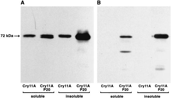
The genes cry11A(His6) and cry11A+p20(His6) were successfully expressed in recombinant E. coli after IPTG induction of the tac promoter, as indicated by protein separation on SDS-PAGE and Western blots using anti-Cry11A rabbit serum (Fig. 1A). Fractionation of the cell extracts by centrifugation showed that Cry11A was present in the fraction extractable with Tris buffer, as well as in the fraction that could be solubilized by 1% SDS in the presence of 8 M urea. As indicated in Fig. 1, the insoluble fraction of Cry11A accumulated in the presence of P20. It is unclear at present why the protein reacting with the anti-Cry11A antibodies is present in multiple bands. The most likely explanation is that Cry11A protein is processed at multiple sites (Fig. 1 and 3). The intensity of multiple bands increases upon coexpression of the p20 gene with the cry11A. This may indicate a different processing of Cry11A in the presence of P20 and is consistent with the conclusion that P20 interacts with Cry11A (see below).
FIG. 1.
Accumulation of Cry11A protein in E. coli and P. putida cells harboring different constructs. Soluble and insoluble fractions, prepared as indicated in Materials and Methods, corresponding to equivalent amount of cells, were loaded onto each lane. SDS-PAGE proteins were transferred to nitrocellulose filters by electroblotting and developed with anti-Cry11A antibodies. (A) E. coli. (B) P. putida.
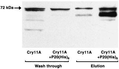
FIG. 3.
Cobinding of Cry11A and P20(His6) proteins to Ni2+ column. Crude extracts of cells, one containing Cry11A and the other containing P20(His6) protein, were mixed and loaded onto Ni2+ spin column (Qiagen). The column was washed with buffer containing 20 mM imidazole, followed by elution with the same buffer containing 200 mM imidazole. The Western blot of the “washing” and “elution” fractions was developed with anti-Cry11A primary antibodies.
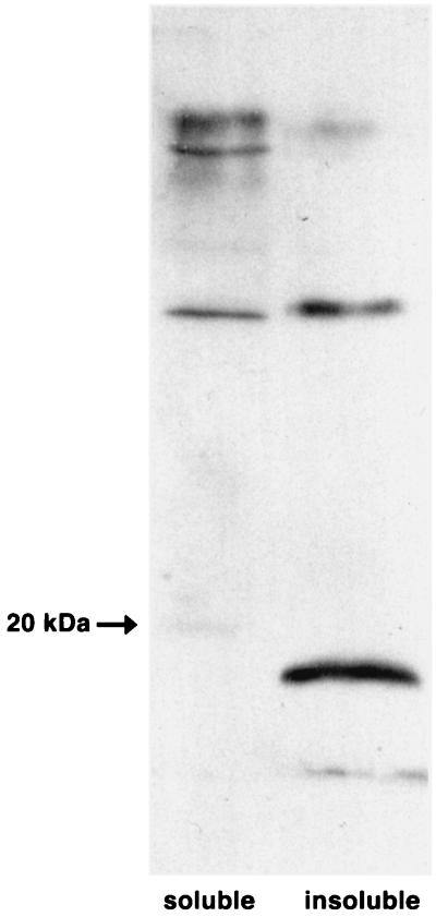
The synthesis of Cry11A(His6) was not detectable when cry11A gene was expressed in P. putida. However, when cry11A and p20 genes were expressed together, significant amounts of Cry11A protein were produced (Fig. 1B). A Western blot using anti-histidine antibodies showed that P20(His6) was present in the insoluble fraction in E. coli (Fig. 2). It runs slightly faster than expected from its predicted molecular weight. It is not known at present whether this is due to anomalous mobility or is a result of processing. Overall, the Western blots in Fig. 1 and 3 show that both recombinant E. coli and P. putida bacteria contained higher levels of Cry11A when the adjacent gene p20 was present on the same DNA fragment.
FIG. 2.
Accumulation of P20 protein in E. coli cells expressing cry11A+p20(His6) genes. Soluble and insoluble fractions were prepared as described in Materials and Methods. P20(His6) protein was detected with monoclonal antibodies against the histidine tag.
Bioassays of recombinant E. coli and P. putida.
Production of the Cry11A(His6) protein conferred measurable larvicidal activity to the recombinant E. coli cells, whether P20 was coproduced or not (Table 2). However, recombinant E. coli producing Cry11A+P20(His6) had more than a 10-fold increase in toxicity compared to recombinant E. coli producing Cry11A(His6) protein without P20 (Table 2). The differences were statistically significant because the 95% confidence intervals bounding the estimated LC50 values from the Cry11A+P20(His6) and Cry11A(His6) bioassays did not overlap (Table 2). Results with P. putida were more striking. When Cry11A(His6) was produced alone, there was no evident larval mortality. However, when cry11A+p20(His6) was expressed as a gene cluster, there was mortality in the same range of LC50 values as in E. coli (Table 2). Mortality in negative controls (i.e., E. coli and P. putida cells containing plasmids with no inserts but induced with IPTG) was negligible and, correspondingly, there were no estimated LC50s for them. Separately, constructs of E. coli DH5α expressing the p20 gene were assayed against Aedes aegypti larvae using the same protocol at a dilution of 7.4 × 10⁷ cells/ml, but there was no observed larval mortality (data not shown). To determine if there were differences in toxicity between strains producing Cry11A(His6) or Cry11A without the His6 extension, E. coli recombinants with one or the other gene were bioassayed as before. Results were as follows: Cry11A(His6), LC50 = 5.45 ± 4.96 to 5.68 (log10 cells/ml); Cry11A, LC50 = 5.67 ± 4.83 to 5.92 (log10 cells/ml). The 95% confidence intervals of these LC50 estimates overlap, indicating no difference in the toxicity of Cry11A(His6) and Cry11A without the His6 extension.
TABLE 2.
Results of bioassays against third-instar Aedes aegypti larvae using recombinant E. coli and P. putida expressing cry11A(His6) or cry11A+p20(His6)
| Strain and protein(s)a | LC50 ± 95% CIb (log10 cells/ml) | Concn range tested (log10 cells/ml) |
|---|---|---|
| E. coli DH5α | ||
| Cry11A | 5.35 ± 5.09 (to 5.55) | 4.52–7.42 |
| Cry11A+P20 | 4.17 ± 2.61 (to 4.74) | 3.18–7.42 |
| Control | NEc | 4.52–7.42 |
| P. putida PB2442 | ||
| Cry11A | NE | 4.52–7.42 |
| Cry11A+P20 | 4.59 ± 2.81 (to 5.33) | 3.18–7.42 |
| Control | NE | 4.52–7.42 |
Control, plasmid without gene cluster.
CI, confidence interval.
NE, no LC50 estimate due to scant mortality.
Coelution of P20 and Cry11A.
Results of the coelution experiment showed that more Cry11A was retained on the column when mixed with P20(His6) (Fig. 3, lanes 3 and 4). Cry11A protein that was not mixed with P20(His6) was mostly washed off the nickel chelate column by 20 mM imidazole (Fig. 3, lane 1), and only a small fraction was retained by the column (Fig. 3, lane 3). When P20(His6) was mixed with the Cry11A extract, demonstrably more Cry11A was retained by the Ni-NTA column (Fig. 3, lane 4) and correspondingly less was present in the wash fraction (Fig. 3, lane 2). Interestingly, the majority of Cry11A retained on the Ni-NTA column has a lower molecular mass than the 72-kDa protein detected in the cell extracts that expressed the cry11A gene (see also Fig. 1). Since the Ni-NTA chromatography was run at room temperature we believe that this band must be the result of processing. We favor processing, rather than nonspecific proteolytic degradation, since the band is very discrete and shows no signs of smearing. It is possible that Cry11A is processed differently upon the interaction with P20 as is also suggested by the results presented in Fig. 1, lanes 1 and 2.
DISCUSSION
The results reported here demonstrate expression of the cry11A gene at high levels in both E. coli and P. putida (when p20 gene was present), levels sufficient so that the Cry11A protein synthesized had retained larvicidal activity against Aedes aegypti at cell concentrations on the order of 104 and 105 cells/ml. It is important to note that Cry11A retained toxic activity when the histidine tag was present; thus, this tag did not interfere with protein activity; our bioassay data comparing strains of recombinant E. coli expressing cry11A or cry11A(His6) support this conclusion. Therefore, other studies in which the histidine tag was present were not confounded. Here, we inserted the coding sequence of the cry11A gene into vectors that provided the strong E. coli promoters (T5-lac) and ribosome-binding sites that are present in pQE and pMMB603 vectors. Previous investigations failed to document toxicity of recombinant E. coli cells carrying cry11A, apparently because of poor gene expression (2, 4, 8, 16). We are unaware of any other studies showing expression of cry11A and resultant larvicidal toxicity of recombinant Pseudomonas sp., although Thanabalu et al. (24) documented toxicity of C. crescentus, another gram-negative bacterium, when expressing cry genes from B. thuringiensis and B. sphaericus, while Liu et al. (15) demonstrated efficient synthesis and toxicity to mosquito larvae of a similar combination of toxins in the gram-negative bacterium A. excentris. Thus, on the basis of our results and those of others, expression of cry toxin genes cloned from B. thuringiensis H-14, B. sphaericus, or other sources and introduced into gram-negative bacteria could provide a new means for delivering these toxins to mosquito larvae. Because gram-negative bacteria are native and ubiquitous in larval mosquito habitats and can draw upon nutrient resources in these habitats through heterotrophic processes, they make suitable candidates toward this end (14, 15, 22). Indeed, our motivation to include P. putida here was that previously we found Pseudomonas sp. to comprise 111 of 830 (13.4%) of bacterial isolates characterized from an Anopheles mosquito habitat in Michigan (22).
Results of our studies comparing expression of cry11A(His6) with the gene cluster cry11A+p20(His6) suggest that Cry11A protein accumulated in bacterial cells in a predominantly insoluble form and that this process was enhanced by the coexpression of the p20 gene located downstream of the cry11A gene. The molecular mechanism of this enhancement is not known, but because these genes were induced simultaneously from the T5-lac promoter, the influence of the P20 protein on accumulation was likely posttranscriptional. Visick and Whiteley (27) documented expression of cry11A (cryIVD) in E. coli when p20 was also expressed; however, the protein observed on Western blots consisted of three products (the 72-kDa protein and two smaller proteins), and there were no accompanying bioassay data to demonstrate toxicity of the recombinants. Consequently, it was unclear from that study if the protein was active. P20 apparently enhances production of the 27-kDa protein Cyt1A, the 72-kDa Cry11A protein, and also β-galactosidase (mutant lacZX90) in E. coli (1, 16, 27). It also enhances production of Cry11A and Cyt1A in B. thuringiensis H-14 (31, 32; but see also reference 5). Thus, P20 may have a posttranscriptional, protein-stabilizing function. Strains of E. coli carrying mutations that reduce proteolytic activity produced amounts of Cyt1A similar to those of recombinants without these mutations but producing both Cyt1A and P20 (27), thus supporting the interpretation that P20 has a generalized stabilizing function. Our coexpression studies in which proteins were separated into soluble and insoluble fractions indicate that the presence of P20 protein increases the insoluble form of the Cry11A and that both proteins occur predominantly in the insoluble form in recombinant cells (Fig. 2 and 3).
Molecular chaperones have been defined as a family of proteins that mediate assembly of other polypeptides but are not components of the final functional protein structure (9). A 29-kDa protein encoded by the open reading frame adjacent to cry2A was suggested to enhance efficient production of the δ-endotoxin and was termed a chaperonin (6). More recently, Ge et al. (12) demonstrated that this 29-kDa protein facilitates formation of Cry2A inclusions, functioning possibly as a scaffolding protein. The coelution experiment reported here suggests that Cry11A bound to P20(His6), which itself was bound to the Ni2+ column, under conditions when the Cry11A lacked the histidine (Fig. 3). This observation supports the idea that Cry11A and P20 physically interacted. Thus, P20 would not function as a chaperone per se, following the above strict definition. Of interest is the difference between E. coli and P. putida recombinants in the apparent production or stabilization of Cry11A when P20 was present or absent. In the former strains, Cry11A was visible in Western blots whether P20 was coproduced or not, and the cells were measurably toxic, albeit those recombinants without P20 had a statistically higher LC50 (i.e., were less toxic) than did those expressing p20. In contrast, there was no visible Cry11A protein on Western blots when P20 was absent in P. putida, and these recombinants exhibited no toxicity to larvae, similar to the blank plasmid controls, compared to recombinants with Cry11A and P20(His6). Although the reasons for these differences are unknown, they indicate that P20 may have strong stabilizing functions in recombinant cells produced from different gram-negative species.
ACKNOWLEDGMENTS
We thank William Donovan for donating plasmid pEG261 DNA and Sarjit Gill for supplying an aliquot of antibodies to Cry11A for our initial protein screening. We also thank Michael Kaufman for review of the manuscript and Alexander Raikhel for donating Aedes aegypti eggs.
This study was supported by grant AI-21884 from the NIH.
REFERENCES
- 1.Adams L F, Visick J E, Whiteley H R. A 20-kilodalton protein is required for efficient production of the Bacillus thuringiensis subsp. israelensis 27-kilodalton crystal protein in Escherichia coli. J Bacteriol. 1989;171:521–530. doi: 10.1128/jb.171.1.521-530.1989. [DOI] [PMC free article] [PubMed] [Google Scholar]
- 2.Ben-Dov E, Boussiba S, Zaritsky A. Mosquito larvicidal activity of Escherichia coli with combinations of genes from Bacillus thuringiensis subsp. israelensis. J Bacteriol. 1995;177:2851–2857. doi: 10.1128/jb.177.10.2851-2857.1995. . (Erratum, 177:6319.) [DOI] [PMC free article] [PubMed] [Google Scholar]
- 3.Casadaban M C, Cohen S N. Analysis of gene control signals by DNA fusion in Escherichia coli. J Mol Biol. 1980;138:179–207. doi: 10.1016/0022-2836(80)90283-1. [DOI] [PubMed] [Google Scholar]
- 4.Chang C, Dai S M, Frutos R, Federici B A, Gill S S. Properties of a 72-kilodalton mosquitocidal protein from Bacillus thuringiensis subsp. morrisoni PG-14 expressed in B. thuringiensis subsp. kurstaki by using the shuttle vector pHT3101. Appl Environ Microbiol. 1992;58:507–512. doi: 10.1128/aem.58.2.507-512.1992. [DOI] [PMC free article] [PubMed] [Google Scholar]
- 5.Chang C, Yu Y M, Dai S M, Law S K, Gill S S. High-level cryIVD and cytA gene expression in Bacillus thuringiensis does not require the 20-kilodalton protein, and the coexpressed gene products are synergistic in their toxicity to mosquitoes. Appl Environ Microbiol. 1993;59:815–821. doi: 10.1128/aem.59.3.815-821.1993. [DOI] [PMC free article] [PubMed] [Google Scholar]
- 6.Crickmore N, Ellar D J. Involvement of a possible chaperonin in the efficient expression of a cloned CryIIA delta-endotoxin gene in Bacillus thuringiensis. Mol Microbiol. 1992;6:1533–1537. doi: 10.1111/j.1365-2958.1992.tb00874.x. [DOI] [PubMed] [Google Scholar]
- 7.Dervyn E, Poncet S, Klier A, Rapoport G. Transcriptional regulation of the cryIVD gene operon from Bacillus thuringiensis subsp. israelensis. J Bacteriol. 1995;177:2283–2291. doi: 10.1128/jb.177.9.2283-2291.1995. [DOI] [PMC free article] [PubMed] [Google Scholar]
- 8.Donovan W P, Dankocsik C, Gilbert M P. Molecular characterization of a gene encoding a 72-kilodalton mosquito-toxic crystal protein from Bacillus thuringiensis subsp. israelensis. J Bacteriol. 1988;170:4732–4738. doi: 10.1128/jb.170.10.4732-4738.1988. [DOI] [PMC free article] [PubMed] [Google Scholar]
- 9.Ellis R J, van der Vies S M. Molecular chaperones. Annu Rev Biochem. 1991;60:321–347. doi: 10.1146/annurev.bi.60.070191.001541. [DOI] [PubMed] [Google Scholar]
- 10.Franklin F C, Bagdasarian M, Bagdasarian M M, Timmis K N. Molecular and functional analysis of the TOL plasmid pWWO from Pseudomonas putida and cloning of genes for the entire regulated aromatic ring meta cleavage pathway Proc. Natl Acad Sci USA. 1981;78:7458–7462. doi: 10.1073/pnas.78.12.7458. [DOI] [PMC free article] [PubMed] [Google Scholar]
- 11.Fürste J P, Pansegrau W, Frank R, Blocker H, Scholz P, Bagdasarian M, Lanka E. Molecular cloning of the plasmid RP4 primase region in a multi-host-range tacP expression vector. Gene. 1986;48:119–131. doi: 10.1016/0378-1119(86)90358-6. [DOI] [PubMed] [Google Scholar]
- 12.Ge B, Bideshi D, Moar W J, Federici B A. Differential effects of helper proteins encoded by the cry2A and cry11A operons on the formation of Cry2A inclusions in Bacillus thuringiensis. FEMS Microbiol Lett. 1998;165:35–41. doi: 10.1111/j.1574-6968.1998.tb13124.x. [DOI] [PubMed] [Google Scholar]
- 13.Harlow E, Lane D. Antibodies: a laboratory manual. Cold Spring Harbor, N.Y: Cold Spring Harbor Laboratory Press; 1988. [Google Scholar]
- 14.Kaufman M G, Walker E D, Smith T W, Merritt R W, Klug M J. Effects of larval mosquitoes (Aedes triseriatus) and stemflow on microbial community dynamics in container habitats. Appl Environ Microbiol. 1999;65:2661–2673. doi: 10.1128/aem.65.6.2661-2673.1999. [DOI] [PMC free article] [PubMed] [Google Scholar]
- 15.Liu J W, Yap W H, Thanabalu T, Porter A G. Efficient synthesis of mosquitocidal toxins in Asticcacaulis excentricus demonstrates potential of gram-negative bacteria in mosquito control. Nat Biotechnol. 1996;14:343–347. doi: 10.1038/nbt0396-343. [DOI] [PubMed] [Google Scholar]
- 16.McLean K M, Whiteley H R. Expression in Escherichia coli of a cloned crystal protein gene of Bacillus thuringiensis subsp. israelensis. J Bacteriol. 1987;169:1017–1023. doi: 10.1128/jb.169.3.1017-1023.1987. [DOI] [PMC free article] [PubMed] [Google Scholar]
- 17.Overbye Michel L, Sandkvist M, Bagdasarian M. Specificity of the protein secretory apparatus: secretion of the heat-labile enterotoxin B subunit pentamers by different species of Gram− bacteria. Gene. 1995;152:41–45. doi: 10.1016/0378-1119(94)00691-k. [DOI] [PubMed] [Google Scholar]
- 18.Poncet S, Deleclus A, Klier A, Rapoport G. Evaluation of synergistic interactions among CryIVA, CryIVB and CryIVD toxic components of Bacillus thuringiensis subsp. israelensis crystals. J Invert Pathol. 1995;66:131–133. [Google Scholar]
- 19.Porter A G, Davidson E W, Liu J W. Mosquitocidal toxins of bacilli and their genetic manipulation for effective biological control of mosquitoes. Microbiol Rev. 1993;57:838–861. doi: 10.1128/mr.57.4.838-861.1993. [DOI] [PMC free article] [PubMed] [Google Scholar]
- 20.Sambrook J, Fritsch E F, Maniatis T. Molecular cloning: a laboratory manual. 2nd ed. Cold Spring Harbor, N.Y: Cold Spring Harbor Press; 1989. [Google Scholar]
- 21.SAS Institute. The PROBIT procedure, SAS User's guide: statistics. 5th ed. Cary, N. C.: SAS Institute; 1985. pp. 639–645. [Google Scholar]
- 22.Smith T W, Walker E D, Kaufman M G. Bacterial density and survey of cultivable heterotrophs in the surface water of a freshwater marsh habitat of Anopheles quadrimaculatus larvae (Diptera: Culicidae) J Am Mosq Control Assoc. 1998;14:72–77. [PubMed] [Google Scholar]
- 23.Tabor S, Richardson C C. A bacteriophage T7 RNA polymerase/promoter system for controlled exclusive expression of specific genes. Proc Natl Acad Sci USA. 1985;82:1074–1078. doi: 10.1073/pnas.82.4.1074. [DOI] [PMC free article] [PubMed] [Google Scholar]
- 24.Thanabalu T, Berry C, Hindley J. Cytotoxicity and ADP-ribosylating activity of the mosquitocidal toxin from Bacillus sphaericus SSII-1: possible roles of the 27- and 70-kilodalton peptides. J Bacteriol. 1993;175:2314–2320. doi: 10.1128/jb.175.8.2314-2320.1993. [DOI] [PMC free article] [PubMed] [Google Scholar]
- 25.Thanabalu T, Hindley J, Brenner S, Oei C, Berry C. Expression of the mosquitocidal toxins of Bacillus sphaericus and Bacillus thuringiensis subsp. israelensis by recombinant Caulobacter crescentus, a vehicle for biological control of aquatic insect larvae. Appl Environ Microbiol. 1992;58:905–910. doi: 10.1128/aem.58.3.905-910.1992. . (Erratum, 58:1794.) [DOI] [PMC free article] [PubMed] [Google Scholar]
- 26.Thiery I, Sinegre G, Tandeau de Marsac N. Occurrence and abundance of cyanobacteria in a brackish marshland and their ingestibility by mosquito larvae. Bull Soc Vector Ecol. 1993;18:164–173. [Google Scholar]
- 27.Visick J E, Whiteley H R. Effect of a 20-kilodalton protein from Bacillus thuringiensis subsp. israelensis on production of the CytA protein by Escherichia coli. J Bacteriol. 1991;173:1748–1756. doi: 10.1128/jb.173.5.1748-1756.1991. [DOI] [PMC free article] [PubMed] [Google Scholar]
- 28.Wirth M C, Federici B A, Walton W E. Cyt1A from Bacillus thuringiensis synergizes activity of Bacillus sphaericus against Aedes aegypti (Diptera: Culicidae) Appl Environ Microbiol. 2000;66:1093–1097. doi: 10.1128/aem.66.3.1093-1097.2000. [DOI] [PMC free article] [PubMed] [Google Scholar]
- 29.Wirth M C, Georghiou G P, Federici B A. CytA enables CryIV endotoxins of Bacillus thuringiensis to overcome high levels of CryIV resistance in the mosquito, Culex quinquefasciatus Proc. Natl Acad Sci USA. 1997;94:10536–10540. doi: 10.1073/pnas.94.20.10536. [DOI] [PMC free article] [PubMed] [Google Scholar]
- 30.Wu D, Chang F N. Synergism in mosquitocidal activity of 26- and 65-kDa proteins from Bacillus thuringiensis subsp. israelensis crystal. FEBS Lett. 1985;190:232–236. [Google Scholar]
- 31.Wu D, Federici B A. A 20-kilodalton protein preserves cell viability and promotes CytA crystal formation during sporulation in Bacillus thuringiensis. J Bacteriol. 1993;175:5276–5280. doi: 10.1128/jb.175.16.5276-5280.1993. [DOI] [PMC free article] [PubMed] [Google Scholar]
- 32.Wu D, Federici B A. Improved production of the insecticidal CryIVD protein in Bacillus thuringiensis using cryIA(c) promoters to express the gene for an associated 20-kDa protein. Appl Microbiol Biotechnol. 1995;42:697–702. doi: 10.1007/BF00171947. [DOI] [PubMed] [Google Scholar]
- 33.Wu D, Johnson J J, Federici B A. Synergism of mosquitocidal toxicity between CytA and CryIVD proteins using inclusions produced from cloned genes of Bacillus thuringiensis. Mol Microbiol. 1994;13:965–972. doi: 10.1111/j.1365-2958.1994.tb00488.x. [DOI] [PubMed] [Google Scholar]


