

Abstract
Purpose
To compare the refractive outcome and residual accommodation with respect to various degrees of iris and skin pigmentation in hypermetropic children using 2 drops of cyclopentolate 1% (C + C) or 1 drop of cyclopentolate 1% and 1 drop of tropicamide 1% (C + T).
Methods
Two hundred fifty‐one hypermetropic children were classified according to iris and skin pigmentation (light, medium, dark) and received randomized and double‐blind C + C or C + T. Refractive error (spherical equivalent, SEQ) was determined using the Retinomax‐K + 3. In 204 subjects, residual accommodation (RA) was determined using the PlusoptiX PowerRefractor.
Results
A linear mixed model with a light‐irided and light skin‐pigmented reference group receiving C + T (mean SEQ +3.10 ± 1.87D) indicated significant less hypermetropia in subjects with a dark iris having a medium‐ and dark‐pigmented skin in C + T, −1.02 ± 0.29 (−1.59/−0.45) and −1.53 ± 0.30 (−2.10/−0.95); and in subjects having a light‐, medium‐ and dark‐pigmented skin in C + C, −0.74 ± 0.34 (−1.41/−0.06), −1.26 ± 0.30 (−1.85/−0.66) and −1.84 ± 0.30 (−2.42/−1.26). Similar findings were present for RA. Our model with a light‐irided and light skin‐pigmented reference group receiving C + T (mean RA +0.84 ± 0.61D) indicated significantly higher RA in dark‐irided subjects with medium‐ and dark‐pigmented skin in C + T, +1.05 ± 0.19 (+0.67/+1.43) and +1.35 ± 0.20 (+0.9/+1.74), and in C + C, +1.13 ± 0.21 (+0.71/+1.55) and +1.90 ± 0.19 (+1.51/+2.28).
Conclusions
We found solid evidence that skin pigmentation rather than iris pigmentation is the decisive factor for effectiveness of cycloplegics. Awareness of the limitations of cycloplegic regimens in dark‐irided/pigmented children is needed. Our study showed that cyclopentolate 1% combined with tropicamide 1% provides more accurate refractive outcomes both statistically and clinically integrating the factor skin pigmentation for dark‐irided subjects.
Keywords: dark iris, cyclopentolate, cycloplegics, hypermetropia, residual accommodation, pigmentation
Introduction
Two doses of cyclopentolate 1% and one drop of cyclopentolate 1% combined with one drop of tropicamide 1% are commonly used regimes to assess the refractive error of children. For both regimes, increasing skin pigmentation and crying were found to significantly affect hypermetropic outcome in children with a dark iris (van Minderhout et al. 2019). It is conceivable that the lower hypermetropic outcome found with increasing skin pigmentation and/or crying will also be reflected in subjects with lighter coloured irises.
Although hypermetropic outcome is primarily of interest in paediatric objective refraction, the amount of residual accommodation, for example depth of cycloplegia, is clinically important as the two objectives are correlated. To ensure optimal refractive outcome, residual accommodation should not exceed 1.75D (Gettes & Belmont, 1961).
Scientific literature agrees that in dark‐irided subjects, cycloplegics are less effective in obtaining paralysis of accommodation (Egashira et al. 1993; Khurana et al. 1988; Miranda & Juan 1972; Nishizawa et al. 1988; Pinheiro & Netto 2000; Siu et al. 1999). Manny et al. (1993) found a residual accommodation as high as 2.50D using cyclopentolate 1% in Black African Americans. In contrast, Ebri et al. (2007) reported a significantly smaller residual accommodation of about 0.63 ± 0.06D after cyclopentolate 1% and 0.63 ± 0.05D in cyclopentolate 1% combined with one drop of tropicamide 1% in Nigerian children. The latter study, however, with 70% of the subjects classified as emmetropic, used dynamic near retinoscopy in uncorrected subjects; at starting point, a considerable amount of accommodation (i.e. 2D at 50 cm) is already demanded. Their values therefore are very likely to be biased. Furthermore, the standard deviations of 0.06D and 0.05D indicate the absence of variation between individuals. This does not reflect the clinical findings we encounter in the general population.
Using only iris colour as a decisive element or predictor for residual accommodation in daily clinical practice would go beyond the main goal; an outcome that reflects the actual refractive value of the child. We observed that with increasing skin pigmentation, higher amounts of residual accommodation were present. It seems that a similar negative relation, that is increased residual accommodation in increasing iris and skin pigmentation, is present.
It is also conceivable that different cycloplegic regimes provide significantly different outcomes, especially when incorporating eye and skin pigmentation and crying upon application. Various studies investigated residual accommodation and/or hypermetropic outcome in a population with various degrees of iris pigmentation but did not incorporate the factor we suspect is most likely important; the factor skin pigmentation. A customized treatment should be made for every child. This can only be done if all relevant factors are known. Therefore, the purpose of this study was to investigate the effectiveness of one drop of cyclopentolate 1% combined with one drop of tropicamide 1% (C + T) and two drops of cyclopentolate 1% (C + C), in terms of (1) refractive outcome and (2) residual accommodation, in general and with respect to iris colour, skin pigmentation and crying upon eye drop application in hypermetropic children.
Methods
Ethical considerations
Ethical approval for this study was received from the Central Committee on Research Involving Human Subjects (CCMO; NL32954.098) and the Medical Research Ethics Committee, the Southwest Netherlands (MREC‐ZWN; 1010‐077). The study was conducted according to the principles of the Declaration of Helsinki and applicable Dutch acts. The study was admitted to the Dutch Trial Register (NL2369).
Study design and selection procedures
The study was a randomized, double‐blind clinical trial with a within‐ and between‐subject design. The study population was recruited at an ophthalmology outpatient clinic in a Dutch metropolitan hospital (HMC Westeinde, The Hague, The Netherlands). In all 7‐ to 12‐year‐old children requiring cycloplegic retinoscopy, the near‐retinoscopy technique of Mohindra (1977) was used to assess the noncycloplegic refractive state. Spherical equivalent was calculated with the formula: spherical equivalent (SEQ) =sphere + ½ cylinder. Subjects with a SEQ of ≥+0.25 dioptres (D) and sufficient accommodation, that is >10D with dynamic retinoscopy using the Nott technique (Antona et al. 2009) and a best‐corrected visual acuity of ≥0.7, were asked to participate. Subjects meeting the selection criteria were included in the study after oral and written consent of the parents.
The number of participants was calculated to detect a mean difference in residual accommodation of 0.50 ± 0.60D at a minimum between C + C and C + T. Calculations were made with an alpha of 0.05 and 90% power, yielding a sample size of 31 subjects for each regime. 75 subjects were added for the additional variables yielding a sample size of 137 subjects (274 eye measurements).
Categorization in eye and skin pigmentation groups
The iris colour of the subjects was classified as either ‘light’, ‘medium’, ‘dark’ or ‘very dark’ using the iris pigmentation reference set of Fransen et al. (2008), where 1–14 represented ‘light’, 15–20 ‘medium’, 21 and 22 ‘dark’ and 23 and 24 ‘very dark’. Constitutional skin colour represents an individual’s baseline, or the colour of skin that has not been altered by sun or other types of ultraviolet (UV) exposure. To determine inter‐observer reliability of the eye colour classification system, this was classified by 2 individual observers (investigator and research assistant). In case of discrepancy, the skin colour classification of the investigator was used for statistical analysis.
Organization of interventions
For this study, unit‐dose minims of cyclopentolate hydrochloride 1% and tropicamide 1% (Bausch & Lomb Ltd) were used. Since both interventions had cyclopentolate 1% as first dose, these minims were unblinded. For the second drop, the minims were blinded by removing the small print on the minims and labelled according to their randomization. Management and randomization of interventions was performed by the hospital pharmacy. The randomizations were not known to the participant and/or parents, research assistant and/or conducting researcher creating a double‐blind set‐up. Participants received either C + C or C + T in a double‐blind randomized manner in both eyes. At first, cyclopentolate 1% was instilled unblinded; right eye first and left eye second. With an interval of 5 min, the blinded second eye drop was administered in a similar way. Crying upon or immediately following eye drop, installation was noted.
Measurements
Residual accommodation was measured objectively using the PlusoptiX S04 PowerRefractor II photorefractor (PlusoptiX GmbH, Nuremberg). At a distance of 1 metre, infrared light of 790 nm is projected into the eye and reflected from the retina. The PowerRefractor performs continuous measurements of accommodation (50 Hz, 0.02 seconds, samples) and requires pupil sizes between 3.7 and 7.8 mm. In case of failure of measurements in large pupils, that is light overexposure, a neutral density (ND) filter was used. Neutral density (ND) filters decrease the intensity of light without selectively affecting specific wavelengths of light from the source. We used ND filter OD 0.04 (Newport Corporation, Irvine, USA), which results in a 91% transmission percentage at the 790‐nm wavelength. Since the infrared light is attenuated twice, encountering the ND filter going towards the eye and encountering the ND filter following reflection from the eye, the amount of light reaching the device was 83%. The ND filter was fitted in a hand‐held frame (Fig. 1). The use of the ND filter was validated prior to use in this research (van Minderhout et al. 2021, submitted).
Fig. 1.
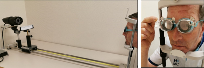
Test set‐up using an adult subject; positioned at 1‐metre distance from the PlusoptiX PowerRefractor, wearing a trial frame with full Retinomax refractive values. A Radner LogMAR reading chart text representing 0.8 decimal was attached directly under the measuring device. Subjects read the text loudly, while the text was slowly moved towards the subject. In case of failure due to a large pupil size, a 0.04 ND filter was used to attenuate the transmitted infrared light.
At 50 min after the first eye drop, the refraction was assessed with the Nikon Retinomax‐K + 3. Immediately thereafter, subjects were positioned in a head and chin rest at 1‐m distance from the measuring device (Fig. 1). Subjects wore a silicon trial frame with the exact Retinomax‐K + 3 values (sphere, cylinder, axis) plus 1D sphere to correct for the measuring distance at 1 metre and thereby obtain a neutral refraction. A Radner LogMAR reading chart text (Maaijwee et al. 2008) representing 0.8 decimal acuity was attached directly under the measuring device. Subjects read the text loudly, while the text was moved towards the subject. Objective video monitoring of right and left eye accommodation took place simultaneously. The PowerRefractor readings of left and right eye in dioptres were noted in 2 decimal points. Spherical equivalent (SEQ) of right and left eye Nikon Retinomax‐K + 3 measurements was calculated. Values were noted up to two decimals.
Outcome measures were 50‐min residual accommodation and SEQ following C + C and C + T, as well as residual accommodation and SEQ with respect to eye and skin pigmentation and crying. The 50‐min SEQ analyses comprised all the subjects included for residual accommodation measurements and all subjects with a 50‐min SEQ of ≥0.25D that were included in an ongoing parallel study, which investigated the recuperation time from cycloplegia.
Statistical analysis
Data were analysed in SPSS 26 for Windows. Differences were considered statistically significant if p < 0.05. A difference in refractive outcome of ≥0.25D was considered clinically significant. Inter‐observer reliability of eye colour classification was determined with the Kappa coefficient of agreement. To correct for correlations between right and left eye measurements, a linear mixed model approach with a between‐ and within‐subject design was used. The Pearson correlation (r) was used to investigate the relation between residual accommodation and iris colour, as well as skin pigmentation.
Results
Two hundred four subjects, 102 C + T and 102 C + C, were included. The study comprised 96 girls, 46 in C + T and 50 in C + C; and 108 boys, 56 in C + T and 52 in C + C. The mean age of the subjects was 8.93 ± 1.49 in C + C and 8.96 ± 1.60 in C + T. In C + T and C + C, 202 and 201 refractive outcomes were generated by the PowerRefractor and analysed, respectively. For residual accommodation measurements, this was 200 in C + T 200 and 197 in C + C 197. The inter‐observer agreement for classification of iris colour and skin pigmentation was excellent: kappa 0.95 (p < 0.001) and kappa 0.92 (p < 0.001). There was a strong association between iris colour and skin pigmentation (r = 0.832, p < 0.001, n = 204). Neither in C + T nor in C + C, subjects cried upon or immediately following eye drop application.
The mean hypermetropia in right and left eye was in C + T +2.50 ± 1.75D and +2.46 ± 1.73D, and in C + C 2.27 ± 1.67D and 2.31 ± 1.73D. There was no statistically nor clinically significant difference in hypermetropic outcome between C + T and C + C; 0.16 SE 0.15D, 95% CI −0.14 to 0.45D, p = 0.297. The mean residual accommodation of right and left eye was in C + T 1.47 ± 1.07D and 1.39 ± 0.99D, and in C + C 1.63 ± 1.21D and 1.56 ± 1.33D. There was no statistically significant difference in residual accommodation between C + T and C + C; −0.16 SE 0.12D, 95% CI −0.39 to 0.07D, p = 0.169.
Hypermetropic outcome
Using subjects with a light‐pigmented iris receiving C + T as the reference group, statistically and clinically significant lower hypermetropia was present in subjects with a dark and very dark iris in both interventions (Table 1A). As a second step, we added the factor skin pigmentation to the interaction model. However, small sample sizes were present, especially in the clinically interesting combinations in darker pigmented eyes in the interaction model between eye colour and skin pigmentation. Small sample sizes cause power issues and adjacent type I and II errors. Therefore, we clustered the iris colour groups Dark and Very Dark (Table 1B). The interaction model between iris and skin colour, using light‐pigmented and light‐irided subjects as the reference group, showed statistically and clinically less hypermetropia in dark‐irided subjects with a dark‐ and a medium‐pigmented skin in both interventions (Table 2A).
Table 1.
(A) Analyses of iris pigmentation as factor in mean refractive outcome, following one dose of cyclopentolate 1% and one dose of tropicamide 1% (C + T; N = 202) and a double dose of cyclopentolate 1% (C + C; N = 201); and a linear mixed model approach with a between‐ and within‐subject design, with subjects with a light iris colour receiving C + T as the reference group. Differences in mean spherical equivalent (SEQ) in dioptres are displayed. (B) Results after clustering iris colour group Dark and Very Dark. (C) Results after adding 47 dark‐irided subjects to the model
|
(A) Spherical equivalent Intervention*Iris colour |
N c |
Mean SEQ ± SD d |
Mean SEQ difference | SE | 95% CI | p | |
|---|---|---|---|---|---|---|---|
| lower bound | Upper bound | ||||||
| C + C a Very Dark | 50 | −1.82 | 0.31 | −2.44 | −1.21 | 0.000 | |
| C + C a Dark | 42 | −1.08 | 0.33 | −1.72 | −0.43 | 0.001 | |
| C + C a Medium | 57 | −0.38 | 0.30 | −0.98 | +0.21 | 0.207 | |
| C + C a Light | 52 | −0.12 | 0.31 | −0.73 | +0.49 | 0.703 | |
| C + T b Very Dark | 38 | −1.64 | 0.34 | −2.30 | −0.97 | 0.000 | |
| C + T b Dark | 58 | −0.79 | 0.30 | −1.38 | −0.19 | 0.010 | |
| C + T b Medium | 50 | −0.39 | 0.31 | −1.00 | +0.24 | 0.225 | |
| C + T b Light | 56 | +3.10 ± 1.87 | 0 e | 0.00 | |||
|
(B) Spherical equivalent Clustered groups dark–very dark Intervention*Iris colour |
N c |
Mean SEQ ± SD d |
Mean SEQ difference | SE | 95% CI | p | |
|---|---|---|---|---|---|---|---|
| Lower bound | Upper bound | ||||||
| C + C a Dark | 92 | −1.48 | 0.28 | −2.02 | −0.94 | 0.000 | |
| C + C a Medium | 57 | −0.38 | 0.31 | −0.99 | +0.22 | 0.212 | |
| C + C a Light | 52 | −0.12 | 0.31 | −0.74 | +0.50 | 0.706 | |
| C + T b Dark | 96 | −1.12 | 0.28 | −1.66 | −0.58 | 0.000 | |
| C + T b Medium | 50 | −0.38 | 0.32 | −1.00 | +0.24 | 0.230 | |
| C + T b Light | 56 | +3.10 ± 1.87 | 0 e | 0.00 | |||
|
C) Spherical equivalent +47 Dark‐irided subjects Intervention*Iris colour |
N c |
Mean SEQ ± SD d |
Mean SEQ Difference | SE | 95% CI | p | |
|---|---|---|---|---|---|---|---|
| Lower Bound | Upper Bound | ||||||
| C + C a Dark | 139 | −1.37 | 0.25 | −1.87 | −0.87 | 0.000 | |
| C + C a Medium | 57 | −0.38 | 0.31 | −0.98 | +0.23 | 0.222 | |
| C + C a Light | 52 | −0.27 | 0.33 | −0.92 | +0.38 | 0.417 | |
| C + T b Dark | 143 | −1.12 | 0.25 | −1.61 | −0.62 | 0.000 | |
| C + T b Medium | 50 | −0.38 | 0.32 | −1.01 | +0.24 | 0.229 | |
| C + T b Light | 56 | +3.10 ± 1.87 | 0 e | 0 | |||
C + C: two drops of cyclopentolate 1%.
C + T: one drop of cyclopentolate 1% and one drop of tropicamide 1%.
Number of measurements.
SEQ ± SD: spherical equivalent and standard deviation in dioptres.
Reference group.
Table 2.
(A) Analyses of eye and skin pigmentation as factor in the mean refractive outcome, following a double dose of cyclopentolate 1% (C + C; N = 201) and one dose of cyclopentolate 1% and one dose of tropicamide 1% (C + T; N = 202); a linear mixed model approach with a between‐ and within‐subject design, with subjects with a light iris colour and light skin pigmentation receiving C + T as the reference group. Difference in mean spherical equivalent (SEQ) in dioptres is displayed. (B) Results after adding 47 dark‐irided subjects to the model
|
A Spherical equivalent Clustered groups Dark–Very Dark Intervention*Iris colour*Skin pigmentation |
N c |
Mean SEQ ± SD d |
Mean SEQ difference | SE | 95% CI | p | |
|---|---|---|---|---|---|---|---|
| Lower bound | Upper bound | ||||||
| C + Ca Dark*Dark | 44 | −1.96 | 0.32 | −2.60 | −1.33 | 0.000 | |
| C + Ca Dark*Medium | 36 | −1.16 | 0.34 | −1.83 | −0.48 | 0.001 | |
| C + Ca Dark*Light | 12 | −0.67 | 0.51 | −1.67 | +0.34 | 0.191 | |
| C + Ca Medium*Medium | 22 | −0.72 | 0.40 | −1.51 | +0.07 | 0.074 | |
| C + Ca Medium*Light | 35 | −0.17 | 0.35 | −0.85 | +0.51 | 0.623 | |
| C + Ca Light*Light | 52 | −0.12 | 0.31 | −0.73 | +0.49 | 0.702 | |
| C + Tb Dark*Dark | 44 | −1.55 | 0.32 | −2.18 | −0.91 | 0.000 | |
| C + Tb Dark*Medium | 46 | −0.90 | 0.32 | −1.53 | −0.27 | 0.005 | |
| C + Tb Dark*Light | 6 | +0.27 | 0.69 | −1.08 | +1.62 | 0.696 | |
| C + Tb Medium*Medium | 22 | −0.64 | 0.40 | −1.44 | +0.15 | 0.112 | |
| C + Tb Medium*Light | 28 | −0.17 | 0.37 | −0.90 | +0.56 | 0.639 | |
| C + Tb Light*Light | 56 | +3.10 ± 1.87 | 0 e | 0.00 | |||
|
B Spherical equivalent +47 Dark‐irided subjects Intervention*Iris colour*Skin pigmentation |
N c |
Mean SEQ ± SD d |
Mean SEQ difference | SE | 95% CI | p | |
|---|---|---|---|---|---|---|---|
| Lower bound | Upper bound | ||||||
| C + Ca Dark*Dark | 56 | −1.84 | 0.30 | −2.42 | −1.26 | 0.000 | |
| C + Ca Dark*Medium | 51 | −1.26 | 0.30 | −1.85 | −0.66 | 0.000 | |
| C + Ca Dark*Light | 32 | −0.74 | 0.34 | −1.41 | −0.06 | 0.032 | |
| C + Ca Medium*Medium | 22 | −0.72 | 0.39 | −1.49 | +0.06 | 0.069 | |
| C + Ca Medium*Light | 35 | −0.18 | 0.34 | −0.84 | +0.49 | 0.599 | |
| C + Ca Light*Light | 52 | −0.12 | 0.30 | −0.71 | +0.47 | 0.693 | |
| C + Tb Dark*Dark | 56 | −1.53 | 0.30 | −2.10 | −0.95 | 0.000 | |
| C + Tb Dark*Medium | 61 | −1.02 | 0.29 | −1.59 | −0.45 | 0.000 | |
| C + Tb Dark*Light | 26 | −0.55 | 0.37 | −1.27 | +0.17 | 0.131 | |
| C + Tb Medium*Medium | 22 | −0.64 | 0.39 | −1.42 | +0.13 | 0.103 | |
| C + Tb Medium*Light | 28 | −0.18 | 0.36 | −0.89 | +0.53 | 0.619 | |
| C + Tb Light*Light | 56 | +3.10 ± 1.87 | 0 e | 0 | |||
C + C: two drops of cyclopentolate 1%.
C + T: one drop of cyclopentolate 1% and one drop of tropicamide 1%.
Number of measurements.
SEQ ± SD: spherical equivalent and standard deviation in dioptres.
Reference group.
Despite clustering, a relatively small sample size was present in the clinically highly interesting group of subjects with a dark iris colour‐ and a light‐pigmented skin. To enhance power, we added 47 dark‐irided noncrying subjects who received C + T and C + C in a previous study. This study compared 5 drops of atropine 0.5% with C + T and C + C in 3‐ to 6‐year‐old hypermetropic children with a dark iris (van Minderhout et al. 2019). Adding these 47 subjects virtually did not change the mean overall difference in the combined groups Dark (Table 1C). The interaction model between iris and skin colour, using light‐pigmented and light‐irided subjects as the reference group, showed virtually no difference in outcomes in dark‐irided subjects with a dark‐ and a medium‐pigmented skin in both interventions (Table 2B). However, in dark‐irided subjects with a light‐pigmented skin, the increase from 12 to 32 subjects had significant influence in intervention C + C, where the p value altered form 0.191 to 0.032. Despite this significance, the 95% confidence outer limit of −0.06 indicated that clinically insignificant difference might exist in the true population.
Residual accommodation
As expected from the previous analysis, we found a highly clinically and statistically significant impact of the factor iris pigmentation on residual accommodation (Table 3A). Integrating skin pigmentation in the model, using light‐pigmented and light‐irided subjects as the reference group, showed significantly increased residual accommodation in subjects with a medium and dark skin pigmentation in subjects with a dark and very dark iris (Table 3B). To overcome small sample sizes in the clinically interesting combinations of eye and skin pigmentations, we clustered the iris colour groups Dark and Very Dark.
Table 3.
(A) Analyses of iris pigmentation as factor in the mean residual accommodation (RA), following one dose of cyclopentolate 1% and one dose of tropicamide 1% (C + T; N = 200) and a double dose of cyclopentolate 1% (C + C; N = 197); and a linear mixed model approach with a between‐ and within‐subject design, with subjects with a light iris colour receiving C + T as the reference group. Difference in mean residual accommodation in dioptres is displayed. (B) Results after adding factor skin pigmentation to the model, with subjects with a light iris colour and light skin pigmentation receiving C + T as the reference group
|
(A) Residual accommodation Intervention *Iris colour |
N a |
Mean RA ± SD d |
Mean RA difference |
SE | 95% CI | p | |
|---|---|---|---|---|---|---|---|
| lower bound | Upper bound | ||||||
| C + C b Very Dark | 50 | +1.64 | 0.19 | +1.26 | +2.02 | 0.000 | |
| C + C b Dark | 42 | +1.20 | 0.20 | +0.80 | +1.60 | 0.000 | |
| C + C b Medium | 53 | +0.26 | 0.19 | −0.11 | +0.63 | 0.172 | |
| C + C b Light | 52 | +0.05 | 0.19 | −0.32 | +0.43 | 0.786 | |
| C + T c Very Dark | 37 | +1.43 | 0.21 | +1.01 | +1.84 | 0.000 | |
| C + T c Dark | 58 | +0.97 | 0.19 | +0.61 | +1.34 | 0.000 | |
| C + T c Medium | 50 | +0.20 | 0.19 | −0.18 | +0.58 | 0.298 | |
| C + T c Light | 55 | 0.84 ± 0.61 | 0 | ||||
|
(B) Residual accommodation Intervention*Iris colour*Skin pigmentation |
N c |
Mean RA ± SD d |
Mean RA difference | SE | 95% CI | p | |
|---|---|---|---|---|---|---|---|
| Lower bound | Upper bound | ||||||
| C + Ca Dark*Dark | 44 | +1.90 | 0.19 | +1.51 | +2.28 | 0.000 | |
| C + Ca Dark*Medium | 36 | +1.13 | 0.21 | +0.71 | +1.55 | 0.000 | |
| C + Ca Dark*Light | 12 | +0.55 | 0.31 | −0.06 | +1.15 | 0.076 | |
| C + Ca Medium*Medium | 18 | +0.45 | 0.29 | −0.12 | +1.02 | 0.120 | |
| C + Ca Medium*Light | 35 | +0.19 | 0.20 | −0.21 | +0.59 | 0.343 | |
| C + Ca Light*Light | 52 | +0.05 | 0.19 | −0.32 | +0.42 | 0.781 | |
| C + Tb Dark*Dark | 43 | +1.35 | 0.20 | +0.96 | +1.74 | 0.000 | |
| C + Tb Dark*Medium | 46 | +1.05 | 0.19 | +0.67 | +1.43 | 0.000 | |
| C + Tb Dark*Light | 6 | +0.34 | 0.42 | −0.12 | +1.76 | 0.300 | |
| C + Tb Medium*Medium | 22 | +0.41 | 0.26 | −0.11 | +0.93 | 0.116 | |
| C + Tb Medium*Light | 28 | +0.08 | 0.22 | −0.34 | +0.51 | 0.701 | |
| C + Tb Light*Light | 55 | 0.84 ± 0.61 | 0 | 0 | |||
C + C: two drops of cyclopentolate 1%.
C + T: one drop of cyclopentolate 1% and one drop of tropicamide 1%.
Number of measurements.
RA ± SD: residual accommodation and standard deviation in dioptres.
Reference group.
Since good cycloplegia is the base for an optimal hypermetropic measurement, we investigated the relation between residual accommodation and the factors eye and skin pigmentation. Controlling for right and left eye measurements and intervention, a highly statistically inverse correlation was found between iris pigmentation and residual accommodation, r = −0.513, p < 0.000 (n = 397). Cohen’s classification indicated a large effect size, with 26.3% of the variance in residual accommodation explained by eye pigmentation. Additional controlling for skin pigmentation altered the relation significantly, r = −0.163, p = 0.001, with eye colour explaining only 2.7% of the variance in residual accommodation, and indicating the significant impact of the factor skin pigmentation.
Discussion
One aim of this study was to investigate the effect of crying in the whole range of eye and skin pigmentations. Earlier research showed significantly less hypermetropia in crying subjects with a dark iris and a pigmented skin (van Minderhout et al. 2019). To our surprise, none of the children in the present study cried or even had resistance towards eye drop application. In the former study, the children were aged 3–6 years. The children who participated in the present study were older and were able to comprehend given information more easily. Therefore, the question whether crying also affects outcome in lighter pigmented subjects remains open. However, due to the absence of this specific confounder true comparisons could be made for the other variables.
It is generally accepted that in darker pigmented eyes, cyclopentolate 1% seems less effective in reaching optimal depth of cycloplegia. Manny et al. (1993) found a mean residual accommodation of 0.99 ± 0.47D in light‐pigmented irises, 1.20 ± 0.86D in dark‐pigmented ones and 2.49 ± 0.62D in the very dark irises of Black African Americans. Similar findings were present in the study of Miranda & Juan (1972); reporting 0.70 ± 0.25D in subjects with a light‐pigmented iris and 2.22 ± 0.96D in subjects with a dark to very dark‐pigmented iris. In a dark‐irided Brazilian (Pinheiro & Netto, 2000) and Korean (Choi et al. 1964) population report, a mean residual accommodation was 1.60 ± 0.58D and 1.21 ± 0.70D.
Less controversy is present for the combination of cyclopentolate 1% with tropicamide 1%. Miranda & Juan (1972) report a mean residual accommodation of 0.66 ± 0.22D in subjects with a light iris colour and 0.77 ± 0.26D in darkly pigmented irises. In the Brazilian study of Pinheiro & Netto (2000), a mean residual accommodation of 0.86 ± 0.56D was present, and in the African American population of Kleinstein et al. (1999), a mean residual accommodation of 0.77 ± 0.61D was found.
The insignificant amount of residual accommodation of 0.63 ± 0.06D (C + C) and 0.36 ± 0.05D (C + T) and having 54% respectively 71% having a residual accommodation of ≤0.50D, found by Ebri et al. (2007), can be explained by the methodology used. The residual accommodation was established using dynamic near retinoscopy in uncorrected subjects. At starting point, a considerable amount of accommodation (i.e. 2.5D at 40 cm) is already demanded. It is likely that a significant percentage of uncorrected hyper‐ and emmetropic subjects were not able to reach the required amount of accommodation. By fully correcting the refraction in our subjects and adding a plus 1D sphere to correct for the measuring distance at 1 m no accommodation effort was demanded, we established a true residual accommodation. Comparable to Manny et al. (1993) and Miranda & Juan (1972), we found statically and clinically significant more residual accommodation in subjects with a dark iris, 1.81D in C + T and 2.04D in C + C; and a very dark iris, 2.27D in C + T and 2.48D in C + C, compared with light‐pigmented irides, 0.84D in C + T and 0.89D in C + C. Anatomic considerations can explain our findings. In general, melanocytes of darker pigmented irises contain more melanin (Albert 2003; Wakamatsu et al. 2006), resulting in an increased pigment binding and thereby less available substance for ciliary receptor blockage. In contrast to the findings of other studies using the cyclopentolate 1% and tropicamide 1% combination, we found virtually no differences compared with a double dose of cyclopentolate 1%.
Solid evidence is found for our hypothesis that skin pigmentation and not iris pigmentation is the decisive factor in the effectiveness of cycloplegics. Besides the significant differences between the consecutive skin pigmentation categories in the subjects with a dark iris, we found after correction for skin pigmentation not 26% but only 2.7% of variance in residual accommodation was explained by iris colour. The underlying base of our findings again can be found in human physiology. The melanocytes of pigmented children contain larger melanosomes and therefore larger amounts of melanin (Wakamatsu et al. 2006). Correspondingly, Wakamatsu et al. (2006) found an increasing amount of uveal melanin with increasing skin pigmentation, meaning that in subjects with a dark iris and a dark‐pigmented skin more binding is present compared to subjects with a dark iris and a light‐pigmented skin. Due to insignificant binding to pigment of tropicamide, the combination with cyclopentolate resulted in a clinically significant smaller residual accommodation for subjects having a dark iris and a dark‐pigmented skin compared with the sole use of cyclopentolate.
In daily clinical practice, not residual accommodation but refractive outcome resulting from the residual accommodation is of interest. As can be expected, similar findings as in residual accommodation were present: significantly less hypermetropia in dark and very dark irises and the combination of cyclopentolate 1% with tropicamide 1% having slightly higher hypermetropic outcome compared with a double dose of cyclopentolate 1% in this specific category of subjects.
A major aim of this study was to compare the refractive outcome in the whole range of iris and skin pigmentations. Despite clustering of iris colour groups Dark and Very Dark, relatively small sample sizes were present in the clinically highly interesting group of subjects having a dark iris and a light‐pigmented skin. Small sample sizes interfere with power, type I and II errors and effect sizes, and thereby replicability of results. We added data of 47 subjects with a dark iris and various grades of skin pigmentation from a former study (van Minderhout et al. 2019) to our present study data.
This decision may be open to discussion. However, we believe that there are no methodological conflicts. These 47 subjects were investigated using the same cycloplegic regimes and methodology as used in the present study. The insufficient sample size for subjects having a dark iris and a light‐pigmented skin was increased by 20 subjects. This altered the outcomes for subjects having a dark iris and a light‐pigmented skin significantly, but only in a double dose of cyclopentolate 1%. Although the significance is perhaps near borderline, the p value indicates that there is only a 3% chance that we found such an association in this specific population due to random error in our sample size. Furthermore, the 95% confidence interval lower limit indicates that a clinically highly significant lower hypermetropia for a double dose of cyclopentolate 1% may exist.
Inspecting the models without and with the additional subjects revealed that outcomes for subjects with (1) a dark iris and (2) a dark iris and medium or darkly pigmented skin virtually did not change after adding these 47 subjects. Clearly, a highly statistically and clinically significant lower hypermetropia, even in the 95% confidence limit upper bound, for these categories of subjects, and in both interventions and in both models is found. In fact, this is the key finding of this study and is of high clinical importance. The present study clearly supports the validity of the outcomes and conclusions of our former study (van Minderhout et al. 2019), that is the impact of increasing skin pigmentation for detecting latent hypermetropia in dark‐irided subjects.
Implications for paediatric ophthalmic professionals and researchers
In daily clinical practice and in scientific research and/or epidemiological studies, the pursued goal in paediatric objective refraction is an outcome that reflects the actual refractive value of the child. Firstly, the paediatric ophthalmology professional must recognize the limitations of the chosen cycloplegics. We showed that in hypermetropic children with a dark iris and a pigmented skin, the amount of residual accommodation and thereby hypermetropic outcome can be at a level that questions the effectiveness of our regimes. In this particular group, there may be a need for additional cycloplegic drops. In particular, in the presence of an (accommodative) esotropia, we established an increased risk for adverse reactions in repeated cyclopentolate 1% eye drops (van Minderhout et al. 2015). Furthermore, cyclopentolate has a significant binding to eye melanin compared with tropicamide 1%. Therefore, we suggest an additional drop of tropicamide 1%. However, for our suggestion the scientific base could be expanded. We recommend further research to determine the optimal regimen for this category of children, taking into account the risk for adverse reactions and the burden of repeated administration of eye drops.
Secondly, crying in eye drop administration must be taken into account. Crying can be a problem in general, but especially in this group of children. We determined crying as a risk factor for lower hypermetropic outcome (van Minderhout et al. 2019). Crying results in a dilution and mechanical removal of the active substance. Crying as a risk factor for effective cycloplegia is also reported by Lim et al. (2019). In their dark‐irided mixed Asian paediatric population in 29.2% of the un‐co‐operative and crying subjects, a pupillary reaction to light was present. They established an odds ratio of 3.13 (1.21–8.23, p = 0.019) for having a reactive pupil because of non‐co‐operation and crying. In dark‐irided pigmented crying children, the need for additional cycloplegic drops is evident. Again, further research is needed.
Finally, it should be realized that the absence of pupillary reaction to light does not reflect an adequate cycloplegia. Loajaroenwanit et al. (2016) found a mean absence of pupillary reaction to light at 50 min (range 30–70 min) using 3 drops of cyclopentolate 1% in Thai children. In their population, a poor correlation between time to the absence of this pupillary reaction and the time of maximum cycloplegia was present (r = −0.07, Spearman’s rank correlation coefficient). Ebri et al report similar findings in their Nigerian population.
Conclusions
Our research provided awareness of the limitations of cycloplegics in children. We found solid evidence for the hypothesis that skin pigmentation and not iris pigmentation is the decisive factor in the effectiveness of cycloplegics. Although in the present study, none of the subjects cried or had resistance towards the eye drop application, it is important to realize that crying, especially in the darker pigmented subjects, results in significantly less hypermetropia in any intervention (van Minderhout et al. 2019). Our study showed that cyclopentolate 1% combined with tropicamide 1% provide both statistically and clinically better outcomes when integrating the factor skin pigmentation for dark‐irided subjects. Incorporating the significant lower risk for adverse reactions (van Minderhout et al. 2015), we recommend the combination of cyclopentolate 1% and tropicamide 1% as the first‐choice regime. In subjects with a dark iris and having a medium to darkly pigmented skin, the amount of residual accommodation is considerable, resulting in a significantly lower hypermetropic outcome. For this particular group of children, additional cycloplegic eye drops, especially in the presence of crying and/or (accommodative) esotropia, are necessary. Further research to determine the optimal regime, taking into account crying during application, the risk for adverse reactions and the burden of repeated administration of eye drops, for this group of children, is necessary.
We thank T.F.H. (Thomas) Vissers for his bibliographical assistance. We thank M.H.L. (Marleen) Vermeulen‐Jongen, B. (Brigitte) Simonsz‐Toth and M. (Marieke) Kwantes for their inquiries in participating patients and thereby enabling our study. We thank the group of research assistants for their excellent work.
HMvM was involved at every stage from the literature search, planning and design of the study, data abstraction, data analysis, data interpretation and writing. MVJ was involved with the study plan and design and writing. DG was involved in data abstraction, data analysis and especially data interpretation and writing. NESD was involved in data interpretation and editing the manuscript for important intellectual content, and she is the guarantor. All authors had full access to the data (including statistical reports and tables) in the study and can take responsibility for the integrity of the data and the accuracy of the data analysis, and prepared the initial manuscript drafts, which were subsequently edited by all authors. All authors agreed to submission.
This study was supported by the Innovatiefonds Zorgverzekeraars Nederland.
References
- Albert DM, Green WR, Zimbric ML et al. (2003): Iris melanocyte numbers in Asian, African American, and Caucasian irides. Trans Am Ophthalmol Soc 101: 217–221. [PMC free article] [PubMed] [Google Scholar]
- Antona B, Sanchez I, Barrio A, Barra F & Gonzalez E (2009): Intra‐examiner repeatability and agreement in accommodative response measurements. Ophthalmic Physiol Opt 29: 606–614. 10.1111/j.1475-1313.2009.00679.x. [DOI] [PubMed] [Google Scholar]
- Choi O, Choo CH & Cho SC (1964): The studies on the residual accommodation of Koreans. II. The residual accommodation under 0.5 percent scopolamine and 1 percent cyclogyl cycloplegia. Yonsei Med J 5: 62–64. [DOI] [PubMed] [Google Scholar]
- Ebri A, Kuper H & Wedner S (2007): Cost‐effectiveness of cycloplegic agents: results of a randomized controlled trial in Nigerian children. Invest Ophthalmol Vis Sci 48: 1025–1031. [DOI] [PubMed] [Google Scholar]
- Egashira SM, Kish LL, Twelker JD et al. (1993): Comparison of cyclopentolate versus tropicamide cycloplegia in children. Optom Vis Sci 1993: 1029–1036. [DOI] [PubMed] [Google Scholar]
- Franssen L, Coppens JE & van den Berg TJTP (2008): Grading of iris color with an extended photographic reference set. J Optometry 1: 36–40. [Google Scholar]
- Gettes BC & Belmont O (1961): Tropicamide: comparative cycloplegic effects. Arch Ophthalmol 66: 336–340. 10.1001/archopht.1961.00960010338007. [DOI] [PubMed] [Google Scholar]
- Khurana AK, Ahluwalia BK & Rajan C (1988): Status of cyclopentolate as a cycloplegic in children: A comparison with atropine and homatropine. Acta Ophthalmol 66: 721–724. [DOI] [PubMed] [Google Scholar]
- Kleinstein RN, Mutti DO, Manny RE, Shin JA & Zadnik K (1999): Cycloplegia in African‐American children. Optom Vis Sci 76: 102–107. [DOI] [PubMed] [Google Scholar]
- Laojaroenwanit S, Layanun V, Praneeprachachon P & Pukrushpan P (2016): Time of maximum cycloplegia after instillation of cyclopentolate 1% in children with brown irises. Clin Ophthalmol 10: 897–902. 10.2147/OPTH.S102611. [DOI] [PMC free article] [PubMed] [Google Scholar]
- Lim J, Chia A, Saffari SE & Handa S (2019): Factors affecting pupil reactivity after cycloplegia in Asian children. Asia Pac J Ophthalmol (Phila) 8: 304–307. 10.1097/APO.0000000000000254. [DOI] [PMC free article] [PubMed] [Google Scholar]
- Maaijwee K, Mulder P, Radner W & van Meurs JC (2008): Reliability testing of the Dutch version of the Radner Reading Charts. Optom Vis Sci 85: 353–358. 10.1097/OPX.0b013e31816bf58b. [DOI] [PubMed] [Google Scholar]
- Manny RE, Fern KD, Zervas HJ et al. (1993): 1% cyclopentolate hydrochloride: another look at the time course of cycloplegia using an objective measure of the accommodative response. Optom Vis Sci 70: 651–665. [DOI] [PubMed] [Google Scholar]
- Miranda MN & Juan S (1972): Residual accommodation; a comparison between cyclopentolate 1% and a combination of cyclopentolate 1% and tropicamide 1%. Arch Ophthalmol 87: 515–517. [DOI] [PubMed] [Google Scholar]
- Mohindra I (1977): A non‐cycloplegic refraction technique for infants and young children. J Am Optom Assoc 48: 518–523. [PubMed] [Google Scholar]
- Nishizawa AR, Orton RB & Cadera W (1988): Comparison of 0.5% cyclopentolate plus 0.5% tropicamide and 1% cyclopentolate alone for mydriasis of dark irides. Can J Ophthalmol 3: 299–300. [PubMed] [Google Scholar]
- Pinheiro RK & Netto AL (2000): Comparative study of residual accommodation after installation of the following eyedrops: 1% tropicamide, 1% cyclopentolate and 1% tropicamide, 1% cyclopentolate. Arch Bras Ophthalmol 63: 475–479. [Google Scholar]
- Siu AW, Sum AC, Lee DT, Tam KW & Chan SW (1999): Prior topical anesthesia reduces time to full cycloplegia in Chinese. Jpn J Ophthalmol 43: 466–471. [DOI] [PubMed] [Google Scholar]
- Van Minderhout HM, Joosse MV, Grootendorst SE & Schalij‐Delfos NE (2015): Adverse reactions following routine anticholinergic eye drops in a paediatric population: an observational cohort study. BMJ Open 5: e008798. 10.1136/bmjopen-2015-008798. [DOI] [PMC free article] [PubMed] [Google Scholar]
- Van Minderhout HM, Joosse MV, Grootendorst SE & Schalij‐Delfos NE (2019): A randomized clinical trial using atropine, cyclopentolate and tropicamide to compare refractive outcome in hypermetropic children with a dark iris; skin pigmentation and crying as significant factors for hypermetropic outcome. Strabismus 24: 1–12. 10.1080/09273972.2019.1629466. [DOI] [PubMed] [Google Scholar]
- Van Minderhout HM, Joosse MV & Schalij‐Delfos NE (2021): Neutral Density filters as tool for cycloplegic photorefractor measurements; a validation study. JOVC Submitted. [Google Scholar]
- Wakamatsu K, Kavanagh R, Kadekaro AL et al. (2006): Diversity of pigmentation in cultured human melanocytes is due to differences in the type as well as quantity of melanin. Pigment Cell Res 19: 154–162. [DOI] [PubMed] [Google Scholar]