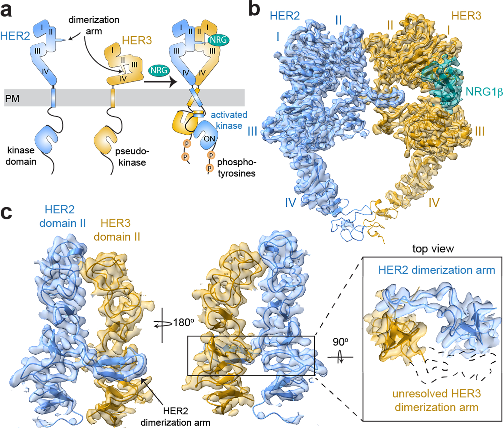
Fig. 1 |. Overall structure of the HER2/HER3/NRG1β extracellular domain dimer complex.

a, Cartoon schematic of the conformational changes that the inactive HER2 and HER3 monomers are predicted to undergo during heterodimerization in the presence of neuregulin (NRG) 1β. PM is plasma membrane. b, Cryo-EM map and the resulting structural model of the HER2/HER3/NRG1β extracellular domain complex, with HER2 shown in light blue, HER3 in gold and NRG1β in teal. Extracellular domains I-IV are marked on the structures. c, Zoomed-in view of the dimerization interface to illustrate lack of density for the HER3 dimerization arm. An outline of the expected location of the HER3 dimerization arm based on previous extracellular domain structures is shown as a dotted path in the top view.
