Abstract
Penetrating brain injury from marble is rare. Marbles, commonly known as “guli” among locals, is a popular children's traditional game in Malaysia. This study discusses two cases of intracranial marble injury, both accidentally shot by children with home-made air guns during the period of Movement Control Order with one elderly patient who passed away. While the diagnosis was uneventful, the management was not straightforward. Strategies of prehospital, operative, postoperative management, and rehabilitation are discussed, including prognostic factors. Because of its rarity, the management of such injuries is complex and nonstandardized.
Keywords: Keywords, penetrating brain injury, low-velocity, intracranial injury
Background
Penetrating brain injury (PBI) is the most life-threatening type of traumatic brain injury (TBI) with a mortality rate of 23 to 93% that increases to 87 to 100% in patients presenting with poor neurological status. 1 Gunshot injuries are the most common type of PBI, accounting for 12% of all TBI cases, and only 10% of patients with these injuries survive to reach the hospital. 1 Half of those who reach the hospital die while the remaining survivors often suffer long-term neurological sequelae. In contrast, there are not many reports on PBI involving marble, except for a case report 2 that describes a toddler with a gunshot wound over the forehead with preseptal cellulitis caused by a marble shot by a slingshot. The toddler survived with minimal neurological sequelae. We report here two cases of PBI by marbles during the period of Malaysia's Movement Control Order for the coronavirus pandemic.
Case Presentation
Case 1: Dangers of Playing with a Gun
History, Physical Examination, and Investigations
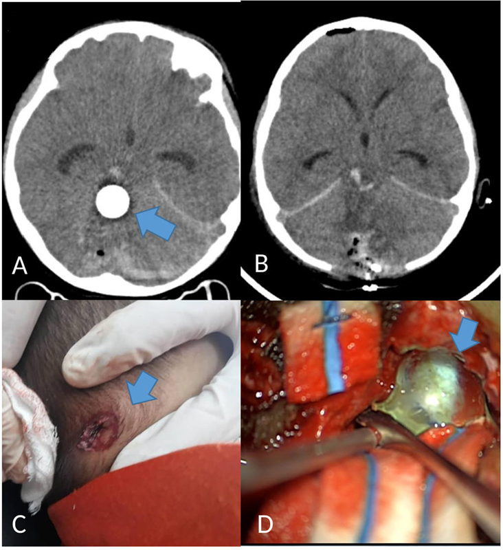
A 5-year-old boy was playing with his older 6-year-old brother outside their house with an air gun, illegally home-made by their father from lead pipe and wood for game hunting. The older boy accidentally shot his younger brother in the head from a distance of 1 m who lost his consciousness. The shot boy was rushed to the nearby community clinic and subsequently reached our neurosurgical center 8 hours after the accident. There were no documented seizures, and he remained hemodynamically stable throughout the transfer. He sustained a deep, circular penetrating wound of 2 cm in diameter at the right suboccipital scalp ( Fig. 1C ). The boy's Glasgow Coma Scale (GCS) was E1V1M3 with pinpoint pupils. He was intubated and given intravenous ceftriaxone, phenytoin, and tranexamic acid. Computed tomography (CT) of the brain ( Fig. 1A ) showed a round hyperdense foreign body at the right posterior fossa abutting the dorsal part of the midbrain. There was fourth ventricular effacement causing obstructive hydrocephalus. Apart from that, right occipital comminuted fractures were seen. A 3-cm long tract with blood and air within was visualized through the swollen right cerebellum. The patient was planned for a cerebral angiogram due to the foreign body's proximity to the galenic complex. However, he desaturated despite being ventilated, probably because of the proximity of the marble to the respiratory center of the brainstem. He was pushed immediately to surgery.
Fig. 1.

( A ) Brain CT showed a round hyperdense foreign body ( blue arrow ) at the right cerebellum, close to the mid brain. A 3-cm long tract with blood and air within was seen along its entry point. ( B ) Postoperation brain CT: less affected fourth ventricle; brain lax; Slyvian fissure opened. ( C ) Entry point of penetrating brain injury: a deep circular wound over the right occipital scalp ( blue arrow ). ( D ) Marble found and removed using tumor holding forceps ( blue arrow ).
Surgery
An emergency external ventricular drain (EVD) insertion, posterior fossa craniectomy, and foreign body removal were performed. EVD insertion served to relieve the hydrocephalus and was high pressure upon cannulation of the ventricle. The surgical microscope was used during the surgery. Suboccipital craniectomy was done to accommodate the oedematous cerebellum. The tract of the contused brain was followed until the foreign body was found. The marble was removed in one piece ( Fig. 1D ). It was unshattered and measured 13 mm in diameter. There was no vascular injury. Hemostasis was secured and washout done with copious amounts of gentamicin saline before closing.
Postoperative Outcome
The boy underwent cerebral resuscitation for 48 hours. Postoperation CT brain: less effaced fourth ventricles. Brain lax, Sylvian fissure opened ( Fig. 1B ). His GCS was E3VtM5 and pupils 3 mm in diameter, reactive to light bilaterally upon weaning off sedation. He was extubated on day 3 postoperation and successfully weaned to room air on day 6. At 10 days posttrauma, his GCS improved to E4V2M6. Neurological examination revealed right-sided limbs and truncal ataxia as evidenced by right intentional tremor, right dysdiadochokinesia, right-sided hypotonia, dysarthria, titubation, and gait ataxia. Intravenous ceftriaxone 50 mg/kg twice a day was administered for a total of 1 week. He was on a nasogastric tube for feeding and was discharged with outpatient physiotherapy, occupational, speech-language therapy in outpatient pediatrics, and neurosurgical follow-up appointments.
Case 2: The Hunting Accident
History, Physical Examination, and Investigations
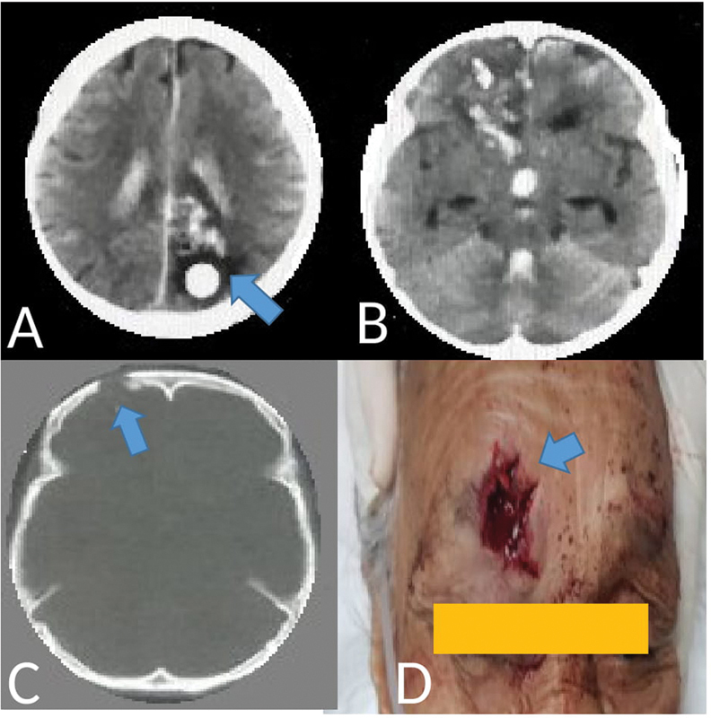
A previously healthy 87-year-old lady was shot in the frontal region of her head by her 15-year-old granddaughter using a self-made air gun during a hunting trip in the forest nearby their longhouse. This air gun was also illegally made for hunting purposes. She lost her consciousness and was brought to the nearest district hospital before arriving at our center 5 hours after the accident. A jagged penetrating wound actively bled over the right frontal region and emitted brain matter ( Fig. 2D ). The patient had a GCS of E2V1M4, and pupils were 2 mm in diameter bilateral and sluggish. She was intubated and administered intravenous ceftriaxone, phenytoin, tetanus, and tranexamic acid. There were no documented seizures.
Fig. 2.

( A ) Brain CT reveals a round hyperdense foreign body at the occipital region ( blue arrow ). ( B ) Foreign body entered at right frontal, crossed the brainstem, and lodged into the contralateral occipital lobe. ( C ) The entry point was traced to the right frontal bone ( blue arrow ). ( D ) Jagged right frontal penetrating wound ( blue arrow ).
It took 3 hours for transfer of the patient from the district polyclinic to our tertiary hospital with neurosurgical facilities. Her systolic blood pressures were maintained normotensive, and SpO2 greater than 95% throughout transfer. The district polyclinic did not have arterial blood gas facilities, hence CO2 monitoring to keep the level between 35 and 40 mm Hg was not possible. Head of the bed was propped up 30 degrees throughout transfer.
Upon our assessment at the neurosurgical center, the patient's GCS dropped to E1VTM1, and pupils became pinpoint. CT of the brain revealed a round hyperdense foreign body at the left occipital region ( Fig. 2A ). The object's entry point was traced to the right frontal bone, with the fracture segments buried in the third ventricle ( Fig. 2C ). The object's tract crossed the midline, lodging in the contralateral left occipital region. Intraventricular hemorrhage (IVH) was seen over all the ventricles ( Fig. 2B ).
Given her critical condition and minimal benefit that surgery would offer due to the multilobar and transventricular involvement, as well as old age, the family agreed for conservative management. The patient passed away the next day.
Discussion
PBI can be classified based on the injury velocity and mechanism of injury. 3 High-velocity injuries create damage beyond the immediate point of contact, while low-velocity injuries cause localized damage along the trajectory of penetrating object. The kinetic energy generated by the penetrating object is equal to the mass times square of its velocity (Ek = ½ mv 2 ). 3
Bullets or missile injuries have less mass, but travel at higher velocities and is accompanied by percussion waves during its transit through brain matter causing significant cavitation, explosive skull fractures, and widespread destruction of neuronal cell membranes that may propagate as far down as the medulla oblongata. 3 Nondeforming projectiles like marbles have a tendency to yaw inside tissue, which increases penetrating and results in a moderate wound cavity. Most nonbullet penetrating objects, such as nails or knives, impart less damage to the skull and brain because they have less kinetic energy to transfer on impact. 3
Still, do not underestimate the marble as ammunition; a marble shot to the head can kill, as illustrated in this case series.
Preoperative Strategies
CT brain is the initial imaging modality of choice given its speed, widespread availability, and nearly 100% sensitivity in detecting surgical lesions for effective surgical planning. 1 Cerebral angiography is recommended when the wound's trajectory is through or near the Sylvian fissure, the supraclinoid carotid artery, the vertebrobasilar vessels, the cavernous sinus region, or major dural venous sinuses, and is 73% sensitive in identifying traumatic intracranial pseudoaneurysm and non-aneurysmal arterial injuries of the first-order branches. 4 The development of otherwise unexplained subarachnoid hemorrhage or delayed hematoma could suggest the presence of a vascular injury, warranting an angiography. In some initially negative angiography, some vascular injuries are delayed in onset, appearing weeks or months after the trauma. A repeat angiography 2 to 3 weeks posttrauma is recommended for suspicious cases. 4 Magnetic resonance imaging (MRI) plays a limited role as it is time-consuming and contraindicated in patients with metallic foreign bodies. 1
However, in our first case, there was insufficient time to perform the angiogram because the patient deteriorated. A postoperative angiography was not done because intraoperatively, there was minimal bleeding, deeming a vascular injury unlikely.
Intraoperative Strategies
The goals of surgery are to remove the foreign object and surrounding debris through its trajectory with minimum injury, adequate wound irrigation for decontamination, secure all active bleeders, and watertight closure of the dura to prevent cerebrospinal fluid leak. Moreover, the marble in case 1 abutted the dorsal midbrain.
Intraoperative ultrasonography is a reliable method to provide real-time neuronavigation during neurosurgery without the error of brain shift that depends on preoperative images and is comparable or even better than image-guided surgery, CT and MRI for landmark visualization. 5 Unfortunately, intraoperative ultrasonography is not available in our center.
Postoperative Management
Primary deficits of the boy who survived in case 1 were whole body ataxia, dysphagia, difficulty communicating, and emotional lability. He demonstrated truncal ataxia resulting in poor sitting balance and required total assistance with upper body and lower body dressing, bathing, toileting, and all bed mobility. Early mobility was performed until conventional neuromuscular reeducation interventions could be tolerated. 6 The physical therapist also provided a home exercise program and fall prevention education to the family and nursing. 6 Six months after the accident, the boy was able to stand with assistance and perform most activities of daily living with standby or set-up assistance.
Throughout the entire rehabilitation process, the boy's mother was actively involved, and that likely facilitated optimum outcomes. 6 Another factor in the patient's recovery was the absence of serious medical complications postsurgically, allowing for consistent, progressive, and aggressive rehabilitation. 6
Psychologically, the act of accidentally killing another person, the feelings of responsibility for the death, and acute grief must be dealt with. 7 Both accidental perpetrators were referred to a counselor and were taught useful strategies for coping. 7 The families also underwent sessions with the counselor, where they were taught strategies to be supportive, and to watch out for any changes in behavior, lack of interest and concentration, and symptoms of depression. 7 None of them developed significant psychiatric issues up to today.
The police deemed both cases as accidents, and no charges were made.
Prognostic Factors
Surgical treatments are being offered to more patients than ever before, and increasingly to high-risk patients because preserving quality of life is a key treatment goal. 8 The elderly patient in case 2 presented with multiple poor prognostic factors of old age, low GCS score, abnormal pupillary light reflex, and multilobar with transventricular injuries. 9 These poor prognostic indicators are generally not good surgical candidates with a minimal chance of favorable outcome, prompting the family to agree for conservative management. IVH is a particularly poor prognostic sign, with expected mortality of 50 to 80%. 10 Other CT of the brain findings associated with poor prognosis includes brainstem, bihemispheric, multilobar, or transventricular injuries, injury to eloquent brain, subarachnoid hemorrhage, large intracerebral hemorrhage, and midline shift. 9
Malaysian Gun Policy
According to the Arms Act No. 206 of 1960, only licensed gun owners in Malaysia may lawfully acquire, possess, or transfer a firearm or ammunition, including fake gun or airgun. 11 Despite the country's stringent gun laws, there are 74,962 unregistered weapons in Malaysia. 11 There ought to be a stricter implementation of the gun laws, and adults who own guns must prohibit child access to firearms.
Take Home Messages
Parents need to tighten supervision and limit a child's access to guns.
Management of traumatic brain injury is best done by a multidisciplinary team that consists of a neurosurgeon, emergency department physician, neurologist, intensivist, neurosurgical nurses, physical therapist, and an internist.
Surgery is just the first step in recovery. Postoperative rehabilitation is a long process that takes months to years, an important aspect not to be neglected.
Footnotes
Conflict of Interest None declared.
References
- 1.Vakil M T, Singh A K. A review of penetrating brain trauma: epidemiology, pathophysiology, imaging assessment, complications, and treatment. Emerg Radiol. 2017;24(03):301–309. doi: 10.1007/s10140-016-1477-z. [DOI] [PubMed] [Google Scholar]
- 2.Vinuthinee N, Azreen-Redzal A, Juanarita J, Zunaina E. Gunshot-like wound caused by sling shot injury – a case report. Int Med Case Rep J. 2015;8:47–50. doi: 10.2147/IMCRJ.S75198. [DOI] [PMC free article] [PubMed] [Google Scholar]
- 3.Van Wyck D W, Grant G A, Laskowitz D T.Penetrating traumatic brain injury: a review of current evaluation & management concepts. [Online]Accessed July 9, 2021 at:https://www.iomcworld.org/open-access/penetrating-traumatic-brain-injury-a-review-of-current-evaluation-andmanagement-concepts-2155-9562-1000336.pdf
- 4.Alpers P, Picard M.Malaysia—Gun Facts, Figures and the Law. Sydney School of Public Health, The University of Sydney; 2020 [Online]. Accessed May 28, 2020 at:https://www.gunpolicy.org/firearms/region/malaysia
- 5.Bagli S Y, Hasari T B.Tek Bir Kurumun Hastaya ‹liflkin Tecrübesi. Traumatic brain injury due to gunshot wounds: a single institution's experience with 442 consecutive patients. [Online]Accessed April 20, 2020 at:http://turkishneurosurgery.org.tr/pdf/pdf_JTN_670.pdf
- 6.Elmesallamy W AEA.Demonstrative study of brain anatomical landmarks by intraoperative ultrasound imaging[Online]. Accessed April 1, 2020 at:https://link.springer.com/article/10.1186/s41984-019-0056-5
- 7.Wilson C M, Mitchell C L, Hebert K M. Cerebellar stroke occupational therapy and physical therapy management from intensive care unit to outpatient: a case report. Cureus. 2017;9(12):e1949. doi: 10.7759/cureus.1949. [DOI] [PMC free article] [PubMed] [Google Scholar]
- 8.Ziegelmann M, Köhler T S, Bailey G C, Miest T, Alom M, Trost L. Surgical patient selection and counseling. Transl Androl Urol. 2017;6(04):609–619. doi: 10.21037/tau.2017.07.19. [DOI] [PMC free article] [PubMed] [Google Scholar]
- 9.Shaw S, Hughes G, Stephens T. Understanding decision making about major surgery: protocol for a qualitative study of shared decision making by high-risk patients and their clinical teams. BMJ Open. 2020;10(05):e033703. doi: 10.1136/bmjopen-2019-033703. [DOI] [PMC free article] [PubMed] [Google Scholar]
- 10.Kazim S F, Shamim M S, Tahir M Z, Enam S A, Waheed S. Management of penetrating brain injury. J Emerg Trauma Shock. 2011;4(03):395–402. doi: 10.4103/0974-2700.83871. [DOI] [PMC free article] [PubMed] [Google Scholar]
- 11.Hinson H E, Hanley D F, Ziai W C.Management of intraventricular hemorrhage[Online]. Accessed May 6, 2020 at:https://www.ncbi.nlm.nih.gov/pmc/articles/PMC3138489/ [DOI] [PMC free article] [PubMed]


