Abstract
Melanotic neuroectodermal tumor of infancy (MNTI) is a rare, pigmented tumor. It is a locally aggressive neoplasm of neural crest origin with a high recurrence rate. It occurs in craniofacial sites in more than 90% of cases and most commonly in the maxilla. It may also occur in epididymis, testis, ovaries, soft tissue, and bones of the extremities. It occurs in infants younger than 1 year of age with a slight male preponderance. We report a rare case of a child presenting with midline frontal pigmented MNTI.
Keywords: melanotic neuroectodermal tumor of infancy, congenital melanocarcinoma, superior sagittal sinus
Introduction
Krompecher first described the tumor as “congenital melanocarcinoma” in 1918. 1
It has been universally accepted by the term “Melanotic Neuroectodermal Tumor of Infancy” (MNTI) described by Borello and Gorlin with slightly more than 500 reported cases to date. 2 A total of 91 cases of cranial MNTI have been described in 78 articles published between 1918 and 2018. 3 The age of diagnosis varied from infancy to 21 years, with an average of 16.4 months. Seventy (80%) patients were diagnosed younger than 1 year. Of the 89 patients, 52 (59.4%) were males and 37 (41.6%) females.
Analysis of the location of the lesion showed 11 (12.1%) patients had lesion involving the midline skull, 14 (15.4%) cases involving anterior fontanelle, 50 (54.9%) cases involving other parts of skull, and 16 (17.6%) cases involving the brain, cerebellum, pineal or ventricular region. 3
Case History
A 5-month-old male child presented with a 6-weeks' history of insidious onset, gradually growing midline scalp swelling with no history of fever, altered sensorium, seizure, ulcer over swelling, or watery discharge. Social milestones and motor development were adequate for age. Maternal, birth, and family history were normal. On examination, a 2.5-cm, hard, nonpulsatile, nontransilluminant bony lesion was noted anterior to anterior fontanelle. Fontanelle was lax.
Computed tomography (CT) and magnetic resonance imaging (MRI) (plain and contrast) brain revealed a well-defined extra-axial lesion in the right para falcine frontal region with invasion into superior sagittal sinus (SSS) with extracranial extension ( Fig. 1 ).
Fig. 1.
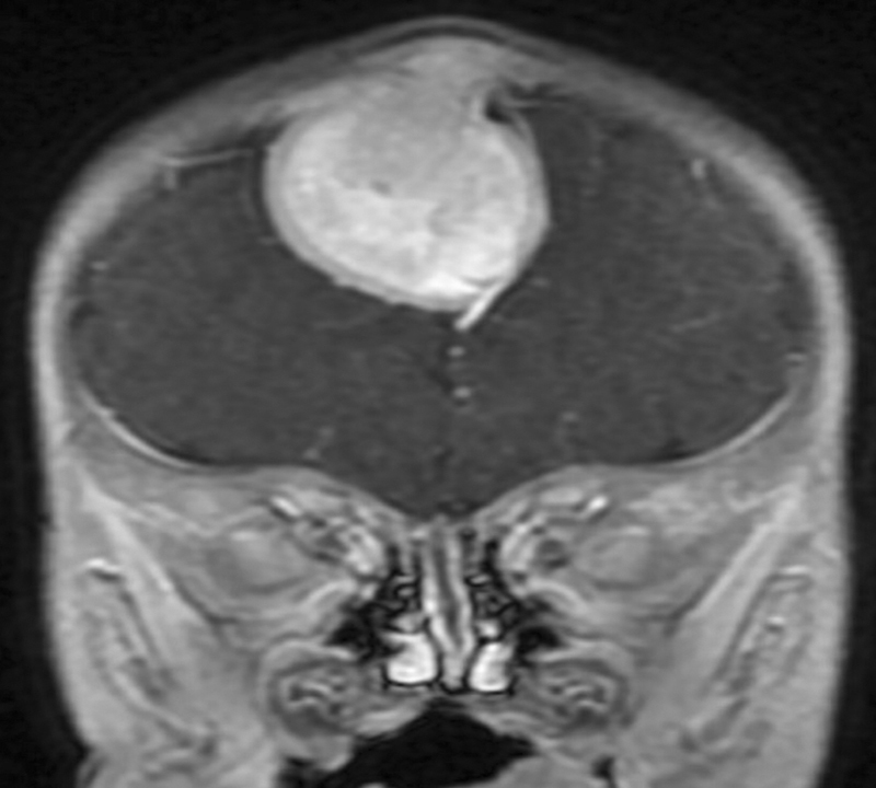
Magnetic resonance imaging brain (contrast) coronal showing falcine lesion occluding superior sagittal sinus extending up to subcutaneous plane.
Patient underwent right frontal craniotomy and subtotal excision of midline frontal lesion. Tumor was arising from precoronal falx, extending more to right and completely occluding SSS up to just in front of coronal suture and transgressed dura to present itself under the bone. Tumor was of mixed consistency, not vascular. A small bit of tumor involving the SSS posteriorly was preserved. Morcellized bone pieces taken from the right parietal bone were placed over the area of bone defect and over the dura. Following discharge, child was lost to follow-up ( Figs. 2 and 3 ).
Fig. 2.
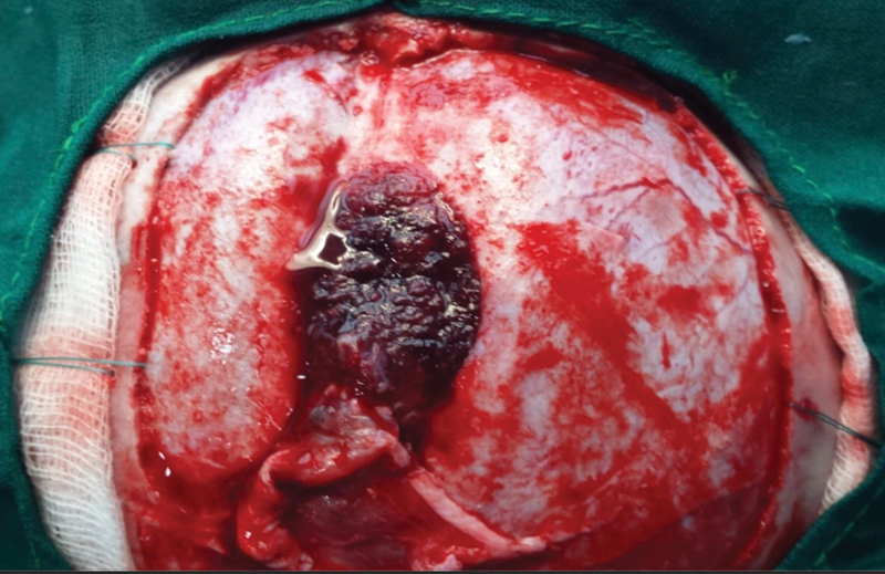
Intraoperative image showing reddish black lesion seen breaching the dura on left side extending across midline.
Fig. 3.
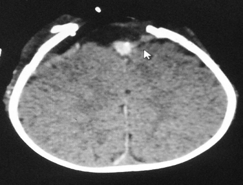
Postoperative scan showing residual tumor that has been attached to sinus.
Histopathology showed cellular neoplasm composed of two populations of cells—small basophilic cells with scant cytoplasm surrounded by desmoplastic stroma and large epithelioid cells arranged in alveolar pattern showing pigmentation.
Immunohistochemistry (IHC) showed the tumor cells were positive for vimentin, neural cells were positive for synaptophysin, epithelioid cells were positive for cytokeratin and HMB-45. K i -67 index was less than 2% ( Fig. 4 ).
Fig. 4.
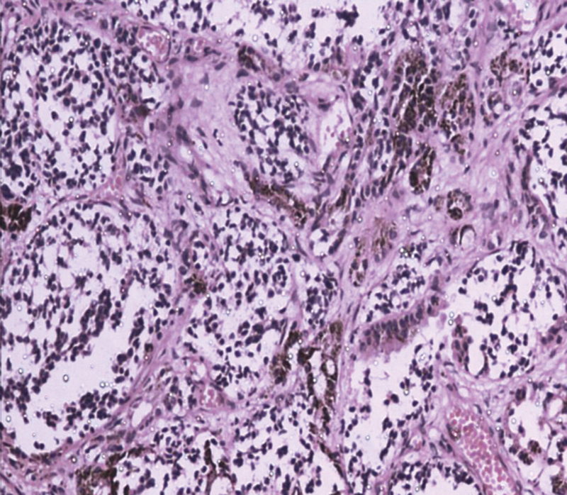
Hematoxylin and eosin, 400× magnification.
Tumor showed biphasic pattern with small neuroblastic cells and large epithelioid melanin containing cells. Background showed dense fibrosis ( Figs. 5 and 6 ).
Fig. 5.
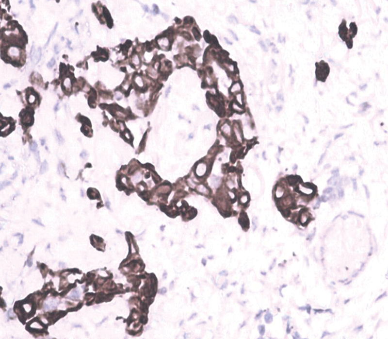
Cytokeratin—positive in epithelioid cells.
Fig. 6.
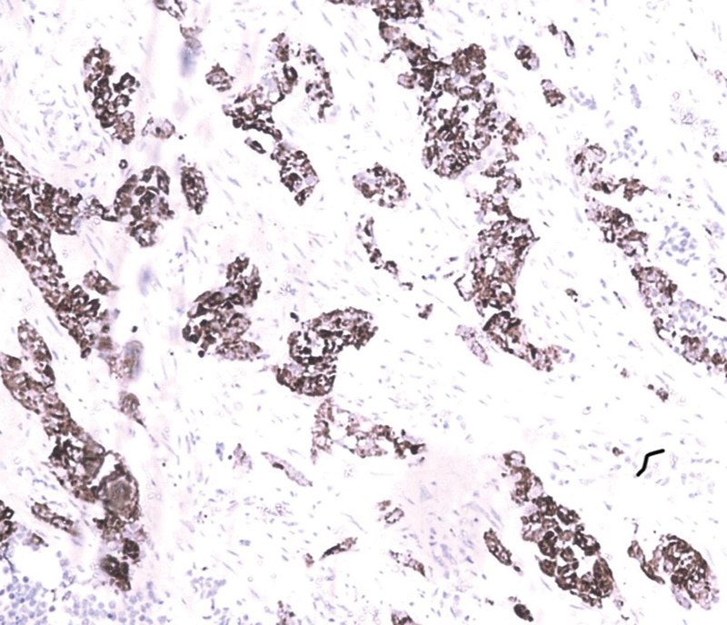
HMB-45 positive.
Child had no deficits postoperatively.
Discussion
MNTI is a rare pigmented craniofacial tumor of infants presenting mainly in the first year of life with a slight male predilection. Patients present with a gradually enlarging, firm, painless swelling underlying an intact skin with blue or black discoloration. It was proposed of neural crest origin, as many patients were found to have increased urinary vanillylmandelic acid (VMA). 2 The measured levels of VMA may return to baseline following surgical resection. 4
CT is the imaging of choice for determining extent of bony involvement by the lesion. On CT, it appears as a hypodense lesion with the melanin component appearing hyperdense. Enlargement of the lesion leads to destruction of the bone causing “sun-burst” appearance. On MRI, it appears as a well-defined hypointense mass on T1 and T2 with melanin appearing hyperintense and helpful in extent of soft tissue involvement. 5
Gross specimens appear as a firm, lobulated, superficially infiltrating well-circumscribed mass with a blue-black hue. Cut surface of the lesion appears gray to black depending on the melanin. Microscopically, MNTI is composed of small, round neoplastic cells with well-vascularized fibrocollagenous stroma. The histologic hallmark of MNTI is the presence of a biphasic population of cells composed of large melanogenic cells and smaller neuroblast-like cells but both populations can also be present in isolation. 6 Neurogenic cells are located centrally and melanogenic cells at the periphery with a high nuclear cytoplasmic ratio with “salt and pepper” chromatin. Mitoses and necrosis are unusual, unless the tumor is malignant.
Definitive diagnosis is by IHC staining. Both neurogenic and melanogenic cells show positivity for neuron-specific enolase and vimentin and typically negative for S100. 6 Melanogenic cells are positive for cytokeratins and markers of melanocyte differentiation—HMB-45 and dopamine β-hydroxylase. They may also be positive for epithelial membrane antigen. 6 Neurogenic cells are positive for synaptophysin. Foci of glial and divergent skeletal muscle differentiation can be seen in some cases and stain with myogenic markers such as desmin, myogenin, and muscle specific actin causing to be misdiagnosed as rhabdomyosarcoma. Molecular or genetic abnormalities have not been identified in MNTI. One report showed that one of three cases had BRAF-V600E mutation.
A recent systematic review analyzing various treatment modalities for MNTI demonstrated that surgical excision appears to be the treatment of choice as it has lower recurrence and morbidity rates. Few patients undergoing neoadjuvant chemotherapy had decreased need for wider surgical resection. A small minority of patients were treated successfully with chemotherapy alone. 7 Metastasis to lymph nodes and central nervous system has been reported in 3% of cases. Recurrence rates were variable ranging from 15 to 27%. 7 It is purported to be due to multicentric growth and incomplete surgical resection, leading to multiple surgeries in some cases. 6
Conclusion
MNTI is a rare, pigmented tumor in infants. Because of its rapid infiltrative growth, high local recurrence, and high risk of malignant transformation, early diagnosis and complete excision are necessary for best clinical outcome.
Funding Statement
Funding None.
Footnotes
Conflict of Interest None declared.
References
- 1.Krompecher E. Zur Histogenese und Morphologie der Adamantinome und sonstiger Kiefergeschwulste. Beitr Pathol Anat Allg Pathol. 1918;64:165–197. [Google Scholar]
- 2.Borello E, Gorlin R. Melanotic neuroectodermal tumor of infancy: a neoplasm of neural crest origin. Cancer. 1966;19(02):196–206. doi: 10.1002/1097-0142(196602)19:2<196::aid-cncr2820190210>3.0.co;2-6. [DOI] [PubMed] [Google Scholar]
- 3.Ren Q, Chen H, Wang Y, Xu J. Melanotic neuroectodermal tumor of infancy arising in the skull and brain: a systematic review. World Neurosurg. 2019;130:170–178. doi: 10.1016/j.wneu.2019.07.017. [DOI] [PubMed] [Google Scholar]
- 4.Kumari T P, Venugopal M, Mathews A, Kusumakumary P. Effectiveness of chemotherapy in melanotic neuroectodermal tumor of infancy. Pediatr Hematol Oncol. 2005;22(03):199–206. doi: 10.1080/08880010590921450. [DOI] [PubMed] [Google Scholar]
- 5.Haque S, McCarville M B, Sebire N, McHugh K. Melanotic neuroectodermal tumour of infancy: CT and MR findings. Pediatr Radiol. 2012;42(06):699–705. doi: 10.1007/s00247-011-2339-1. [DOI] [PMC free article] [PubMed] [Google Scholar]
- 6.Mendis B R, Lombardi T, Tilakaratne W M. Melanotic neuroectodermal tumor of infancy: a histopathological and immunohistochemical study. J Investig Clin Dent. 2012;3(01):68–71. doi: 10.1111/j.2041-1626.2011.0086.x. [DOI] [PubMed] [Google Scholar]
- 7.Woessmann W, Neugebauer M, Gossen R, Blütters-Sawatzki R, Reiter A. Successful chemotherapy for melanotic neuroectodermal tumor of infancy in a baby. Med Pediatr Oncol. 2003;40(03):198–199. doi: 10.1002/mpo.10135. [DOI] [PubMed] [Google Scholar]


