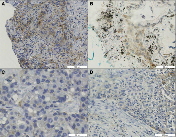
Figure 6.

Difficult‐to‐score features. A, Neoplastic tissue surrounded by benign programmed death‐ligand 1 (PD‐L1)‐positive cells. B, PD‐L1 staining in neoplastic cells: partly nuclear, partly cytoplasmic, and partly membranous (anthracosis and ink). C, Low‐intensity PD‐L1 staining. D, PD‐L1‐positive immune cells infiltrating neoplastic tissue. [Colour figure can be viewed at wileyonlinelibrary.com]
