Abstract
Background
Haemophilic knee arthropathy presents functional and structural alterations and chronic pain. Self‐induced myofascial release aims to treat fascial restrictions and improve functionality.
Aim
This study investigated the safety and effectiveness of a self‐induced myofascial release protocol in patients with haemophilic knee arthropathy.
Methods
Twenty‐five patients with bilateral haemophilic knee arthropathy were recruited (n = 50 knees). The patients followed an intervention protocol, with daily exercises for 8 weeks. The dependent variables were: safety of the technique (periodic telephone monitoring), joint state (Haemophilia Joint Health Score), pain intensity (visual analogue scale), pressure pain threshold (pressure dynamometer), range of motion (universal goniometer) and hamstring flexibility (Fingertip‐To‐Floor test). The resulting values were measured at baseline (T0) and after the intervention (T1). Paired t‐test compared the means between the assessments. Effect size was obtained using Cohen's d mean difference formula. The minimum detectable change of each variable was calculated.
Results
There were no cases of joint bleeding either during or after the procedure. The results showed improvements after the experimental period in joint state (Mean difference [MD]: 1.38; 95% confidence interval [95%CI]: .94;1.81), pain intensity (MD: 1.19; 95%CI: .70;1.67), pressure pain threshold (MD: ‐23.25; 95%CI: ‐26.25;‐19.84), flexion (MD: ‐4.36; 95%CI: ‐5.70;‐3.01), loss of extension (MD: 4.10; 95%CI: 3.01;5.18) and hamstring flexibility (MD: 3.54; 95%CI: 2.61;4.46).
Conclusions
Myofascial self‐release using a foam roller is safe in patients with haemophilic knee arthropathy. A myofascial self‐release protocol can improve perceived pain, range of motion and knee joint status, as well as hamstring flexibility in patients with haemophilic knee arthropathy.
Keywords: haemophilic knee arthropathy, joint pain, range of motion, self‐induced myofascial release
1. BACKGROUND
Haemophilia is a congenital coagulopathy characterized by a deficiency of a clotting factor. Haemophilia A is characterized by a deficiency of clotting factor VIII, while factor IX is missing in haemophilia B. Depending on the percentage of blood clotting factor there are three phenotypes: severe (< 1%), moderate (1–5%) and mild (> 5–40%). 1 Although it is a haematological disease, haemophilia is characterized by bleeding in the locomotor system. The joints are affected in 80% of cases (hemarthrosis), with bleeding in knees, elbows and ankles. 2
The recurrence of hemarthrosis induces biochemical and enzymatic alterations, and hypertrophy of the synovial membrane. Such recurrence leads to degenerative joint injury (haemophilic arthropathy). 3 Arthropathy represents degenerative, progressive and irreversible intraarticular damage. Haemophilic arthropathy is characterized by the loss of joint mobility, chronic pain, muscle atrophy and impaired proprioception. This leads patients to reduce their activities and social participation, thereby decreasing their perceived quality of life. 4
Injuries due to trauma and microtrauma by repeated and sustained overload predispose the conjunctive system to changes. These patterns favour pathological changes in the tissue that can cause mechanical and functional alterations as a consequence of modifications in the concentration and orientation of the collagen fibres of the conjunctival matrix. 5 In this way, healthy tissue in the vicinity of the site of injury or trauma can also be compromised and its function altered. 6
The fascial system, which is a heterogeneous network of connective tissue, 7 is built on a tensegrity‐based architecture that provides stability to cells and the organism as well as mobility. 8 The mechanical changes undergone by the tensegrity system in order to adapt to the loads and to environmental factors, condition part of the physiological responses generated in the cells and the extracellular matrix.
Physiotherapy techniques using moderate mechanical loads have shown positive effects in terms of reducing fibrosis and swelling in patients with fascial tissue injury. 6 Through the fascia mechanoreceptors 9 fascial therapy attempts to change the orientation of the collagen fibres and alter the tone of the associated motor units. 10
A number of studies have reported the safety and efficacy of physiotherapy programs based on manual therapy using myofascial techniques in patients with haemophilic arthropathy of the ankle 11 , 12 and elbow. 13 , 14 Such studies noted a decrease in the frequency of hemarthrosis during the follow‐up period, achieving improvements in range of motion and disability in patients with haemophilic arthropathy. Goto et al. 15 described that monitoring the perception of pain combined with use of a prophylactic treatment are key aspects in order to reduce the frequency of bleeding and pain, thus improving joint function. Acute and chronic processes lead to a shortening of the periarticular structures, mainly the soft tissues and flexor muscles. The improved joint condition reported after the intervention period may justify the effectiveness of this intervention.
Self‐induced myofascial release is a physiotherapy technique able to treat soft tissue restrictions with the help of a foam roller. Self‐induced myofascial release involves patients using their own body weight, plus materials such as a foam roller, to exert pressure on the soft tissues affected. 16 Various studies have analysed the effectiveness of this technique in healthy athletes, achieving to reduce stress and muscle stiffness, as well as pain and swelling, while increasing range of motion in the hip, knee and ankle, when applied to the quadriceps, hamstrings, and plantar flexor muscles. 17
The direct pressure exerted with the patient's weight when using the foam roller on the treated region produces local mechanical and global neurophysiological effects that relax the tissues and attenuate the perceived pain in the treated region and surrounding tissues. 18 Self‐induced myofascial release can improve the range of knee motion 19 and the pressure pain threshold at trigger points after a single session in healthy subjects. 20 Similarly, it has been noted that the decreased intensity of knee pain in athletes with chronic pain, after an 8‐week self‐induced myofascial release program, was maintained at 2 months follow‐up. 21
The study hypothesis was that a self‐induced myofascial release protocol is able to improve the perception of pain, range of knee motion, hamstring flexibility, and joint status in patients with haemophilic knee arthropathy. The aim of this study was to assess the safety and efficacy of a self‐induced myofascial release protocol using a foam roller in patients with haemophilic knee arthropathy.
2. METHODS
2.1. Study design
Prospective cohort study to assess the safety and effectiveness of a physiotherapy protocol using self‐induced myofascial release with a foam roller in patients with haemophilic knee arthropathy.
2.2. Patient recruitment and selection
The patients were recruited from the Spanish Federation of Haemophilia and the Galician Association of Haemophilia between September and December 2020.
Inclusion criteria to participate in the study were: patients with a medical diagnosis of haemophilia A or B; with a severe phenotype of haemophilia (< 1% FVIII/FIX); with a medical diagnosis of bilateral haemophilic knee arthropathy and more than three points on the Haemophilia Joint Health Score 22 ; being over 18 years old; and no scheduled orthopaedic surgeries during the study phase.
Exclusion criteria were: patients with hemarthrosis in the month before the beginning of the study; patients unable to walk; severe functional alterations of the upper limb that prevented exercise; and failure to sign the informed consent document.
Patients with on‐demand or prophylaxis treatment and those with antibodies against clotting factor concentrates (inhibitors) were included.
2.3. Ethical considerations
Prior to the start of the study, the principal researcher explained the objectives, and possible risks and benefits of the intervention to the patients. After the oral presentation, they were given an information sheet with the data and characteristics of the study. All patients signed the informed consent document, in accordance with the Helsinki statement. This study was approved by the Research Ethics Committee of the University of Murcia (ID: 2428/2019). The Spanish Medicines Agency of the Spanish Ministry of Health classified the study as an “Observational Non‐drug study” (Resolution S‐201901700000731). Before starting the experimental phase, the research project was registered with the International Clinical Trials Registry (www.clinicaltrials.gov; NCT03914287).
2.4. Measurement instruments
The patients were evaluated prior to the intervention and at 8 weeks, after the experimental phase, reproducing the initial evaluation under the same conditions. All evaluations were performed by the same physiotherapist, blinded to the study conditions.
The primary variable for measuring the safety of the intervention was the development of knee hemarthrosis. During the experimental phase, weekly telephone monitoring was carried out to evaluate any possible development of knee hemarthrosis or other complications (hematomas).
The six secondary variables were: joint health, pain intensity, pressure pain threshold, range of motion in knee flexion and extension, and hamstring muscle flexibility.
Joint state was measured on the Haemophilia Joint Health Score (HJHS). 22 This measuring instrument, specific for patients with haemophilia, assesses the joint condition in knees, ankles and elbows. This scale includes eight items (joint swelling, duration of swelling, muscle atrophy, strength, crepitus on motion, flexion and extension loss, and pain) and scores range from 0 to 20 points per joint (the higher the score, the greater the degree of joint deterioration).
The intensity of joint pain was assessed using the visual analogue scale (VAS). 23 This scale rates knee joint pain with scores from zero to 10 points (from no pain to the maximum perceived pain).
The pressure pain threshold was measured using an algometer (Wagner FPIXTM Digital Algometer). Fischer's measurement protocol was followed. 24 The instrument was attached to a hard rubber tip 1.1 cm in diameter, with a dial calibrated in kg/cm 2 and .1 kg/cm 2 divisions. The method described by Hogeweg et al. was used. 25 Before taking the measurements, patients were instructed to say “yes” as soon as pressure exerted by the algometer becomes slightly painful. With the instrument placed perpendicular to the skin surface, the pressure was increased at an even rate. 26 The unit of measurement is kg/cm 2 (the higher the score, the higher the pressure pain threshold).
The range of knee motion was measured with a universal goniometer, with one‐degree increments. The protocol described by the American Academy of Orthopaedic Surgeons 27 was used. Mobility was measured in the sagittal plane, under no load and without pain, with the fibula being the distal reference point. Degrees are the unit of measurement (the higher the degree of flexion and the lower the degree of extension, the better the range of motion).
Hamstring muscle flexibility was measured with the Fingertip‐To‐Floor test. 28 With this test, the distance between the fingertip and the floor was calculated with the patient performing maximum hip flexion and maximum knee extension. Measurements are taken in centimetres (the greater the distance, the poorer the flexibility).
Similarly, the pre‐treatment evaluation included the main clinical, anthropometric and sociodemographic variables.
2.5. Intervention
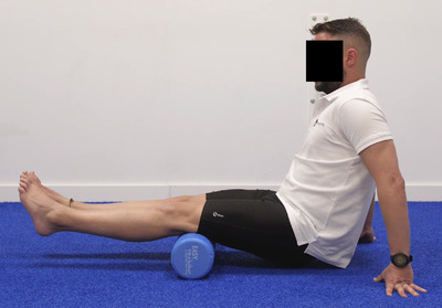
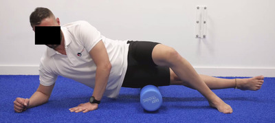
This study followed the protocol designed by Meroño‐Gallut et al. 29 for patients with haemophilic arthropathy. For its implementation, a 30‐cm long 15‐cm diameter foam roller was used, and an 8‐cm diameter ball. The total duration of the intervention was 15 min, with seven sessions per week over a period of 8 weeks. The physiotherapist who supervised the intervention followed up on all patients by telephone on a weekly basis to check for the absence of hemarthrosis during the intervention and to detect the possible need for protocol adaptations depending on the clinical situation of each patient. Each patient was individually provided with instructions as to how perform the exercises during the pre‐treatment evaluation, customizing the program if necessary. All patients had access to a mobile application, designed ad hoc for the research study (He‐Foam), where they could learn about the exercises through demonstration videos. Table 1 shows the main methodological characteristics of the intervention. Figures 1 and 2 show the release of the hamstring muscle group and abductor muscles.
TABLE 1.
Treatment protocol characteristics
| Maneuver | Region being treated | Material | Patient's position | Action | Slide |
|---|---|---|---|---|---|
| Release of posterior leg region |
‐ Achilles tendon ‐ Calf and soleus muscles |
FR | Sitting with knees extended and hands resting on the floor. | Longitudinal slides, crossing one leg over the leg being stimulated, with simultaneous toe flexion‐extension. | 15 repetitions (bilateral) |
| Release of anterior leg region | Extensor retinaculum of the ankle and anteroexternal muscles | FR | In a quadruped position, with the FR on the anterior part of the leg, adapting hip rotation to focus on the site to be treated. | Longitudinal sliding with simultaneous flexion‐extension of toes. | 15 repetitions (bilateral) |
| Release of the hamstrings muscle group | Hamstrings | FR | Sitting on the floor with the FR under the hamstrings, knees extended and hands resting on the floor. | Longitudinal slides on both hamstrings together (bilateral) and on a single hamstring. | 15 repetitions |
| Release of adductor muscles | Adductor | FB | Sitting on a chair with the PF between the thighs, adjusting work on the muscles by bringing legs together. | Circular slides with hip flexion‐extension in the distal, middle and proximal third of adductors | Five movements (bilateral) |
| Release of abductor muscles | Abductors | FR | Lateral decubitus with the FR on the side of the thigh. | Longitudinal slides on the lateral region of the thigh. | 15 repetitions (bilateral) |
| Pelvitrochanteric muscle release | Gluteal | FR | Sitting on the FR, adapted to one side region, dropping body weight towards that side. | Slides over the gluteal area. | 15 repetitions (bilateral) |
Abbreviations: FB, Foam Ball; FR, Foam Roller.
FIGURE 1.

Release of the hamstring muscle group
FIGURE 2.

Release of abductor muscles
2.6. Sample size
The sample size was calculated using the statistical package G * Power (version 3.1.9.2; Heinrich‐Heine‐Universität Düsseldorf, Germany). Assuming a mean effect size (d = .70), with an alpha level (type I error) of .05 and a statistical power of 95% (1‐β = .95), a sample size of 24 patients with haemophilic knee arthropathy was estimated. Patients were recruited from three Spanish regions (Madrid, Murcia and Malaga), through the Spanish Federation of Haemophilia.
Twenty‐five adult patients (n = 50 knees) with haemophilia were recruited. The median age was 36 (IR: 10) years. Most patients were diagnosed with haemophilia A (72%) and were on prophylactic treatment (80%). All patients had a severe disease phenotype (< 1% FVIII/FIX) and only 14% had a history of antibodies to clotting factor concentrates (inhibitors). Table 2 shows the descriptive characteristics of the patients included in the study.
TABLE 2.
Descriptive characteristics of patients with haemophilia at baseline
| Variables | Median | IR |
|---|---|---|
| Age (years) | 36 | 10.00 |
| Weight (kg) | 83 | 15.5 |
| Height (cm) | 173 | 5.50 |
| n | % | |
| Type of haemophilia | ||
| A | 18 | 72 |
| B | 7 | 28 |
| Type of treatment | ||
| Prophylaxis | 20 | 80 |
| On demand | 5 | 20 |
| Development of inhibitors | ||
| Yes | 6 | 24 |
| No | 19 | 76 |
Abbreviation: IR, interquartile range.
2.7. Statistical analysis
The statistical analysis was carried out with version 19.0 of the statistical package SPSS for Windows (IBM Company, Armonk, NY, USA). The main descriptive statistics of central tendency and dispersion (median and interquartile range) of the independent variables have been calculated. Changes after the intervention were analysed with the Student's t‐test for related samples. The effect size of the changes observed after the intervention was calculated using Cohen's d typified mean difference formula (1998), 30 being classified as large (d > .80), medium (d > .50) or small (d > .20). The minimum detectable change (MDC) was calculated by estimating the standard error of measurement (SEM). SEM was calculated using the formula: SEM = SDpre ∗ √1‐intraclass correlation coefficient (ICC). Based on SEM, MDC was obtained (MDC = Z‐score ∗ √ 2 ∗ SEM). The confidence level was set at 95% (Z score = 1.96).
An intent‐to‐treat analysis was performed to analyse the results. The selected significance level was .016 (α = .05 / 3).
3. RESULTS
Through the periodic telephone follow‐up over the 8 weeks of intervention, it was noted that none of the patients with haemophilia experienced intra‐articular haemorrhagic events in the knee joint, derived from the intervention, or secondary to trauma.
After the intervention we observed changes (P < .001) in joint state (t = 6.34; gl = 24), pain intensity (t = 4.94; gl = 24), pressure pain threshold (t = ‐13.70; gl = 24), knee flexion (t = ‐6.53; gl = 24) and extension (t = 7.61; gl = 24), and hamstring muscle flexibility (t = 7.71; gl = 24).
A large effect size was observed in the changes in knee pain intensity (d = .93), while the effect size was medium in loss of knee extension (d = .72) and pressure pain threshold (d = .51). The minimum detectable change in all dependent variables was calculated. The difference in means was greater than the minimum detectable change in the variables for pressure pain threshold (‐23.25 vs. 6.81), and knee flexion (‐4.36 vs. 4.36) and extension (4.1 vs. 4.04). The proportion of minimum detectable change in pressure pain threshold was 68%, while in knee flexion and extension the proportions of minimum detectable change were 38% and 50%. Tables 3 shows changes after the intervention and the results of calculating the effect size and the proportion of minimum detectable change.
TABLE 3.
Means (and standard deviations), effect size and minimal detectable change of joint status, joint pain, range of motion and hamstring flexibility evaluated in the different assessments
| Variables | T0 | T1 | 95% CI | MD | ES | ICC | SEM | MDC (MDCp) |
|---|---|---|---|---|---|---|---|---|
| Haemophilia Joint Health Score (0–20) | 5.42 (4.40) | 4.04 (3.09) | .94 – 1.81 | 1.38** | .36 | .958 | .90 | 2.632 (28) |
| Intensity of joint pain (0–10) | 1.79 (1.38) | .60 (1.16) | .70 – 1.67 | 1.19** | .93 | .742 | .70 | 2.32 (22) |
| Pressure pain threshold (Kg / cm 2 ) | 101.06 (44.99) | 124.31 (46.42) | −26.65–19.84 | −23,25** | −.51 | .982 | 6.03 | 6.81 (88) |
| Flexion (degrees) | 130.04 (16.32) | 134.40 (14.88) | −5.70–3.01 | −4.36** | .27 | .977 | 2.47 | 4.36 (36) |
| Loss of extension (degrees) | 7.08 (5.94) | 2.98 (5.33) | 3.01 – 5.18 | 4.10* | .72 | .872 | 2.12 | 4.04 (52) |
| Range of motion (degrees) | 120.60 (23.60) | 128.64 (22.05) | −10.55–5.52 | −8.04** | −.35 | .982 | 3.16 | 4.93 (68) |
| Hamstring flexibility (cm) | 8.84 (9.49) | 5.30 (7.09) | 2.61 – 4.46 | 3.54** | .42 | .961 | 1.87 | 3.79 (56) |
Outcome measures at baseline (T0) and after the 8‐week period of interventions (T1); 95% CI: 95% confidence interval.
Abbreviations: MD, means difference; ES, effect size; ICC, intraclass correlation coefficient; SEM, standard error of measurement; MDC, minimal detectable change; MDCp, proportion of minimal detectable change.
*Significant difference between assessments (P < .05).
**Significant difference between assessments (P < .01).
4. DISCUSSION
The aim of this study was to evaluate the safety and effectiveness of a self‐induced myofascial release protocol in the treatment of patients with haemophilic knee arthropathy. Similarly, the results obtained after the study period suggest that a self‐induced myofascial release protocol can be effective in improving joint state, pain, knee mobility, and hamstring flexibility in patients with haemophilic knee arthropathy. Haemophilic arthropathy is characterized by loss of range of motion, chronic pain, muscle atrophy, and impaired proprioception. This leads to a restriction of the patient's functional capacities that limits their normal daily activities, social participation and their perceived quality of life. Decreased pain and improved range of motion in the knee reported subsequent to applying the self‐induced myofascial release protocol described herein could improve the functional capacity in patients with haemophilic arthropathy.
Implementation of the self‐induced myofascial release program has not caused knee hemarthrosis during the intervention period. Self‐induced myofascial release can decrease the stresses of the periarticular connective system by inducing a reduction in mechanical pressure and joint overload generated by haemophilic arthropathy.
Lab tests have evidenced how prolonged strain applied by mechanical stimuli have an impact on the yield stress of connective tissue in vitro and in vivo. Its plastic elongation phase requires an effective elasticity of 3–8% elongation of the fibre. Beyond this deformation, the tissue tears or inflammation appears. 31 However, by applying prolonged interventions at up to 1‐h intervals with low fibre elongation, permanent deformation can be achieved without inducing breakage or inflammation with mechanical stress using an applied force of 24–115 kg. 31
Safety of this therapy has already been reported in previous articles where myofascial release techniques were applied to patients with haemophilic ankle 11 , 12 and elbow 13 , 14 , 32 arthropathy. This joint release process can decrease the mechanical joint factor which can favour a reduction in the frequency of hemarthrosis. 33
After the intervention, we noted a significant decrease in the intensity of pain and a significant increase in the pressure pain threshold. Chronic pain is one of the most disabling clinical manifestations in patients with haemophilia, affecting their functional capacity and their perception of quality of life. The overall neurophysiological effect that roller pressure can cause by relaxing muscle tissue, reducing local and surrounding pain, has been described. This process is carried out through the afferent inputs of the central nervous system from the Golgi tendon reflexes, reducing the activity rate of the motor unit causing subsequent muscle relaxation. Likewise, the action of mechanoreceptors and nociceptors, by stimulation of the nervous system or by inhibition of the H reflex, could generate muscle relaxation. 34 The improvement in pain intensity is consistent with the efficacy reported in the use of manual therapy techniques aimed at joint decompression and stretching of the joint capsule in patients with haemophilic knee arthropathy. 35
The study intervention employed a grid foam roller noting improvement in the pressure pain threshold of the knee. Scott et al. 34 compared the efficiency of three roller models having the same density and different surfaces. The use of a multilevel and three‐dimensional foam roller showed greater efficiency than the use of a smooth surface roller. This effect is due to the textured grid surface design of these rollers that provides greater local myofascial deformation, increasing the neurophysiological effects of the technique.
The mechanical effect produced by the pressure exerted by the foam roller, can change the viscoelastic properties of the myofascia. Thixotropy (decreased viscosity), can reduce myofascial restriction, as a result of fluid changes and cell responses. Applying a mechanical stress that is maintained over time with a high applied force can affect plasticity 31 and induce a gel‐like fascial state. These changes can lead to greater soft tissue compliance and increased joint range of motion. In our study, mechanical stress was maintained over 8 weeks, 15 min daily, with a high applied force (average body mass of 83 kg). The combination of applying mechanical stress with body mass and using the Foam Roller 31 can induce a gel‐like fascial state, which would produce greater compliance of the soft tissues by increasing the range of the knee joint.
From a clinical standpoint, these changes can be seen as increased tolerance to the stretching of muscles and surrounding tissues, as measured by changes in range of joint motion. 33 Our study reports changes in knee flexion and extension movements. The use of foam rollers has also shown to improve knee mobility in healthy subjects.
MacDonad et al. 16 reported an improvement in the range of motion of the knee from 9° to 11° in 11 healthy subjects after applying four 2‐min sessions with a Foam Roller in the quadriceps, at 24–48‐h intervals. Cheatham and Stull 33 described an enhanced knee range of motion after a 2‐min daily protocol for 1 month, using a Foam Roller on the quadriceps.
Depending on the density, the surface and the diameter of the foam roller, the pressure exerted on the tissue varies and, thus the therapeutic effect. 36 The use of grid surface rollers, as in our study, has shown effectiveness in improving knee mobility. 34 This improvement may be due to the fact that the textured surface provides greater local myofascial deformation that increases the mechanical effects of the technique.
The protocol administered in this study by self‐induced myofascial release involves direct pressure of the roller on the muscles. This can produce local mechanical and global neurophysiological effects that relax the tissues and relieve pain in the treatment site and surrounding tissue. 37 Similarly, it can reduce arterial stiffness, increasing arterial tissue perfusion and improving vascular endothelium functions related to tissue relaxation. 38 These changes can improve muscle flexibility, as reported for hamstring flexibility in our study. These results are consistent with the increase in hamstring flexibility in healthy subjects. 39
Self‐induced myofascial release appears to be effective for the improvement of joint state in patients with knee arthropathy. Joints in which haemophilic arthropathy is present are exposed to an early degenerative process. Repeated joint bleeding, synovitis and chronic pain are elements that activate the inflammatory response and tissue repair mechanisms. Pain and chronic inflammatory processes generate changes in the connective structure. 40 Increased range of motion, improved flexibility of the hamstring muscles and lower stresses on the connective components are clinical factors underlying the changes noted on the Haemophilia Joint Health Score.
4.1. Limitations of the study
The main limitation of this study is its design. Given the low prevalence of the disease, cohort studies in patients with haemophilia can be used mainly to evaluate the safety of the intervention. However, a clinical trial is needed to confirm our findings regarding the efficacy of myofascial self‐release using a foam roller. The absence of measurements of lower limb functionality variables prevents us from assessing the relationship of this variable with the biomechanical function of the knee joint. This study does not show long‐term results, so it is unclear how long the benefits achieved will last. Because this is a prospective cohort study without a control group, we are unable to comment on the effectiveness of myofascial self‐release using a foam roller in the management of patients with haemophilic arthropathy.
4.2. Recommendations for future research
Randomized clinical studies are needed to confirm the results reported by this study. Likewise, methodological quality measures should be implemented, such as follow‐up period, sample randomization and intent‐to‐treat analysis. The evaluation of variables such as muscle activation and muscle strength could provide information on the action of foam roller‐based myofascial self‐release in the muscles of the treated regions.
5. CONCLUSIONS
Myofascial self‐release using a foam roller is safe in patients with haemophilic knee arthropathy. The intensity of pain and the pressure pain threshold may improve after following the myofascial self‐release protocol using a foam roller in patients with haemophilic arthropathy. A myofascial self‐release protocol can improve range of motion, knee joint status, and hamstring flexibility in patients with haemophilia and knee arthropathy. Further randomized clinical studies should confirm the findings on safety and efficacy reported by this study.
ACKNOWLEDGEMENTS
The authors are especially grateful to the Spanish Federation of Haemophilia and the Galician Association of Haemophilia for their help in recruiting sample subjects. The work was supported by an Investigator‐Initiated Research grant (no. IIR‐ES‐002614) from Baxalta GmbH, now part of Takeda group of companies.
Pérez‐Llanes R, Donoso‐Úbeda E, Meroño‐Gallut J, Ucero‐Lozano R, Cuesta‐Barriuso R. Safety and efficacy of a self‐induced myofascial release protocol using a foam roller in patients with haemophilic knee arthropathy. Haemophilia. 2022;28:326–333. 10.1111/hae.14498
DATA AVAILABILITY STATEMENT
The data that support the findings of this study are available on request from the corresponding author. The data are not publicly available due to privacy or ethical restrictions.
REFERENCES
- 1. Srivastava A, Santagostino E, Dougall A, et al. WFH guidelines for the management of hemophilia, 3rd edition. Haemophilia. 2020;26(S6):1‐158. [DOI] [PubMed] [Google Scholar]
- 2. Valentino LA. Blood‐induced joint disease: the pathophysiology of hemophilic arthropathy: blood‐induced joint disease. J Thromb Haemost. 2010;8(9):1895‐1902. [DOI] [PubMed] [Google Scholar]
- 3. Valentino LA, Hakobyan N, Enockson C. Blood‐induced joint disease: the confluence of dysregulated oncogenes, inflammatory signals, and angiogenic cues. Semin Hematol. 2008;45(2 Suppl 1):S50‐57. [DOI] [PubMed] [Google Scholar]
- 4. Gouw SC, Timmer MA, Srivastava A, et al. Measurement of joint health in persons with haemophilia: A systematic review of the measurement properties of haemophilia‐specific instruments. 10. [DOI] [PMC free article] [PubMed]
- 5. Bouffard NA, Cutroneo KR, Badger GJ, et al. Tissue stretch decreases soluble TGF‐beta1 and type‐1 procollagen in mouse subcutaneous connective tissue: evidence from ex vivo and in vivo models. J Cell Physiol. 2008;214(2):389‐395. [DOI] [PMC free article] [PubMed] [Google Scholar]
- 6. Zügel M, Maganaris CN, Wilke J, et al. Fascial tissue research in sports medicine: from molecules to tissue adaptation, injury and diagnostics: consensus statement. Br J Sports Med. 2018;52(23):1497. [DOI] [PMC free article] [PubMed] [Google Scholar]
- 7. Schleip Robert, Jäger Heike, Klingler Werner. What is “fascia”? A review of different nomenclatures. J Bodyw Mov Ther. 2012;16(4):496‐502. [DOI] [PubMed] [Google Scholar]
- 8. Sharkey J. Fascia and tensegrity the quintessence of a unified systems conception. Int J Anat Appl Physiol. 2021:174‐178. [Google Scholar]
- 9. DeStefano LA. Greenman's Principles of Manual Medicine. Wolters Kluwer; 2017. Fifth edition. [Google Scholar]
- 10. Schleip R. Fascial plasticity–a new neurobiological explanation: part 1. J Bodyw Mov Ther. 2003;7(1):11‐19. [Google Scholar]
- 11. Donoso‐Úbeda E, Meroño‐Gallut J, López‐Pina JA, Cuesta‐Barriuso R. Safety and effectiveness of fascial therapy in adult patients with hemophilic arthropathy. A pilot study. Physiother Theory Pract. 2018;34(10):757‐764. [DOI] [PubMed] [Google Scholar]
- 12. Donoso‐Úbeda E, Cuesta‐Barriuso R, Meroño‐Gallut J, et al. Safety and effectiveness of fascial therapy in adult patients with hemophilic arthropathy of ankle. A cohort study. Haemophilia. 2018;24(1):41‐44. [Google Scholar]
- 13. Cuesta‐Barriuso R, Pérez‐Llanes R, Donoso‐Úbeda E, López‐Pina JA, Meroño‐Gallut J. Effects of myofascial release on frequency of joint bleedings, joint status, and joint pain in patients with hemophilic elbow arthropathy: a randomized, single‐blind clinical trial. Medicine (Baltimore). 2021;100(20):e26025. [DOI] [PMC free article] [PubMed] [Google Scholar]
- 14. Cuesta‐Barriuso R, Pérez‐Llanes R, López‐Pina JA, Donoso‐Úbeda E, Meroño‐Gallut J. Manual therapy reduces the frequency of clinical hemarthrosis and improves range of motion and perceived disability in patients with hemophilic elbow arthropathy. A randomized, single‐blind, clinical trial. Disabil Rehabil. 2021:1‐8. [DOI] [PubMed] [Google Scholar]
- 15. Goto M, Takedani H, Nitta O, Kawama K. Joint function and arthropathy severity in patients with hemophilia. J Jpn Phys Ther Assoc Rigaku Ryoho. 2015;18(1):15‐22. [DOI] [PMC free article] [PubMed] [Google Scholar]
- 16. Macdonald GZ, Penney MDH, Mullaley ME, et al. An acute bout of self‐myofascial release increases range of motion without a subsequent decrease in muscle activation or force. J Strength Cond Res. 2013;27(3):812‐821. [DOI] [PubMed] [Google Scholar]
- 17. Beardsley C, Škarabot J. Effects of self‐myofascial release: a systematic review. J Bodyw Mov Ther. 2015;19(4):747‐758. [DOI] [PubMed] [Google Scholar]
- 18. Cheatham SW, Kolber MJ. Does roller massage with a foam roll change pressure pain threshold of the ipsilateral lower extremity antagonist and contralateral muscle groups? An exploratory study. J Sport Rehabil. 2018;27(2):165‐169. [DOI] [PubMed] [Google Scholar]
- 19. Debruyne DM, Dewhurst MM, Fischer KM, Wojtanowski MS, Durall C. Self‐mobilization using a foam roller versus a roller massager: which is more effective for increasing hamstrings flexibility? J Sport Rehabil. 2017;26(1):94‐100. [DOI] [PubMed] [Google Scholar]
- 20. Cheatham SW, Kolber MJ, Cain M. Comparison of video‐guided, live instructed, and self‐guided foam roll interventions on knee joint range of motion and pressure pain threshold: a randomized controlled trial. Int J Sports Phys Ther. 2017;12(2):242‐249. [PMC free article] [PubMed] [Google Scholar]
- 21. Li L, Huang F, Huang Q, et al. Compression of myofascial trigger points with a foam roller or ball for exercise‐induced anterior knee pain: a randomized controlled trial. Altern Ther Health Med. 2020;26(3):16‐23. [PubMed] [Google Scholar]
- 22. Fischer K, De Kleijn P. Using the Haemophilia Joint Health Score for assessment of teenagers and young adults: exploring reliability and validity. Haemophilia. 2013;19(6):944‐950. [DOI] [PubMed] [Google Scholar]
- 23. Hawksley H. Pain assessment using a visual analogue scale. Prof Nurse Lond Engl. 2000;15(9):593‐597. [PubMed] [Google Scholar]
- 24. Fischer AA. Pressure algometry over normal muscles. Standard values, validity and reproducibility of pressure threshold. Pain. 1987;30(1):115‐126. [DOI] [PubMed] [Google Scholar]
- 25. Hogeweg JA, Kuis W, Oostendorp RAB, Helders PJM. General and segmental reduced pain thresholds in juvenile chronic arthritis. Pain. 1995;62(1):11‐17. [DOI] [PubMed] [Google Scholar]
- 26. Reeves JL, Jaeger B, Graff‐Radford SB. Reliability of the pressure algometer as a measure of myofascial trigger point sensitivity. Pain. 1986;24(3):313‐321. [DOI] [PubMed] [Google Scholar]
- 27. Greene WB, Heckman JD. American Academy of Orthopaedic Surgeons. The Clinical Measurement of Joint Motion. 1st ed.. American Academy of Orthopaedic Surgeons; 1994. [Google Scholar]
- 28. Perret C, Poiraudeau S, Fermanian J, Colau MML, Benhamou MAM, Revel M. Validity, reliability, and responsiveness of the fingertip‐to‐floor test. Arch Phys Med Rehabil. 2001;82(11):1566‐1570. [DOI] [PubMed] [Google Scholar]
- 29. Meroño‐Gallut AJ, Cuesta‐Barriuso R, Pérez‐Llanes R, Donoso‐Úbeda E, López‐Pina J‐A. Self‐myofascial release intervention and mobile app in patients with hemophilic ankle arthropathy: protocol for a randomized controlled trial. JMIR Res Protoc. 2020;9(7):e15612. [DOI] [PMC free article] [PubMed] [Google Scholar]
- 30. Cohen J. Statistical Power Analysis for the Behavioral Sciences. 2nd ed.. L. Erlbaum Associates; 1988. [Google Scholar]
- 31. Threlkeld AJ. The effects of manual therapy on connective tissue. Phys Ther. 1992;72(12):893‐902. [DOI] [PubMed] [Google Scholar]
- 32. Pérez‐Llanes R, Meroño‐Gallut J, Donoso‐Úbeda E, et al. Safety and effectiveness fascial therapy in the treatment of adult patients with hemophilic elbow arthropathy: a pilot study. Physiother Theory Pract. 2020:1‐10. [DOI] [PubMed] [Google Scholar]
- 33. Cheatham SW, Stull KR. Roller massage: difference in knee joint range of motion and pain perception among experienced and nonexperienced individuals after following a prescribed program. J Sport Rehabil. 2020;29(2):148‐155. [DOI] [PubMed] [Google Scholar]
- 34. Cheatham SW, Stull KR. Roller massage: comparison of three different surface type pattern foam rollers on passive knee range of motion and pain perception. J Bodyw Mov Ther. 2019;23(3):555‐560. [DOI] [PubMed] [Google Scholar]
- 35. Cuesta‐Barriuso R, Trelles‐Martínez RO. Manual therapy in the treatment of patients with hemophilia B and inhibitor. BMC Musculoskelet Disord. 2018;19(1):26. [DOI] [PMC free article] [PubMed] [Google Scholar]
- 36. Couture G, Karlik D, Glass SC, Hatzel BM. The effect of foam rolling duration on hamstring range of motion. Open Orthop J. 2015;9(1):450‐455. [DOI] [PMC free article] [PubMed] [Google Scholar]
- 37. Grabow L, Young JD, Alcock LR, et al. Higher quadriceps roller massage forces do not amplify range‐of‐motion increases nor impair strength and jump performance. J Strength Cond Res. 2018;32(11):3059‐3069. [DOI] [PubMed] [Google Scholar]
- 38. Okamoto T, Masuhara M, Ikuta K. Acute effects of self‐myofascial release using a foam roller on arterial function. J Strength Cond Res. 2014;28(1):69‐73. [DOI] [PubMed] [Google Scholar]
- 39. De Souza A, Sanchotene CG, Lopes CMD, et al. Acute effect of 2 self‐myofascial release protocols on hip and ankle range of motion. J Sport Rehabil. 2019;28(2):159‐164. [DOI] [PubMed] [Google Scholar]
- 40. Langevin HM, Sherman KJ. Pathophysiological model for chronic low back pain integrating connective tissue and nervous system mechanisms. Med Hypotheses. 2007;68(1):74‐80. [DOI] [PubMed] [Google Scholar]
Associated Data
This section collects any data citations, data availability statements, or supplementary materials included in this article.
Data Availability Statement
The data that support the findings of this study are available on request from the corresponding author. The data are not publicly available due to privacy or ethical restrictions.


