Abstract
Background
Adipose tissue (AT) is one of the most important mesenchymal stem cell (MSC) sources because of its high quantities, availability and ease of collection. After being collected samples, they should be transported to a laboratory for stem cell (SC) isolation, culture and expansion for future clinical application. Usually, laboratories are distant from animal husbandry centers; therefore, it is necessary to provide suitable conditions for adipose tissue transportation, such that adipose-derived MSCs are minimally affected. In the current study, the impact of tissue maintenance under different conditions on MSCs derived from these tissues was evaluated. We aimed at finding suitable and practical transportation methods in which ASCs go through the slightest changes.
Results
In the current study, after being collected, equine AT was randomized into eight groups: four samples were maintained in stem cell culture media at 25 οC and 4 οC for 6 and 12 hrs. as transportation via SC media groups. Three samples were frozen at three different temperatures (− 20, − 75 and − 196 οC) as cryopreserved groups; these samples were defrosted 1 week after cryopreservation. Fresh and unfrozen AT was evaluated as a control group. The tissue samples were then initiated into enzymatic digestion, isolation and the culturing of SCs. Cells at passage three were used to evaluate the ability to form colonies, proliferation rate, plotting of the cell growth curve, and viability rate. All experiments were performed in triplicate. Stem cell isolation was successful in all groups, although purification of SCs from the first series of cryopreservation at − 196 οC and two series of − 20 οC was unsuccessful. There was no significant difference between the surface area of colonies in all groups except for − 20 οC. The growth rate of transportation via stem cell media at 25 οC for 6 hrs. was similar to that of the control group. MTT analysis revealed a significant difference between 25 οC 12 hrs. Group and other experimental groups except for control, 4 οC 12 hrs. and − 196 οC group.
Conclusion
Data have shown freezing at − 75 οC, transportation via stem cell media at 4 οC for 12 hrs. and 25 οC for 6 hrs. are acceptable tissue preservation and transportation methods due to minor effects on MSCs features.
Supplementary Information
The online version contains supplementary material available at 10.1186/s12917-022-03379-1.
Keywords: Equine, Mesenchymal stem cell, Adipose tissue
Background
Over the last few years, stem cells have come to the researchers’ attention [1–3]. Mesenchymal stem cells are regarded as one of the most favorable cell types for regenerative medicine because of their properties and therapeutic prospects, such as differentiation potential into the various mesenchymal lineage, anti-inflammatory, modulation of immune responses, angiogenic characteristics and low immunogenicity, which makes them ideal for allogenic cell-based therapies [4–9]. Currently, MSCs are utilized to address musculoskeletal disorders in veterinary medicine [10–15]. Moreover, the cell-based treatment seems to be a promising future therapy for a wide variety of different disorders like autoimmune, neurological, cardiovascular, ophthalmological and hepatic disorders and skin wound healing [16–21]. Mesenchymal stem cells originate from various sources, mainly bone marrow-derived MSCs and adipose-derived stromal/stem cells (ASC) have been extensively utilized to treat different animal diseases [1, 22]. Adipose tissue constitutes a good and available source of MSCs in equine medicine [11, 23, 24]. In comparison with the other sources of SCs, AT can be obtained easier, which is the main benefit of this tissue [23, 25]. Furthermore, the frequency of ASCs in AT is greater than the frequency of bone marrow-derived stem cells [26].
To use autologous ASCs for clinical application and treatment of ligament and tendon disorders and joint diseases in horses, the fat sample is extracted and sent to the laboratory for stem cell isolation and expansion. Processed samples are returned to a veterinarian for therapeutic application [27]. Hence utilization of ASCs for the clinical application requires finding a suitable method to maintain the collected tissue, as well as providing appropriate conditions for tissue transportation between animal farms, laboratories, animal medical centers and even between cities.
Commercial tissue transportation is affected by several factors, such as the distance between farms and laboratories, the way of shipment, the amount of transported tissue and the transportation temperature. This study aimed to provide appropriate and feasible conditions for tissue maintenance that ASCs undergo the slightest changes and cells isolated from preserved tissues can be used in future clinical practice. The current study evaluated the effect of temperature and time on ASCs proliferation and viability. The study design was based on the possibility of implementing these methods in the field. Nevertheless, more evaluations and tests, including cell stability validation, chromosomal stability and microbiological tests should be performed in order to utilize isolated cells from transported tissue in animals.
Results
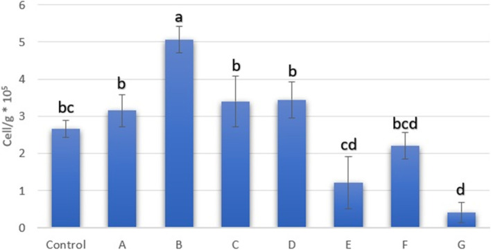
After mechanical dissociation and enzymatic digestion, adipose-derived nucleated cells were quantified. The cell number is presented in Fig. 1. We isolated SCs from all groups (control, 4 οC 6 hrs. (A), 25 οC 6 hrs. (B), 4 οC 12 hrs. (C), 25 οC 12 hrs. (D) and − 75 οC (F)) except for the first series of cryopreservation at -196οC (G) and the first and second series of cryopreservation at − 20 οC (E).
Fig. 1.
The number of nucleated cells isolated from digested adipose tissue (cell/g × 105); Different superscripts differ significantly (p < 0.05)
Morphological analysis
Adherent cells were observed at specific times. On day three after seeding, sporadic adherent cells were observed (Fig. 2-A). On the days four to five after seeding, the number of adherent cells increased and cells varied in morphology (round cells, cells with short cytoplasmic appendages and spindle-shaped cells) (Fig. 2-B). In the cryopreserved groups, cells formed colonies. About 7–12 days after seeding, cells achieved about 70% confluency; at this time, most cells had a fibroblast-like shape (Fig. 2-C).
Fig. 2.
Morphologies of passage 0 ASCs. A single adherent cells at day three after seeding (control group). B Heterogeneous adherent cells on day five after seeding (group F). C On day seven after seeding, nodular aggregation of cells with 90% confluence (control group)
Colony-forming assay
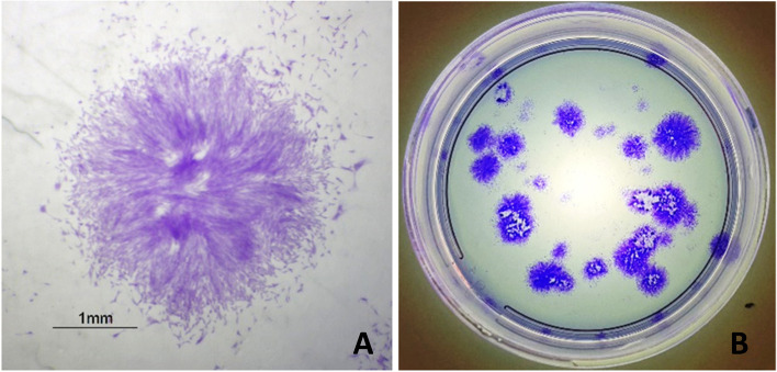
Stem cell colonies are illustrated in Fig. 3. The statistical analysis of the number of colonies and their surface areas is shown in Table 1. In terms of the number of colonies, there was no significant difference. The control group had the highest number of colonies at 52.77 ± 15.32 colonies/100 seeded cells, followed by group G at 44.16 ± 39.83 colonies/100 seeded cells. Group E stood last at 1.66 colonies/100 seeded cells. Analysis of the surface area revealed a significant difference between the group E and the other seven experimental groups. The colony surface area of groups C and A were approximately similar at 29.91 ± 15.37 × 104 pixels and 29.78 ± 13.71 × 104 pixels.
Fig. 3.
A Microscopic illustration of a colony from group C. B Macroscopic illustration of colonies formed ten days after seeding from group B
Table 1.
Colony-forming assay results
| Experimental group | Petri dish number | Colonies number/35 mm Petri dish | Surface area (×104) (pixel) |
|---|---|---|---|
| Mean ± SEM | |||
| Control | 9 | 52.77 ± 15.32 | 16.62 ± 8.74 a |
| A | 9 | 32.33 ± 7 | 29.78 ± 13.71 a |
| B | 9 | 34.77 ± 6 | 25.17 ± 14.09 a |
| C | 9 | 21.22 ± 5.28 | 29.91 ± 15.37 a |
| D | 9 | 23.11 ± 9.79 | 18.03 ± 14.43 a |
| E | 3 | 1.66 | 2.15 b |
| F | 9 | 33.88 ± 16.45 | 12.61 ± 8.97 ac |
| G | 6 | 44.16 ± 39.83 | 3.62 ± 1.26 bc |
Different superscripts differ significantly (p < 0.05)
Determining cell proliferation
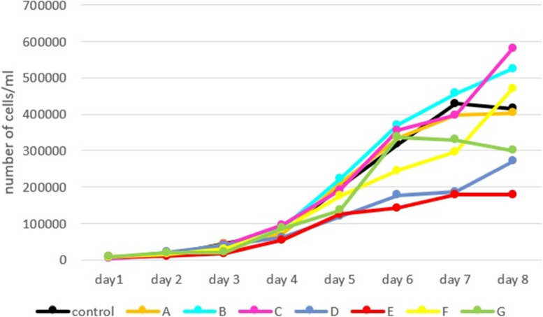
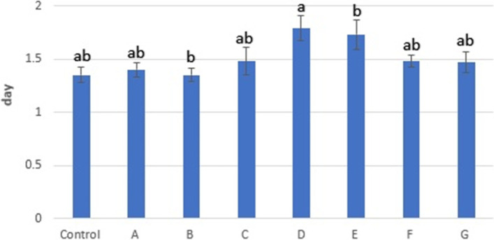
After cell numbers were recorded for 8 days, the growth curve was plotted using these data; the growth curve is illustrated in Fig. 4. Statistical analysis revealed no significant difference between the number of cells on different days except for day seven. Based on the growth curve, the stationary phase of the control group was considered day seven, so doubling time (DT) was calculated for all groups at this time. Statistical analyses of DT are presented in Fig. 5. These analyses showed that there was a significant difference between the group D and the other experimental groups, except for the E. Our analyses showed that the Control and group B had the highest growth rate at ≈ 1.35 ± 0.07 days, whereas D had the lowest at 1.79 ± 0.12.
Fig. 4.
Growth curve of third passaged ASCs, almost all groups follow the same growth pattern. The growth curve includes a latent phase (about day one to two), a logarithmic phase (about days three to six) and a stationary or plateau phase (about days seven to eight). There was a significant difference between the groups B and D, and between B and E, on day seven. There was also a significant difference between C and D
Fig. 5.
Doubling time results, Different superscripts differ significantly (p < 0.05)
MTT assay
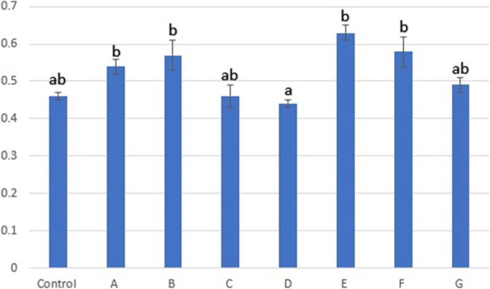
This method is a colorimetric assay stand for cellular metabolic activity and proliferation rate. As shown in Fig. 6, there was a significant difference between the mean absorbance at 560 nm (AB560) of group D and the other experimental groups except for the control, C and G. Based on AB560, the highest absorbance was recorded for group E at 0.63 ± 0.02 which means group E had the highest proliferation rate or cell metabolism. In contrast, the absorbance of group D stood last at 0.44 ± 0.01.
Fig. 6.
Spectrophotometrically absorbance 560 nm, Different superscripts differ significantly (p < 0.05)
Discussion
The first step of SC clinical application is appropriate sampling and sample transportation. Almost all farms are distant from laboratories, so there is a need for establishing a method to transfer samples. Different factors affect transportation, such as the distance between farms and labs, the transportation route, such as by car or airplane, the box in which tissue is transported, the volume of transported material, the transportation time and temperature, etc. In the current study, the impacts of the latter two factors are evaluated. It is also noteworthy that tissue samples were excised through a surgery operation rather than liposuction, so the cryoprotectant effect was not studied in the cryopreserved groups.
In the present study, transportation of fat samples at room temperature (25 οC) or by using a refrigerator (4 οC), freezer (− 20 οC), dry ice (− 75 οC), or liquid nitrogen (− 196 οC) was assessed. Note that it is feasible to implement these methods on farms.
Heterogeneous morphology of adherent cells was seen four to 5 days after primary seeding. Vidal et al. reported a heterogeneous appearance [28]. Grzesiak et al. described the diversity of cells as being due to different cell population presence, including MSCs, preadipocytes, fibroblasts, smooth muscle and endothelium cells [23].
Arnhold et al. claimed that the CFU assay is a well-founded assay to measure MSCs’ stemness and this method confirms whether a cell population is appropriate for clinical application [29]. The CFU number in the present study was 1.66–52.77 colonies/100 seeded cells. After the control group, the highest number of colonies was recorded in G, while E had the lowest number of colonies. Vidal et al. performed the CFU assay for zero, second and fourth passage ASCs and the number of colonies was reported about 15–43 [28]. In a study by Alipour et al., the number of colonies for third passage equine ASCs was reported 5.33–5.37% [30]. The results of these two studies are consistent with the current study results. However, in a survey by Meirelles et al., the CFU assay was performed for the equine ASCs obtained from stromal vascular fraction; they reported the number of colonies per 100 and 1000 seeded cells were 1–7 and 13–52, respectively. Meirelles and colleagues stated that there is a significant difference between different biological samples [31]. Therefore, the difference in the number of colonies between the current study and the study by Meirelles et al. can be explained. Suga et al. determined the colony area of human pluripotent SCs and the number of fluorescently stained nuclei. They reported a linear relationship between the area of colonies and the cell number. They also claimed that determining the colony areas is a good method to discover cells and colonies’ growth rate, whereas counting cells during passage leads to cell wasting and damage [32]. Orozco-Fuentes et al. showed that the number of cells with smaller nuclei increases when a colony grows and intracellular distance declines [33]. Consequently, colony area analysis reveals the cell proliferation rate. In the current study, cryopreserved groups had smaller colony surface areas in comparison with the control group, while the other four groups’ surface areas were larger.
According to the statistical analysis of the present study, DT was 1.35–1.79 days. In a study by Pall et al. on the first passage equine MSCs, DT was reported at 4.1 ± 0.19. Alipour et al. reported a DT 40–46 hrs. for P3 equine MSCs [4, 30]. Vidal et al. in a study on equine ASCs reported that DT for the third passage cells was about 2.4 days [28]. Kim et al. reported different growth rates between cells of various passages [34]. Alipour et al. stated that different culture conditions affect DT [30].
Evaluation of the growth curve showed that the lag phase was short. The logarithmic phase started on day three and continued for about 4 days. It seems that the growth rate of the control, A, E and G groups had declined and the stationary phase of cells started on day seven, but for the rest of the experimental groups, the stationary phase was not seen after 8 days. These results were consistent with the results from the study by Alipour et al. [30].
MTT assay was used to evaluate the cell metabolism. Ghasemi et al. reported that the MTT assay depends on many factors and shows the rate of cell proliferation and metabolism [35]. In the current study, the highest absorbance was recorded in the group E, followed by F. Among groups transported via stem cell media, B had the largest absorbance and A ranked second. Group D had the lowest absorbance. Comparing these data with DT and colony surface area results, which were used to measure cell proliferation rate, showed that although group E had the lowest proliferation rate, it had the highest metabolic activity. In contrast group D have the lowest metabolic activity and low proliferation rate. Consequently, none of these methods are suggested for tissue transportation.
Roato et al., after collecting human AT using liposuction and freezing lipoaspirates at − 20 οC, − 80 οC and − 196 οC, evaluated MSCs viability by MTT assay. They reported that the MSCs viability of cryopreserved samples at − 80 οC and − 196 οC was greater than cryopreserved samples at − 20 οC. Their histological analysis showed that the tissue structures were well preserved. There was no proof of tissue degeneration or necrosis in cryopreserved samples at − 80 οC and − 196 οC, comparable to the fresh AT. The analysis of cell mortality after enzymatic digestion revealed that the high number of dead cells in the cryopreserved group had a significant difference with low quantities of dead cells in fresh AT. They showed the protective effect of AT on MSCs-derived tissue in cryopreserved samples [36]. Moscatello et al., after collecting human adipose tissue through liposuction, the lipoaspirates were frozen at − 20 οC or fat was resuspended in MCDB 201 medium and cryopreserved with or without cryoprotectant at − 80 οC using a control rate freezing method (1 οC/min), following samples were preserved at − 196 οC. Cryopreserved samples were thawed and cultured in plastic cell culture flasks to determine stromal vascular cell viability after cryopreservation. They reported that there were no viable stromal vascular cells in the groups that were frozen without adding cryoprotectant, while they managed to isolate stromal vascular cells in the groups that were frozen with cryoprotectant. They conclude by using a cryoprotectant, utilizing a control-rate freezing method and storage at − 196 οC provide an appropriate condition that maintains ASCs viability [37]. Lie et al. preserved AT from liposuction procedure at − 20 and − 80 οC with or without a cryoprotective agent before thawing and assessing the existence of ASCs through seeding stromal vascular fraction. They reported that no ASCs were isolated from frozen samples without cryoprotectant. In contrast, they isolated ASCs from samples with cryoprotectant [38]. Although no cryoprotectant was added in the current study, the isolation of ASCs from cryopreserved ATs was successful. As the cryopreserved samples were collected through a surgical operation rather than liposuction, the ASCs might be protected by adipose tissue.
In the current study, group E had the lowest number of nucleated cells after enzymatic digestion. Group F had higher nucleated cells than G. Li et al. described that intracellular ice formation and osmotic stress following cryopreservation caused cell damage [39]. Wang et al. reported that tissue freezing at − 20 οC caused ASCs degeneration and apoptosis [40]. Wolter et al. explained that cells had metabolic activity at − 20 οC. There was no cellular activity at temperatures below − 130 οC, but freezing cells below − 130 οC is harmful. They reported that − 80 οC could be a suitable cryopreservation temperature [41]. These studies are consistent with our current cryopreservation results.
Conclusion
The current study results have shown that freezing at − 20 οC is not a suitable method for AT cryopreservation due to poor SCs isolation. The difference between the results of the − 75 οC and − 196 οC groups was not significant, although cryopreservation at − 75 οC is more efficient than at-196 οC; As in − 75 οC group SCs isolation was successful in all three series of sampling, while that for − 196 οC was only in 2 series. Therefore, cryopreservation at − 75 οC is the best method for adipose tissue transportation among cryopreserved groups.
Comparing other groups revealed that maintaining AT at room temperature for 12 hrs. is not an effective method for tissue transferring. Tissue maintenance at 4 οC for 6 and 12 hrs. and 25 οC for 6 hrs. Are acceptable methods due to having minor effects on ASCs.
Methods
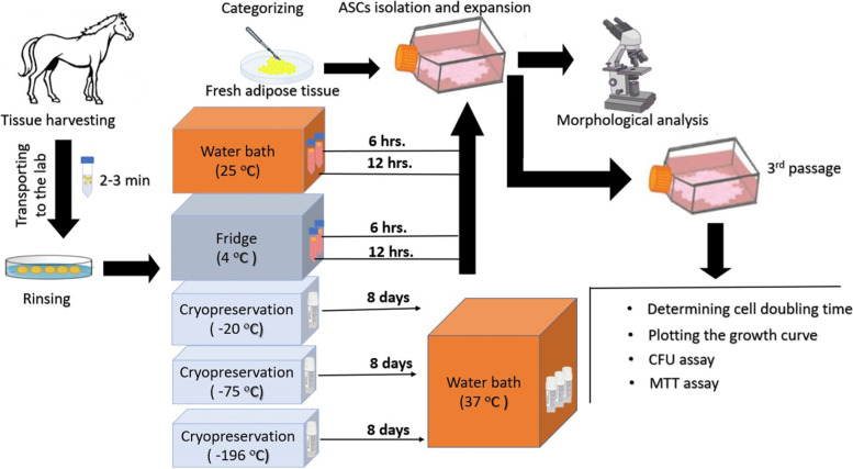
Figure 7 illustrates the different stages of the study.
Fig. 7.
Graphic illustration of methods
Tissue harvesting
Three subcutaneous AT samples were taken from horses ranging from three to 5 years. The project underwent ethical review and was approved by the local Ethics Committee of Ferdowsi University of Mashhad (IR.UM.REC.1400.284). The care and use of experimental animals complied with local animal welfare laws, guidelines and policies.
The procedure was performed on the standing animal in a chute while sedated (Detomidine hydrochloride (Ceva, France) 0.02 mg/kg, IV and Xylazine (Alfasan, Netherland) 0.5 mg/kg, IV). The region above the dorsal gluteal muscles was clipped and anesthesia was provided by a line block (20 ml of Lidocaine Hydrochloride (Nasr, Iran) 1 ml/cm). The surgical area was prepared for aseptic surgery. A horizontal incision was made along the spinal column. Blunt dissection was made through the subcutaneous tissue down to the AT. Tissue was excised and placed in a 50 ml sterile tube filled with 40 ml phosphate buffer saline (PBS) (Biowest, France) containing amphotericin B (1 mg/ml), penicillin (100 IU/ml) and streptomycin (100 mg/ml) (Atocel, Austria) and immediately transported to the laboratory.
Skin closure was performed. After a topical antibiotic was applied, a bandage was placed on the surgical site and changed daily for 3 days. Postoperative systemic antibiotic and nonsteroidal anti-inflammatory were administered for 3 days.
Experimental groups
The samples were washed three times with PBS supplemented with antibiotic and antifungal in the laboratory before being weighed and divided into eight groups (the amount of tissue for all experimental groups was 600 mg):
Control group: Fresh adipose tissue
Transportation via stem cell media groups:
Two specimens of AT were placed in two conical tubes filled with 6 ml stem cell media (low glucose Dulbecco’s Modified Eagle’s Medium (DMEM) (Biosera, France)) and each was maintained at 4 οC and 25 οC for 6 hrs. (A and B, respectively).
Two specimens of AT were placed in two conical tubes filled with 6 ml stem cell media and each was maintained at 4 οC and 25 οC for 12 hrs. (C and D, respectively).
-
3-
Cryopreserved groups: Three specimens of AT were placed in three cryotubes and stored at − 20, − 75 and − 196 οC for 8 days E, F and G, respectively). Then, tubes were placed in a water bath and tissues were thawed at 37 οC for 15 minutes.
Isolation and expansion of MSCs
The isolation of the MSCs procedure was based on the technique previously reported [30]. AT was washed, minced with a scalpel blade, then digested in 3.5 ml digestion media containing 0.1% collagenase type I (Bioidea, Iran) with continuous shaking at 37 οC for 60 minutes. During the last 5 min of enzymatic digestion, 0.001% DNase enzyme (Cinnage, Iran) was added to the suspension. Digested contents were filtered through a 70 μm cell strainer; an extra 3 mL of DMEM was poured through the cell strainer to collect any remaining cells from the strainer. The enzyme was neutralized with DMEM containing 10% fetal bovine serum (FBS) (Biochrom, UK). The suspension was centrifuged at 550 g for 5 minutes and the supernatant was removed. Stromal vascular fraction pellet was resuspended in DMEM. The cell concentration was calculated with a hemocytometer and a standard trypan blue assay was performed to analyze cell viability.
Finally, cells were seeded in 25 cm2 cell culture flasks. The flasks were incubated in a humidified chamber at 37 οC and 5% CO2. The cells were identified as passage (P0). After 3 days, unattached cells were aspirated and the media was changed every 48 hrs. Until a minimum of 70% confluency of the flasks was achieved. After being washed using PBS solution with 1% antibiotic, cells were passaged using 0.5% trypsin enzyme (Biowest, France). Cells were suspended in stem cell media, quantified and subcultured at a density of 80,000 cells/flask. Cells were also cryopreserved in DMEM with 10% DMSO (dimethyl sulfoxide) (Sigma, USA), 20% FBS and 1% antibiotic penicillin (100 IU/ml) and streptomycin (100 mg/ml) and stored at -196οC for future studies. Flasks were maintained until the third passage. Then cells were utilized for subsequent evaluations.
Morphological analysis
Cells at P0 were observed daily using an inverted microscope and morphological changes were recorded.
Colony-forming assay
Cells were plated in a 35 mm cell culture dish at a density of 100 cells/dish suspended in stem cell media in three replications. Dishes were incubated in a humidified chamber at 37 οC and 5% CO2 for 10 days. Next, stem cell media was removed and dishes were rinsed with PBS; then 1 mL of Crystal violet 5% (Sigma, USA) was added to each plate. After 10 min of incubation at room temperature, dishes were washed with distilled water. Finally, stained colonies were fixed in 1 ml formalin 10% for an hour, formalin was aspirated and the plate was washed with distilled water and dried at room temperature. Colonies were observed under a stereomicroscope and photographed with a digital camera. The pictures were analyzed by image-processing software (image j, version 1.50 b) for colony number and surface evaluation.
Determining cell doubling time and plotting the growth curve
Cells were seeded in a 24-well plate at a density of 10,000 cells per well and incubated at 37 οC and 5% CO2 in stem cell media. Adherent cells of three wells were detached by trypsinization and counted daily. The average number of three wells was calculated and recorded from days one to eight after seeding to plot the growth curve.
After the growth curve was plotted and the stationary phase of the control group was identified, cell DT was calculated for cells at this time according to the following formula:
CD: cell doubling time, Nf: cell number at the end of culture, Ni: cell number at the beginning of culture.
DT = CT/CD.
CD: cell doubling time, CT: cell culture time.
MTT assay
The MTT (3-(4,5-dimethylthiazol-2-yl)-2,5-diphenyltetrazolium bromide) assay is used to measure cell viability. This evaluation was performed for all groups simultaneously; therefore, the frozen cells of the second passage of all groups were defrosted and seeded in a 35 mm cell culture dish. The dishes were incubated at 37 οC and 5% CO2 for 24 hrs.. Following this, cells were trypsinized and cultured in a 96-well plate at a density of 10,000 cells per well in three replicas and incubated in a humidified chamber at 37 οC and 5% CO2 in stem cell media for 24 hrs.. The culture media was aspirated, adherent cells were washed in PBS and 100 uL MTT 10X solution (Atocel, Austria) (5 mg/ml solution in PBS following continuous shaking at 4 οC for 24 hrs., filter sterilization was performed) was added. Three unseeded wells were considered as blank samples and all steps were treated like the other wells. The plate was incubated at 37 οC and 5% CO2 for 4 hrs.. Afterward, 100 μL Isopropanol hydrochloride was added with continuous shaking for 30 minutes at room temperature. Finally, the spectrophotometric absorbance (AB560) was measured using a plate reader.
Statistical analysis
Data were statistically analyzed using Sigma Plot software. First, the normality of data was checked using the Shapiro-Wilk test (P < 0.05). Then, a one-way repeated measures analysis of variance was used to compare the mean in each group and between all groups. In this test, p values lower than 0.05 were considered statistically significant. All data are reported as mean ± SEM.
Supplementary Information
Acknowledgments
The authors would like to thank to all of the colleagues at the Center of Excellence in Ruminant Abortion and National Mortality of Ferdowsi University of Mashhad for all their support.
Abbreviations
- SC
Stem cell
- AT
Adipose tissue
- MSC
Mesenchymal stem cell
- ASC
adipose-derived stromal/stem cells
- P
Passage
- CFU
Colony-forming unit
- DT
Doubling time
- AB560
Absorbance at 560 nm
- IV
Intra-venous
- PBS
Phosphate buffer saline
- FBS
Fetal bovine serum
- DMEM
low glucose Dulbecco’s Modified Eagle’s Medium
- CD
Cell doubling time
- Nf
Cell number at the end of culture
- Ni
Cell number at the beginning of culture
- CT
Cell culture time
Authors’ contributions
PM was the supervisor, designed the study and analyzed data. FR, SK and SG contributed to collecting the samples. FR and SK carried out the experiments and assembled data. FR and PM contributed to writing, reviewing and editing the final manuscript. AP was involved in planning the work. All authors read and approved the final manuscript.
Funding
This work was financially supported by the funds granted (Grant No. 48136) by the Applied Research Center, Vice-chancellor for Research of Ferdowsi University of Mashhad.
Availability of data and materials
All data generated or analyzed during this study are included in this published article and its supplementary information files.
Declarations
Ethics approval and consent to participate
The project underwent ethical review and was approved by the local Ethics Committee of Ferdowsi University of Mashhad (IR.UM.REC.1400.284). The care and use of experimental animals complied with local animal welfare laws, guidelines and policies. The study was also carried out in compliance with the ARRIVE guidelines [42].
Consent for publication
Not applicable.
Competing interests
The authors declare that they have no competing interests.
Footnotes
Publisher’s Note
Springer Nature remains neutral with regard to jurisdictional claims in published maps and institutional affiliations.
References
- 1.Sultana T, Lee S, Yoon H-Y, Lee JI. Current status of canine umbilical cord blood-derived mesenchymal stem cells in veterinary medicine. Stem Cells Int. 2018;2018:8329174. doi: 10.1155/2018/8329174. [DOI] [PMC free article] [PubMed] [Google Scholar]
- 2.Zakrzewski W, Dobrzyński M, Szymonowicz M, Rybak Z. Stem cells: past, present, and future. Stem Cell Res Ther. 2019;10(1):1–22. doi: 10.1186/s13287-019-1165-5. [DOI] [PMC free article] [PubMed] [Google Scholar]
- 3.Markoski MM. Advances in the use of stem cells in veterinary medicine: from basic research to clinical practice. Scientifica. 2016;2016:4516920. doi: 10.1155/2016/4516920. [DOI] [PMC free article] [PubMed] [Google Scholar]
- 4.Pall E, Toma S, Crecan C, Cenariu M, Groza I. Isolation and functional characterization of equine adipos tissue derived mesenchymal stem cells. Agri Agricult Scie Procedia. 2016;10:412–416. doi: 10.1016/j.aaspro.2016.09.083. [DOI] [Google Scholar]
- 5.Han Y, Li X, Zhang Y, Han Y, Chang F, Ding J. Mesenchymal stem cells for regenerative medicine. Cells. 2019;8(8):886. doi: 10.3390/cells8080886. [DOI] [PMC free article] [PubMed] [Google Scholar]
- 6.Fan X-L, Zhang Y, Li X, Fu Q-L. Mechanisms underlying the protective effects of mesenchymal stem cell-based therapy. Cell Mol Life Sci. 2020;77(14):2771–2794. doi: 10.1007/s00018-020-03454-6. [DOI] [PMC free article] [PubMed] [Google Scholar]
- 7.Hill ABT, Bressan FF, Murphy BD, Garcia JM. Applications of mesenchymal stem cell technology in bovine species. Stem Cell Res Ther. 2019;10(1):1–13. doi: 10.1186/s13287-019-1145-9. [DOI] [PMC free article] [PubMed] [Google Scholar]
- 8.Voga M, Adamic N, Vengust M, Majdic G. Stem cells in veterinary medicine—current state and treatment options. Front Vet Sci. 2020;7:278. doi: 10.3389/fvets.2020.00278. [DOI] [PMC free article] [PubMed] [Google Scholar]
- 9.Zhang J, Huang X, Wang H, Liu X, Zhang T, Wang Y, et al. The challenges and promises of allogeneic mesenchymal stem cells for use as a cell-based therapy. Stem Cell Res Ther. 2015;6(1):1–7. doi: 10.1186/scrt535. [DOI] [PMC free article] [PubMed] [Google Scholar]
- 10.Dias IE, Pinto PO, Barros LC, Viegas CA, Dias IR, Carvalho PP. Mesenchymal stem cells therapy in companion animals: useful for immune-mediated diseases? BMC Vet Res. 2019;15(1):1–14. doi: 10.1186/s12917-019-2087-2. [DOI] [PMC free article] [PubMed] [Google Scholar]
- 11.Arévalo-Turrubiarte M, Olmeo C, Accornero P, Baratta M, Martignani E. Analysis of mesenchymal cells (MSCs) from bone marrow, synovial fluid and mesenteric, neck and tail adipose tissue sources from equines. Stem Cell Res. 2019;37:101442. doi: 10.1016/j.scr.2019.101442. [DOI] [PubMed] [Google Scholar]
- 12.Conze P, Van Schie HT, Rv W, Staszyk C, Conrad S, Skutella T, et al. Effect of autologous adipose tissue-derived mesenchymal stem cells on neovascularization of artificial equine tendon lesions. Regen Med. 2014;9(6):743–757. doi: 10.2217/rme.14.55. [DOI] [PubMed] [Google Scholar]
- 13.Romero A, Barrachina L, Ranera B, Remacha A, Moreno B, De Blas I, et al. Comparison of autologous bone marrow and adipose tissue derived mesenchymal stem cells, and platelet rich plasma, for treating surgically induced lesions of the equine superficial digital flexor tendon. Vet J. 2017;224:76–84. doi: 10.1016/j.tvjl.2017.04.005. [DOI] [PubMed] [Google Scholar]
- 14.Mousaei Ghasroldasht M, Matin MM, Kazemi Mehrjerdi H, Naderi-Meshkin H, Moradi A, Rajabioun M, et al. Application of mesenchymal stem cells to enhance non-union bone fracture healing. J Biomed Mater Res A. 2019;107(2):301–311. doi: 10.1002/jbm.a.36441. [DOI] [PubMed] [Google Scholar]
- 15.Yuan J, Cui L, Zhang WJ, Liu W, Cao Y. Repair of canine mandibular bone defects with bone marrow stromal cells and porous β-tricalcium phosphate. Biomaterials. 2007;28(6):1005–1013. doi: 10.1016/j.biomaterials.2006.10.015. [DOI] [PubMed] [Google Scholar]
- 16.Glenn JD, Whartenby KA. Mesenchymal stem cells: emerging mechanisms of immunomodulation and therapy. World J Stem Cells. 2014;6(5):526. doi: 10.4252/wjsc.v6.i5.526. [DOI] [PMC free article] [PubMed] [Google Scholar]
- 17.Mount NM, Ward SJ, Kefalas P, Hyllner J. Cell-based therapy technology classifications and translational challenges. Philos Trans R Soc B Biol Sci. 2015;370(1680):20150017. doi: 10.1098/rstb.2015.0017. [DOI] [PMC free article] [PubMed] [Google Scholar]
- 18.Nishimura T, Takami T, Sasaki R, Aibe Y, Matsuda T, Fujisawa K, et al. Liver regeneration therapy through the hepatic artery-infusion of cultured bone marrow cells in a canine liver fibrosis model. PLoS One. 2019;14(1):e0210588. doi: 10.1371/journal.pone.0210588. [DOI] [PMC free article] [PubMed] [Google Scholar]
- 19.Lanci A, Merlo B, Mariella J, Castagnetti C, Iacono E. Heterologous Wharton's jelly derived mesenchymal stem cells application on a large chronic skin wound in a 6-month-old filly. Front Vet Sci. 2019;6:9. doi: 10.3389/fvets.2019.00009. [DOI] [PMC free article] [PubMed] [Google Scholar]
- 20.Aly RM. Current state of stem cell-based therapies: an overview. Stem Cell Investig. 2020;7:8. doi: 10.21037/sci-2020-001. [DOI] [PMC free article] [PubMed] [Google Scholar]
- 21.Nawab K, Bhere D, Bommarito A, Mufti M, Naeem A. Stem cell therapies: a way to promising cures. Cureus. 2019;11(9):e5712. doi: 10.7759/cureus.5712. [DOI] [PMC free article] [PubMed] [Google Scholar]
- 22.Raoufi M, Tajik P, Dehghan M, Eini F, Barin A. Isolation and differentiation of mesenchymal stem cells from bovine umbilical cord blood. Reprod Domest Anim. 2011;46(1):95–99. doi: 10.1111/j.1439-0531.2010.01594.x. [DOI] [PubMed] [Google Scholar]
- 23.Grzesiak J, Marycz K, Czogala J, Wrzeszcz K, Nicpon J. Comparison of behavior, morphology and morphometry of equine and canine adipose derived mesenchymal stem cells in culture. Int J Morphol. 2011;29(3):1012–1017. doi: 10.4067/S0717-95022011000300059. [DOI] [Google Scholar]
- 24.Bravo M, Moraes J, Dummont C, Filgueiras R, Hashimoto H, GODOY R. Isolation, expansion and characterization of equine adipose tissue derived stem cells/Isolamento, expansão e caracterização de células-tronco do tecido adiposo de equinos. Ars Vet. 2012;28(2):066–074. [Google Scholar]
- 25.Miana VV, González EAP. Adipose tissue stem cells in regenerative medicine. Ecancermedicalscience. 2018;12:822–834. doi: 10.3332/ecancer.2018.822. [DOI] [PMC free article] [PubMed] [Google Scholar]
- 26.Alonso-Goulart V, Ferreira LB, Duarte CA, de Lima IL, Ferreira ER, de Oliveira BC, et al. Mesenchymal stem cells from human adipose tissue and bone repair: a literature review. Biotechnol Res Innov. 2018;2(1):74–80. doi: 10.1016/j.biori.2017.10.005. [DOI] [Google Scholar]
- 27.Alderman D, Alexander R. Advances in stem cell therapy: application to veterinary medicine. Today's veterinary. Practice. 2011;1(1):23–29. [Google Scholar]
- 28.Vidal MA, Kilroy GE, Lopez MJ, Johnson JR, Moore RM, Gimble JM. Characterization of equine adipose tissue-derived stromal cells: adipogenic and osteogenic capacity and comparison with bone marrow-derived mesenchymal stromal cells. Vet Surg. 2007;36(7):613–622. doi: 10.1111/j.1532-950X.2007.00313.x. [DOI] [PubMed] [Google Scholar]
- 29.Arnhold S, Elashry MI, Klymiuk MC, Geburek F. Investigation of stemness and multipotency of equine adipose-derived mesenchymal stem cells (ASCs) from different fat sources in comparison with lipoma. Stem Cell Res Ther. 2019;10(1):1–20. doi: 10.1186/s13287-019-1429-0. [DOI] [PMC free article] [PubMed] [Google Scholar]
- 30.Alipour F, Parham A, Mehrjerdi HK, Dehghani H. Equine adipose-derived mesenchymal stem cells: phenotype and growth characteristics, gene expression profile and differentiation potentials. Cell J (Yakhteh) 2015;16(4):456. doi: 10.22074/cellj.2015.491. [DOI] [PMC free article] [PubMed] [Google Scholar]
- 31.da Silva ML, Sand TT, Harman RJ, Lennon DP, Caplan AI. MSC frequency correlates with blood vessel density in equine adipose tissue. Tissue Eng A. 2009;15(2):221–229. doi: 10.1089/ten.tea.2007.0327. [DOI] [PMC free article] [PubMed] [Google Scholar]
- 32.Suga M, Kii H, Niikura K, Kiyota Y, Furue MK. Development of a monitoring method for nonlabeled human pluripotent stem cell growth by time-lapse image analysis. Stem Cells Transl Med. 2015;4(7):720–730. doi: 10.5966/sctm.2014-0242. [DOI] [PMC free article] [PubMed] [Google Scholar]
- 33.Orozco-Fuentes S, Neganova I, Wadkin LE, Baggaley AW, Barrio RA, Lako M, et al. Quantification of the morphological characteristics of hESC colonies. Sci Rep. 2019;9(1):1–11. doi: 10.1038/s41598-019-53719-9. [DOI] [PMC free article] [PubMed] [Google Scholar]
- 34.Kim H-R, Lee J, Byeon JS, Gu N-Y, Lee J, Cho I-S, et al. Extensive characterization of feline intra-abdominal adipose-derived mesenchymal stem cells. J Vet Sci. 2017;18(3):299. doi: 10.4142/jvs.2017.18.3.299. [DOI] [PMC free article] [PubMed] [Google Scholar]
- 35.Ghasemi M, Turnbull T, Sebastian S, Kempson I. The MTT assay: utility, limitations, pitfalls, and interpretation in bulk and single-cell analysis. Int J Mol Sci. 2021;22(23):12827. doi: 10.3390/ijms222312827. [DOI] [PMC free article] [PubMed] [Google Scholar]
- 36.Roato I, Alotto D, Belisario DC, Casarin S, Fumagalli M, Cambieri I, et al. Adipose derived-mesenchymal stem cells viability and differentiating features for orthopaedic reparative applications: banking of adipose tissue. Stem Cells Int. 2016;2016:4968724. doi: 10.1155/2016/4968724. [DOI] [PMC free article] [PubMed] [Google Scholar]
- 37.Moscatello DK, Dougherty M, Narins RS, Lawrence N. Cryopreservation of human fat for soft tissue augmentation: viability requires use of cryoprotectant and controlled freezing and storage. Dermatol Surg. 2005;31:1506–1510. doi: 10.2310/6350.2005.31235. [DOI] [PubMed] [Google Scholar]
- 38.Lee JE, Kim I, Kim M. Adipogenic differentiation of human adipose tissue–derived stem cells obtained from cryopreserved adipose aspirates. Dermatol Surg. 2010;36(7):1078–1083. doi: 10.1111/j.1524-4725.2010.01586.x. [DOI] [PubMed] [Google Scholar]
- 39.Li B-W, Liao W-C, Wu S-H, Ma H. Cryopreservation of fat tissue and application in autologous fat graft: in vitro and in vivo study. Aesthet Plast Surg. 2012;36(3):714–722. doi: 10.1007/s00266-011-9848-z. [DOI] [PubMed] [Google Scholar]
- 40.Wang WZ, Fang X-H, Williams SJ, Stephenson LL, Baynosa RC, Wong N, et al. The effect of lipoaspirates cryopreservation on adipose-derived stem cells. Aesthet Surg J. 2013;33(7):1046–1055. doi: 10.1177/1090820X13501690. [DOI] [PubMed] [Google Scholar]
- 41.Wolter TP, von Heimburg D, Stoffels I, Groeger A, Pallua N. Cryopreservation of mature human adipocytes: in vitro measurement of viability. Ann Plast Surg. 2005;55(4):408–413. doi: 10.1097/01.sap.0000181345.56084.7d. [DOI] [PubMed] [Google Scholar]
- 42.Du Sert NP, Ahluwalia A, Alam S, Avey MT, Baker M, Browne WJ, et al. Reporting animal research: explanation and elaboration for the ARRIVE guidelines 2.0. PLoS Biol. 2020;18(7):e3000411. doi: 10.1371/journal.pbio.3000411. [DOI] [PMC free article] [PubMed] [Google Scholar]
Associated Data
This section collects any data citations, data availability statements, or supplementary materials included in this article.
Supplementary Materials
Data Availability Statement
All data generated or analyzed during this study are included in this published article and its supplementary information files.


