Abstract
The potential use of Bacillus anthracis as a weapon of mass destruction poses a threat to humans, domesticated animals, and wildlife and necessitates the need for a rapid and highly specific detection assay. We have developed a real-time PCR-based assay for the specific detection of B. anthracis by taking advantage of the unique nucleotide sequence of the B. anthracis rpoB gene. Variable region 1 of the rpoB gene was sequenced from 36 Bacillus strains, including 16 B. anthracis strains and 20 other related bacilli, and four nucleotides specific for B. anthracis were identified. PCR primers were selected so that two B. anthracis-specific nucleotides were at their 3′ ends, whereas the remaining bases were specific to the probe region. This format permitted the PCR reactions to be performed on a LightCycler via fluorescence resonance energy transfer (FRET). The assay was found to be specific for 144 B. anthracis strains from different geographical locations and did not cross-react with other related bacilli (175 strains), with the exception of one strain. The PCR assay can be performed on isolated DNA as well as crude vegetative cell lysates in less than 1 h. Therefore, the rpoB-FRET assay could be used as a new chromosomal marker for rapid detection of B. anthracis.
Bacillus anthracis is a causal agent of anthrax, a serious and often fatal infection of livestock and humans. It is considered one of the most effective biological weapons of mass destruction because of its highly pathogenic nature and spore-forming capability and has attracted attention due to its potential use as a biological warfare agent (2). This bacterium can infect humans by cutaneous, gastrointestinal, or respiratory routes. The standard laboratory method of identification takes advantage of the lytic nature of the B. anthracis-specific gamma bacteriophage (9). Anthrax bacilli are often distinguished on the basis of time-consuming morphological or phenotypic characteristics, such as gram-positive staining, spore-forming capability, nonhemolytic reaction on sheep blood agar, sensitivity to penicillin, nonmotile nature, and inability to ferment salicin (11). B. anthracis is distinguished from the other members of the closely related Bacillus cereus group of bacteria by the presence of the toxin-encoding pXO1 (19, 24) and capsule-encoding pXO2 plasmids (14, 23, 34). Both plasmids are needed for virulence; thus, the absence of either plasmid results in attenuation.
B. anthracis, Bacillus thuringiensis, B. cereus, and Bacillus mycoides, are members of the B. cereus group of bacilli. These closely related bacteria are pathogens of mammals (B. anthracis and B. cereus) and insects (B. thuringiensis). The B. cereus group is one of the most taxonomically ambiguous group of bacilli (27). In fact, DNA-DNA hybridization (30) and pulsed-field gel electrophoresis (15) have shown great homology among B. anthracis, B. thuringiensis, and B. cereus. A recent multilocus enzyme electrophoresis study has concluded that the members of this group belong to one species (16).
Although specific assays are available for the detection of pathogenicity-related plasmids (18, 28), chromosomal markers in conjuction with plasmid markers should be used for complete genotyping of B. anthracis strains. Such a combined approach will provide insight into the chromosomal backbone or genetic background and indicate the pathogenic nature of the strain. Plasmids are more unstable than chromosomal DNA, and isolates lacking either or both plasmids have been found to exist in nature (33). Also, pXO2 has been successfully transferred into other bacilli, and toxin genes, such as lef and cya, have been expressed in heterologous systems (4, 5, 20). Thus, naturally occurring as well as genetically modified B. anthracis strains cannot be characterized without ambiguity. Moreover, chromosomal markers are stable targets for detection and are important for accurate identification of B. anthracis in outbreaks (26) as well as during the analysis of ancient samples (C. Redmond, M. J. Pearce, R. J. Manchee, and B. P. Berdal, Letter, Nature 393:747–748).
Several chromosomal markers are currently available for B. anthracis detection, such as the vrrA gene (1, 17), Ba813 marker (25), and SG-850 marker (10). These marker assays suffer from being time consuming or labor intensive or having limited specificity. For instance, the SG-850 assay involves PCR amplification of the SG-749 locus, followed by enzymatic digestion with AluI and gel analysis. The vrrA marker can group B. anthracis isolates into several categories based on the number of repeat units of this sequence, which requires post-PCR analysis (17). Recently some B. cereus and B. thuringiensis isolates have been found to contain the Ba813 marker (26); hence its use as a B. anthracis-specific target is questionable (29). The 16S rRNA gene also does not provide sufficient polymorphism to differentiate B. anthracis from closely related bacilli (3). Thus, no absolutely specific chromosomal marker is presently available for the detection of B. anthracis.
The rpoB gene, which codes for the β-subunit of RNA polymerase, has served as a signature sequence for bacterial identification as well as a locus for phylogenetic analysis (21). Moreover, rpoB is a highly conserved housekeeping gene, and at least one copy is present in all bacteria because of its essential role in cellular metabolism. This gene, along with rpoC, which encodes for the β′-subunit, constitutes the catalytic center of the pentameric bacterial RNA polymerase (6). Due to its discriminatory power, the rpoB gene has been used to develop probes for specific detection and phylogenetic analysis of Coxiella burnetti, Rickettsia, and Yersinia pestis (12, 13, 22).
Bacterial strains and DNA preparation.
A total of 144 B. anthracis strains from different geographical locations (Table 1), 29 B. cereus strains, 49 B. thuringiensis strains, 73 Bacillus spp. Ba813+ strains (29), a strain each of B. mycoides, B. subtilis, and B. megaterium, and 22 unknown bacilli were used to test the specificity of the assay. Sixteen B. anthracis strains and a total of 20 other bacilli strains of B. cereus (n = 6), B. thuringiensis (n = 6), B. mycoides (n = 1), B. subtilis (n = 1), and other bacilli (n = 6) were used for the determination of variable region 1 of the rpoB gene sequence (Table 2). All strains used in this study were analyzed for plasmid content by a multiplex PCR assay (28). DNA was extracted by a method outlined by Schraft and Griffiths (32) with modifications as described elsewhere (8). For preparing crude vegetative cell lysates, a sterile toothpick was used to transfer a portion of a fresh colony into 300 μl of distilled water. The cell suspension was boiled at 100°C for 15 min and then centrifuged at 8,000 × g for 5 min. The supernatant was transferred to a fresh microcentrifuge tube and stored at −20°C until further use. For isolating the rifampin-resistant mutant, an individual fresh colony of the rifampin-sensitive B. anthracis 7700 was streaked out on a brain heart infusion agar containing 25 μg of rifampin (Sigma Chemical Co., St. Louis, Mo.) per ml and was incubated overnight at 37°C. Rifampin-resistant colonies were plated out a second time onto a plate containing 50 μg of rifampin/ml in order to confirm this phenotype.
TABLE 1.
B. anthracis strains used in the rpoB-FRET PCR assays
| Strain | pXO1a | pXO2a | Origin | |
|---|---|---|---|---|
| 7700 | − | − | Africa | |
| 7702 | + | − | Africa | |
| Sterne | + | − | Africa | |
| AC1 | + | − | S. America | |
| AC2 | + | − | S. America | |
| AC3 | + | + | S. America | |
| Texas 0077 | + | − | N. America | |
| Ames | + | + | N. America | |
| Δ Ames | − | − | N. America | |
| Vollum | + | + | Europe | |
| 4229 | − | + | Europe | |
| RA3 | + | + | Europe | |
| RA3R | + | − | Europe | |
| RA4 | + | + | Europe | |
| 7611 | + | + | Europe | |
| 7611R | + | − | Europe | |
| 10517 | + | + | Europe | |
| 6183 | + | + | Europe | |
| 6042 | + | + | Europe | |
| BC 575 | + | + | Europe | |
| 9066 | + | + | Europe | |
| 7193 | + | + | Europe | |
| 6681 | + | + | Europe | |
| 8403 | + | + | Europe | |
| 9240 | + | + | Europe | |
| 8490 | + | + | Europe | |
| 6291 | + | + | Europe | |
| A3 | − | + | Europe | |
| A4 | − | + | Europe | |
| A5 | + | + | Europe | |
| A6 | − | + | Europe | |
| A7 | + | + | Europe | |
| A8 | + | + | Europe | |
| A9 | + | + | Europe | |
| A10 | + | + | Europe | |
| A11 | + | + | Europe | |
| A12 | + | + | Europe | |
| A16 | + | + | Europe | |
| A18 | − | + | Europe | |
| A19 | − | + | Europe | |
| A22 | + | + | Europe | |
| A23 | − | + | Europe | |
| A24 | + | + | Europe | |
| A25 | + | + | Europe | |
| A28 | + | + | Europe | |
| A29 | + | + | Europe | |
| A30 | + | + | Europe | |
| A32 | + | + | Europe | |
| A33 | + | + | Europe | |
| A34 | + | + | Europe | |
| A35 | + | + | Europe | |
| A36 | + | + | Europe | |
| A37 | + | + | Europe | |
| A38 | + | + | Europe | |
| A39 | + | + | Europe | |
| A40 | + | + | Europe | |
| A41 | + | + | Europe | |
| A42 | + | + | Europe | |
| A43 | + | + | Europe | |
| A44 | + | + | Europe | |
| A45 | + | + | Europe | |
| A46 | − | + | Europe | |
| A47 | + | + | Europe | |
| A49 | + | + | Europe | |
| A58 | − | − | Europe | |
| A59 | + | + | Europe | |
| A60 | − | + | Europe | |
| A61 | + | + | Europe | |
| A62 | + | + | Europe | |
| A63 | + | + | Europe | |
| A64 | + | + | Europe | |
| A65 | + | + | Europe |
| A66 | + | + | Europe |
| A67 | + | + | Europe |
| A68 | + | + | Europe |
| A69 | + | + | Europe |
| A72 | + | + | Europe |
| A73 | − | + | Europe |
| A74 | − | − | Europe |
| 001 | + | + | Asia |
| 002 | + | + | Asia |
| 044 | + | − | Asia |
| 103 | + | + | Asia |
| 104 | + | + | Asia |
| 105 | + | + | Asia |
| 108 | + | + | Asia |
| 116 | + | + | Asia |
| 125 | + | − | Asia |
| 126 | + | + | Asia |
| 128 | + | − | Asia |
| 129 | + | + | Asia |
| 136 | + | + | Asia |
| 137 | + | + | Asia |
| 139 | + | + | Asia |
| 140 | + | + | Asia |
| 141 | + | + | Asia |
| 142 | + | − | Asia |
| 143 | + | − | Asia |
| 144 | + | + | Asia |
| 145 | + | + | Asia |
| 146 | + | + | Asia |
| 147 | + | + | Asia |
| 148 | + | + | Asia |
| 149 | + | + | Asia |
| 150 | + | + | Asia |
| 151 | + | + | Asia |
| 152 | + | + | Asia |
| 153 | + | + | Asia |
| 154 | + | + | Asia |
| 155 | + | + | Asia |
| 156 | + | + | Asia |
| 157 | + | + | Asia |
| 158 | + | + | Asia |
| 159 | + | + | Asia |
| 160 | + | + | Asia |
| 161 | + | + | Asia |
| 162 | + | + | Asia |
| 163 | + | + | Asia |
| 164 | + | − | Asia |
| 165 | + | + | Asia |
| 166 | + | + | Asia |
| 167 | + | + | Asia |
| 168 | + | + | Asia |
| 169 | + | + | Asia |
| 170 | + | + | Asia |
| 171 | + | + | Asia |
| 172 | + | + | Asia |
| 173 | + | + | Asia |
| 174 | + | + | Asia |
| 175 | + | + | Asia |
| 176 | + | + | Asia |
| 177 | + | + | Asia |
| 178 | + | + | Asia |
| 179 | + | + | Asia |
| 180 | + | + | Asia |
| 181 | + | + | Asia |
| 182 | + | + | Asia |
| 183 | + | + | Asia |
| Ba107 | + | + | ND |
| ΔUM-2311 | − | − | ND |
| ΔANR-1099 | − | − | ND |
| 1014 | − | + | ND |
| ACB | + | + | ND |
| 0074 | + | − | ND |
Plasmids were detected by PCR (28). +, detected; −, not detected; ND, no data.
TABLE 2.
rpoB Bacillus species sequences analyzed in this study
| Bacillus species | Strain ID | pXO1a | pXO2a | Derivation or location | Accession no. |
|---|---|---|---|---|---|
| B. anthracis | Vollum | − | + | Europe | AF205319 |
| B. anthracis | Ames | + | + | N. America | AF205320 |
| B. anthracis | ΔAmes | − | − | N. America | AF205321 |
| B. anthracis | ΔUM-2311 | − | − | Unknown | AF205322 |
| B. anthracis | Sterne | + | − | Africa | AF205323 |
| B. anthracis | Texas 0077 | + | − | N. America | AF205324 |
| B. anthracis | 001 | + | + | Asia | AF205325 |
| B. anthracis | 002 | + | + | Asia | AF205326 |
| B. anthracis | 044 | + | − | Asia | AF205327 |
| B. anthracis | A58 | − | − | Europe | AF205328 |
| B. anthracis | A74 | − | − | Europe | AF205329 |
| B. anthracis | A7 | + | + | Europe | AF205330 |
| B. anthracis | AC1 | + | − | S. America | AF205331 |
| B. anthracis | 7193 | + | + | Europe | AF205333 |
| B. anthracis | 8403 | + | + | Europe | AF205334 |
| B. anthracis | RA3 | + | + | Europe | AF205335 |
| B. cereus | 14579 | − | − | ATCCb 14579 | AF205336 |
| B. cereus | 229 | − | − | Unknown | AF205337 |
| B. cereus | 27877 | − | − | ATCC 27877 | AF205338 |
| B. cereus | 49069 | − | − | ATCC 49069 | AF205339 |
| B. cereus | 776 | − | − | ATCC 19637 | AF205341 |
| B. cereus | 23261 | − | − | ATCC 23261 | AF205342 |
| B. mycoides | 6462 | − | − | ATCC 6462 | AF205343 |
| B. thuringiensis | T07-005 | − | − | IEBCc T07-005 | AF205344 |
| B. thuringiensis | T07-146 | − | − | IEBC T07-146 | AF205345 |
| B. thuringiensis | T07-202 | − | − | IEBC T07-202 | AF205346 |
| B. thuringiensis | 10 | − | − | Asia | AF205347 |
| B. thuringiensis | 35646 | − | − | ATCC 35646 | AF205348 |
| B. thuringiensis | B8 | − | − | Unknown | AF205349 |
| Bacillus spp. | Ba813_11 (9594/3) | − | − | Europe | AF205350 |
| Bacillus spp. | Ba813_12 (S8553/2) | − | − | Europe | AF205351 |
| Bacillus spp. | Ba813_31 (IB/A) | − | − | Middle East | AF205352 |
| Bacillus sp. | AX16 | − | − | Unknown | AF205353 |
| Bacillus sp. | N52 | − | − | Unknown | AF205354 |
| Bacillus sp. | V770 | − | − | Unknown | AF205355 |
| B. subtilis | 6051 | − | − | ATCC 6051 | AF205356 |
Ramisse et al., 1996 (28). +, detected; −, not detected; ND, no data.
American Type Culture Collection, Manassas, Va.
International Entomopathogenic Bacillus Centre Collection, Pasteur Institute, Paris, France.
Low-stringency PCR amplification and sequence analysis.
The alignment of the amino acid sequences of the RNA polymerase β-subunits of Bacillus subtilis and Escherichia coli permitted the identification of two conserved regions. The conserved region found near the N terminus was RVIVSQ, spanning amino acid residues 132 to 137 of B. subtilis and residues 143 to 148 of E. coli (6). The C terminus conserved region was DDIDHL, and it was found at positions 399 to 404 of B. subtilis and positions 443 to 448 of E. coli.
The sequences of primer rpoB1 (5′-CGTGTTATCGTTTCCCAGC-3′) and rpoB2 (5′-AAGATGATCGATATCATCTG-3′) were derived from the two conserved regions and correspond to nucleotides (nt) 1482 to 1500 and 2281 to 2300 of the B. subtilis rpoB gene (GenBank accession no. L24376). The PCR reaction mixture of 50 μl consisted of 10 mM Tris-HCl (pH 8.3), 75 mM KCl, 3.5 mM MgCl2, 0.2 mM dNTPs (Boehringer Mannheim Corp., Indianapolis, Ind.), 1 μM (each) primers rpoB1 and rpoB2, 0.05 U of AmpliTaq DNA polymerase (Perkin Elmer Corp., Foster City, Calif.)/μl, and 100 ng of DNA template. Amplification was performed in a GeneAmp PCR System 2400 (Perkin-Elmer Corp., Norwalk, Conn.), and the cycling conditions were as follows: initial denaturation at 94°C for 5 min, followed by 35 cycles of 94°C for 1 min, 45°C for 1 min, and 72°C for 1 min, with a final extension of 72°C for 7 min. The amplicons were detected in 2% (wt/vol) SeaKem GTG agarose (FMC Bioproducts, Rockland, Maine) with 40 mM Tris-acetate–1mM EDTA (pH 8.3) as a running buffer and visualized by ethidium bromide staining.
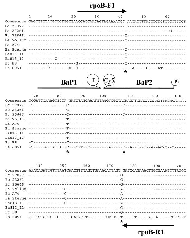
Low-stringency amplification of the variable region of the rpoB gene from different Bacillus species and strains yielded the expected amplicons with a size of 819 bp. Bands of the expected size were excised from the gel, and the DNA was extracted using a QIAquick Gel Extraction kit (QIAGEN Inc., Valencia, Calif.). The PCR products were cloned into vector pCR 2.1 (Invitrogen Corp., Carlsbad, Calif.) and transformed into E. coli. Recombinant plasmids were prepared using the QIAGEN Plasmid Mini Kit. Three clones from each ligation reaction were sequenced in duplicate with the M13 forward and reverse primers using the Applied Biosystems model 373A automated sequencer and the BigDye terminator ready reaction kit (Perkin-Elmer Applied Biosystems). The nucleotide sequences were edited and assembled with the Sequencing Analysis 3.0 and AutoAssembler 3.1.2 programs, respectively; translation into amino acids was accomplished using the Sequence Navigator 3.0.1 program (Perkin-Elmer Applied Biosystems). These 36 sequences were aligned using the Clustal W program (32) from BioNavigator (eBioinformatics Pty Ltd: http://www.ebioinformatics.com/), and four bases specific for B. anthracis were identified. Figure 1 shows the alignment and nucleotide differences of 10 representative strains, including 3 strains of B. anthracis, 2 strains of B. cereus, 2 strains of B. thuringiensis, 2 Ba813+ strains of Bacillus sp. (29), and a single strain of B. subtilis. Thus, nucleotides C (position 42), T (position 84), C (position 108), and A (position 174) are unique to B. anthracis, with the exception of Ba813_11 (Fig. 1). The region of the rpoB gene described in this study appears to be the only region within the rpoB gene that shows variation among different species of bacteria (6).
FIG. 1.
Alignment of the nucleotide sequences from 10 representative Bacillus strains using Clustal W (32). The strains are the following: Bc 27877, Bacillus cereus 27877; Bc 23261, Bacillus cereus 23261; Bt 35646, Bacillus thuringiensis 35646; Ba Vollum, Bacillus anthracis Vollum; Ba A74, Bacillus anthracis A74; Ba Sterne, Bacillus anthracis Sterne; Ba813_11, Bacillus sp. strain Ba813_11; Ba813_12, Bacillus sp. strain Ba813_12; Bt B8, Bacillus thuringiensis BtB8; Bs 6051, Bacillus subtilis 6051. The locations of primers and probes are shown (F, fluorescein; Cy5, cyanine 5; P, phosphate group); the presence of an asterisk denotes a mismatch, a dash indicates identity with the consensus sequence, and nucleotide letters indicate positions showing polymorphism.
Nucleotide sequence accession number.
The nucleotide sequences of the portion of the rpoB gene (variable region 1) described in this study were submitted to GenBank, and the accession numbers are listed in Table 2.
The translation of the nucleotide sequences showed that four bases specific for B. anthracis were in the third positions of the codons and did not change the amino acid sequence. The positions of the amino acids in the β-subunit were alanine at 251, tyrosine at 265, tyrosine at 273, and valine at 295. Thus, although there are differences in the nucleotide sequences, no differences were found in the primary sequence of the RpoB proteins for B. anthracis, B. cereus, and B. thuringiensis.
FRET-PCR assay.
The primers rpoBF1 and rpoBR1 (Table 3; Fig. 1) were selected for high-stringency PCR amplification using Oligo 6 software (National Biosciences Inc., Plymouth, Minn.). The probes BaP1 (3′ end labeled with Fluorescein) and BaP2 (5′ end labeled with Cy5 and 3′ blocked with a phosphate group) were placed 1 bp apart within the PCR product (Fig. 1) and had Tms (7) at least 10°C higher than those of the amplification primers (Table 3).
TABLE 3.
Primers and probes used in the FRET-PCR assay
| Primer or probe | Positiona | Tm (°C)b | Sequence (5′→3′)c |
|---|---|---|---|
| rpoBF1 primer | (1821–1841) | 64.6 | CCA CCA ACA GTA GAA AAT GCC |
| rpoBR1 primer | (1973–1995) | 64.2 | AAA TTT CAC CAG TTT CTG GAT CT |
| BaP1 probe | (1871–1897) | 74.4 | TCC AAA GCG CTA TGA TTT AGC AAA TGT-F |
| BaP2 probe | (1899–1928) | 74.1 | Cy5-GGT CGC TAC AAG ATC AAC AAG AAG TTA CAC-P |
Based on the B. subtilis rpoB gene (GenBank accession no. L24376).
Nearest neighbor method.
F, fluorescein; Cy5, cyanine 5; P, phosphate.
The PCR mixture (10 μl) consisted of 10 mM Tris-HCl (pH 8.3), 50 mM KCl, 2.5 mM MgCl2, 0.2 mM dNTPs, 250 μg of bovine serum albumin/ml (Roche Molecular Biochemicals, Indianapolis, In), 1 μM (each) primers rpoBF1 and rpoBR1, 0.2 μM probe BaP1, 0.4 μM probe BaP2, 0.8U of DNA polymerase KlenTaq1 (Ab Peptides, St. Louis, Mo.), and 50 ng of DNA template or 2 μl of crude vegetative cell lysate. The amplification was performed on a Light-Cycler (Idaho Technology, Idaho Falls, Idaho), which is a rapid, forced-air thermocycler with an integrated fluorimeter for real-time monitoring of PCR reactions (35). The amplification was accomplished by initial denaturation at 95°C for 30 s, followed by 35 cycles of 95°C for 0 s, 63°C for 15 s, and 72°C for 5 s. Once the capillaries were placed in the thermocycler, amplification could be completed in less than 30 min. Detection of the amplification products is accomplished by hybridization of a pair of probes to the amplicons as they are formed, resulting in a fluorescence resonance energy transfer (FRET) (35). Fluorescence was measured once every cycle at the annealing step using the F2/F1 filter to monitor amplification in real time. F1 corresponds to the baseline fluorescein fluorescence, while F2 indicates FRET from fluorescein to Cy5, resulting in the ratio of Cy5/fluorescein fluorescence (F2/F1). The increase in fluorescence is proportional to the amount of PCR product generated (7, 35) and is displayed on the computer screen in the real-time mode. The reactions showing an increase in fluorescence by a minimum of 0.05 fluorescence units (y axis) were scored as positive amplification reactions. The PCR products were also visualized by 2% (wt/vol) gel electrophoresis.
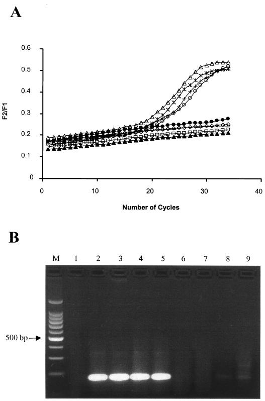
The FRET assay was performed on 144 B. anthracis strains, harboring any combination of the two plasmids and isolated from different geographical locations (Table 1). All these strains tested positive in the FRET assay, since they displayed an increase in fluorescence as well as the presence of the expected PCR product by agarose gel analysis. Another 175 closely related strains, including the B. cereus group and Ba813+ strains, were tested as negative controls to check the specificity of the assay. All related strains, with the exception of Ba813_11, were scored as negative because they did not exhibit an increase in fluorescence. Figure 2 shows the results of the FRET-PCR assay and the electrophoresis of the PCR amplicons using genomic DNA samples from representative strains. B. cereus, B. thuringiensis, Bacillus sp. strain Ba813_12, and B. subtilis did not show amplification. The rpoB FRET-PCR assay is extremely specific for B. anthracis because of the high-stringency PCR conditions coupled with the unique nature of the primers and probes. This specificity occurs at two different levels. The first is at the primer level, as seen in the case of Bacillus sp. strain Ba813_12. In this instance PCR products were not generated due to single base-pair difference at 3′ end of both primers, and as a result an increase in fluorescence was not observed in spite of 100% homology of the probe region with the B. anthracis sequence. The second level of specificity is at the probe level, since 100% base-pairing of probes with target sequence is required for FRET to occur. A single base-pair mismatch between either of the probe sequences with the target region stops the FRET process, indicating a negative result (unpublished data).
FIG. 2.
Results of the FRET-PCR assay using genomic DNA. (A) Fluorescence ratio (F2/F1) is plotted against the number of PCR cycles. The samples are the following: 1, □, negative control (no DNA); 2, ▵, Bacillus anthracis A74; 3, ○, Bacillus anthracis Sterne; 4,  , Bacillus sp. strain Ba813_11; 5, +, Bacillus anthracis Vollum; 6, ●, Bacillus cereus 27877; 7, ◊, Bacillus cereus 23261; 8, −, Bacillus sp. strain Ba813_12; 9, ▴, Bacillus thuringiensis BtB8. (B) Gel electrophoresis of the PCR products. Lanes, M: 100 bp DNA ladder; 1, negative control (no DNA); samples 2 to 9 are the same as in panel A.
The amplicon derived from Bacillus sp. strain Ba813_11 was sequenced, and it was found to have a nucleotide sequence identical to that of B. anthracis (Fig. 1). Consequently, the FRET-PCR assay reported here cannot distinguish Bacillus sp. strain Ba813_11 from B. anthracis strains. The remaining 71 Bacillus spp. Ba813+ strains have sequence identical to that of Bacillus sp. strain Ba813_12 in primer and probe binding regions, and Bacillus sp. strain Ba813_11 appears to be an exception. This strain, Bacillus sp. strain Ba813 (9594/3), was isolated from a station effluent in the Alps in 1997 and was designated a transitional strain because it could not be assigned to a particular species (26, 29). According to the SG-749 locus signature, Bacillus sp. strain Ba813_11 belongs to the B. cereus group (data not shown) and was shown to contain the Ba813+ marker (26). In contrast to the phenotypic characteristics of B. anthracis, the Bacillus sp. strain Ba813_11 is hemolytic, motile, and resistant to penicillin, although it has an rpoB variable region 1 identical to that of B. anthracis.
The assay was not affected by the presence of exogenously added E. coli or mixed Bacillus species DNA (25 ng of B. anthracis DNA + 1,000 ng of exogenously added DNA) representing a mixed microbial community at the ratio of 1:40 (data not shown). The sensitivity of the FRET-PCR assay was examined using different concentrations of exogenously added DNA. Positive fluorescence signals and amplification, as shown by gel electrophoresis, were noticed even when as little as 1 pg of pure genomic DNA was used.
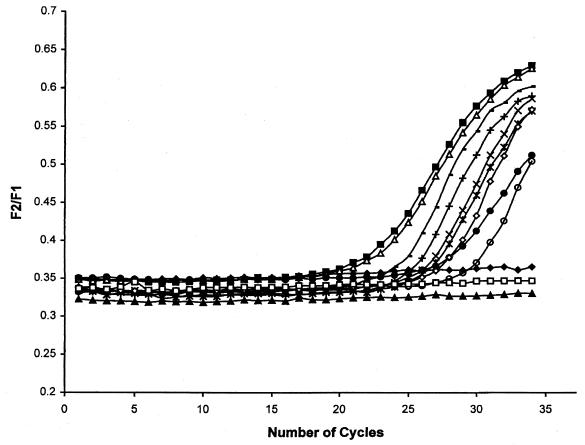
The FRET-PCR assay was also performed on crude vegetative cell lysates from B. anthracis and related bacilli. Figure 3 shows the results of the assay on selected strains. Only B. anthracis displayed an increase in fluorescence and the presence of the expected amplification product. The increase in fluorescence was observed after 22 to 30 cycles. The magnitude of increase in fluorescence is dependent on the quantity of template DNA or the copy number of the gene target in the reaction (35). It should be noted that the DNA amount was not normalized in the different cell lysates because the number of vegetative cells used in different samples was not identical. Thus, the assay can be directly used on freshly grown cultures for rapid identification of B. anthracis strains in less than 1 h. A rifampin-resistant colony exhibited positive results when tested by the FRET-PCR assay because the position of the mutation was found to be outside the rpoB target region, as is the case with B. subtilis (6). However, if new hotspots are found in the primer and/or probe binding sites, it may not be possible to use the assay for rifampin-resistant B. anthracis strains.
FIG. 3.
Results of the FRET-PCR assay using crude vegetative cell lysates. The fluorescence ratio (F2/F1) is plotted against the number of PCR cycles. The sample are the following: ▴, negative control (no DNA); ■, Bacillus anthracis AC3; ▵, Bacillus anthracis 7702; ×, Bacillus anthracis ΔUM-2311;  , Bacillus anthracis A74; ●, Bacillus anthracis 0074; +, Bacillus anthracis Texas 0077; −, Bacillus anthracis ΔANR-1099; ○, Bacillus anthracis 7700; ◊, Bacillus anthracis A58; □, Bacillus cereus 14579; ♦, Bacillus thuringiensis 10792.
The FRET-PCR assay was able to clearly identify and distinguish B. anthracis from other closely related bacilli, signifying that the target of this assay is conserved in all strains of B. anthracis used in this study and that the detection of B. anthracis is independent of the plasmid content. These strains have been isolated from a wide variety of geographic locations, which gives us a reason to believe that this chromosomal marker will continue to be specific to B. anthracis even on further investigation. Extensive testing of strains of the B. cereus group has shown that the FRET-PCR assay is virtually free of cross-reactivity (99.4% specificity), with the exception of the case of Bacillus sp. strain Ba813_11. This assay can be used on endospore suspensions if PCR-amplifiable DNA is released from the spores.
The FRET-PCR assay has several advantages over standard molecular identification techniques. The amplification is monitored in real time, and reactions can be scored as positive or negative without time-consuming routine gel analysis. Moreover, the assay is rapid and highly sensitive when extracted DNA is used as a template for PCR. Using a DNA intercalating fluorescent dye such as SYBR Gold, the specificity of the reaction is evaluated at the end of the PCR amplification (18). The presence of contaminating DNA does not affect the results of the assay, and hence it can be applied for detection of B. anthracis in epidemiological studies and suspected bioterrorist attacks and when analyzing ancient samples. Recent reevaluation of one of the B. anthracis strains (Zimbabwe) that was originally determined to be rpoB FRET positive has confirmed that it is in fact rpoB FRET negative. Moreover, using a newly described technique known as long-range repetitive-element polymorphism–PCR (8), we now have strong evidence suggesting that this strain needs to be regarded as a potential transitional B. anthracis strain. We are presently exploring this possibility, and in the meantime we have removed any mention of this particular strain from this report.
ACKNOWLEDGMENTS
Y.Q. was supported by research grant no. DE-FG02-98-ER62592 from the Department of Energy.
We thank G. Bolus, M. L. Ferguson, and T. Horn for their valuable technical assistance for the sequencing. The assistance of M. A. Wagner and M. Brumlik in critically reading the manuscript and Valerie Taylor and R. Spalletta in editing the manuscript is greatly appreciated. We also thank W. Beyer, R. Böhm, T. N. Brahmbhatt, J. Burans, A. Cataldi, J. Ezzel, Z. Liu, M. Mock, C. L. Turnbough, J. Vaissaire, and R. J. Zabransky for kindly offering Bacillus strains.
REFERENCES
- 1.Andersen G L, Simchock J M, Wilson K H. Identification of a region of genetic variability among Bacillus anthracis strains and related species. J Bacteriol. 1996;178:377–384. doi: 10.1128/jb.178.2.377-384.1996. [DOI] [PMC free article] [PubMed] [Google Scholar]
- 2.Anonymous. Bioterrorism alleging use of anthrax and interim guidelines for management—United States, 1998. Centers for Disease Control and Prevention JAMA. 1999;281:787–789. [PubMed] [Google Scholar]
- 3.Ash C, Farrow J A, Dorsch M, Stackebrandt E, Collins M D. Comparative analysis of Bacillus anthracis, Bacillus cereus, and related species on the basis of reverse transcriptase sequencing of 16S rRNA. Int J Syst Bacteriol. 1991;41:343–346. doi: 10.1099/00207713-41-3-343. [DOI] [PubMed] [Google Scholar]
- 4.Battisti L, Green B D, Thorne C B. Mating system for transfer of plasmids among Bacillus anthracis, Bacillus cereus, and Bacillus thuringiensis. J Bacteriol. 1985;162:543–550. doi: 10.1128/jb.162.2.543-550.1985. [DOI] [PMC free article] [PubMed] [Google Scholar]
- 5.Bernardi L, Vitale G, Montecucco C, Musacchio A. Expression, crystallization and preliminary X-ray diffraction studies of recombinant Bacillus anthracis lethal factor. Acta Crystallogr D Biol Crystallogr. 2000;56:1449–1451. doi: 10.1107/s0907444900010027. [DOI] [PubMed] [Google Scholar]
- 6.Boor K J, Duncan M L, Price C W. Genetic and transcriptional organization of the region encoding the beta subunit of Bacillus subtilis RNA polymerase. J Biol Chem. 1995;270:20329–20336. doi: 10.1074/jbc.270.35.20329. [DOI] [PubMed] [Google Scholar]
- 7.Breslauer K J, Frank R, Blocker H, Marky L A. Predicting DNA duplex stability from the base sequence. Proc Natl Acad Sci USA. 1986;83:3746–3750. doi: 10.1073/pnas.83.11.3746. [DOI] [PMC free article] [PubMed] [Google Scholar]
- 8.Brumlik M J, Szymajda U, Zakowska D, Liang X, Redkar R J, Patra G, DelVecchio V G. Use of long-range repetitive element polymorphism-PCR to differentiate Bacillus anthracis strains. Appl Environ Microbiol. 2001;67:3021–3028. doi: 10.1128/AEM.67.7.3021-3028.2001. [DOI] [PMC free article] [PubMed] [Google Scholar]
- 9.Cowles P B. A bacteriophage for B. anthracis. J Bacteriol. 1931;21:161–166. doi: 10.1128/jb.21.3.161-166.1931. [DOI] [PMC free article] [PubMed] [Google Scholar]
- 10.Daffonchio D, Borin S, Frova G, Gallo R, Mori E, Fani R, Sorlini C. A randomly amplified polymorphic DNA marker specific for the Bacillus cereus group is diagnostic for Bacillus anthracis. Appl Environ Microbiol. 1999;65:1298–1303. doi: 10.1128/aem.65.3.1298-1303.1999. [DOI] [PMC free article] [PubMed] [Google Scholar]
- 11.Dixon T C, Meselson M, Guillemin J, Hanna P C. Anthrax. N Engl J Med. 1999;341:815–826. doi: 10.1056/NEJM199909093411107. [DOI] [PubMed] [Google Scholar]
- 12.Drancourt M, Aboudharam G, Signoli M, Dutour O, Raoult D. Detection of 400-year-old Yersinia pestis DNA in human dental pulp: an approach to the diagnosis of ancient septicemia. Proc Natl Acad Sci USA. 1998;95:12637–12640. doi: 10.1073/pnas.95.21.12637. [DOI] [PMC free article] [PubMed] [Google Scholar]
- 13.Drancourt M, Raoult D. Characterization of mutations in the rpoB gene in naturally rifampin-resistant Rickettsia species. Antimicrob Agents Chemother. 1999;43:2400–2403. doi: 10.1128/aac.43.10.2400. [DOI] [PMC free article] [PubMed] [Google Scholar]
- 14.Green B D, Battisti L, Koehler T M, Thorne C B, Ivins B E. Demonstration of a capsule plasmid in Bacillus anthracis. Infect Immun. 1985;49:291–297. doi: 10.1128/iai.49.2.291-297.1985. [DOI] [PMC free article] [PubMed] [Google Scholar]
- 15.Harrell L J, Andersen G L, Wilson K H. Genetic variability of Bacillus anthracis and related species. J Clin Microbiol. 1995;33:1847–1850. doi: 10.1128/jcm.33.7.1847-1850.1995. [DOI] [PMC free article] [PubMed] [Google Scholar]
- 16.Helgason E, Økstad O A, Caugant D A, Johansen H A, Fouet A, Mock M, Hegna I, Kolstø A B. Bacillus anthracis, Bacillus cereus, and Bacillus thuringiensis—one species on the basis of genetic evidence. Appl Environ Microbiol. 2000;66:2627–2630. doi: 10.1128/aem.66.6.2627-2630.2000. [DOI] [PMC free article] [PubMed] [Google Scholar]
- 17.Jackson P J, Walthers E A, Kalif A S, Richmond K L, Adair D M, Hill K K, Kuske C R, Andersen G L, Wilson K H, Hugh-Jones M, Keim P. Characterization of the variable-number tandem repeats in vrrA from different Bacillus anthracis isolates. Appl Environ Microbiol. 1997;63:1400–1405. doi: 10.1128/aem.63.4.1400-1405.1997. [DOI] [PMC free article] [PubMed] [Google Scholar]
- 18.Lee M A, Brightwell G, Leslie D, Bird H, Hamilton A. Fluorescent detection techniques for real-time multiplex strand specific detection of Bacillus anthracis using rapid PCR. J Appl Microbiol. 1999;87:218–223. doi: 10.1046/j.1365-2672.1999.00908.x. [DOI] [PubMed] [Google Scholar]
- 19.Mikesell P, Ivins B E, Ristroph J D, Dreier T M. Evidence for plasmid-mediated toxin production in Bacillus anthracis. Infect Immun. 1983;39:371–376. doi: 10.1128/iai.39.1.371-376.1983. [DOI] [PMC free article] [PubMed] [Google Scholar]
- 20.Mock M, Labruyere E, Glaser P, Danchin A, Ullmann A. Cloning and expression of the calmodulin-sensitive Bacillus anthracis adenylate cyclase in Escherichia coli. Gene. 1988;64:277–284. doi: 10.1016/0378-1119(88)90342-3. [DOI] [PubMed] [Google Scholar]
- 21.Mollet C, Drancourt M, Raoult D. rpoB sequence analysis as a novel basis for bacterial identification. Mol Microbiol. 1997;26:1005–1011. doi: 10.1046/j.1365-2958.1997.6382009.x. [DOI] [PubMed] [Google Scholar]
- 22.Mollet C, Drancourt M, Raoult D. Determination of Coxiella burnetii rpoB sequence and its use for phylogenetic analysis. Gene. 1998;207:97–103. doi: 10.1016/s0378-1119(97)00618-5. [DOI] [PubMed] [Google Scholar]
- 23.Okinaka R, Cloud K, Hampton O, Hoffmaster A, Hill K, Keim P, Koehler T, Lamke G, Kumano S, Manter D, Martinez Y, Ricke D, Svensson R, Jackson P. Sequence, assembly and analysis of pX01 and pX02. J Appl Microbiol. 1999;87:261–262. doi: 10.1046/j.1365-2672.1999.00883.x. [DOI] [PubMed] [Google Scholar]
- 24.Okinaka R T, Cloud K, Hampton O, Hoffmaster A R, Hill K K, Keim P, Koehler T M, Lamke G, Kumano S, Mahillon J, Manter D, Martinez Y, Ricke D, Svensson R, Jackson P J. Sequence and organization of pXO1, the large Bacillus anthracis plasmid harboring the anthrax toxin genes. J Bacteriol. 1999;181:6509–6515. doi: 10.1128/jb.181.20.6509-6515.1999. [DOI] [PMC free article] [PubMed] [Google Scholar]
- 25.Patra G, Sylvestre P, Ramisse V, Therasse J, Guesdon J L. Isolation of a specific chromosomic DNA sequence of Bacillus anthracis and its possible use in diagnosis. FEMS Immunol Med Microbiol. 1996;15:223–231. doi: 10.1111/j.1574-695X.1996.tb00088.x. [DOI] [PubMed] [Google Scholar]
- 26.Patra G, Vaissaire J, Weber-Levy M, Le Doujet C, Mock M. Molecular characterization of Bacillus strains involved in outbreaks of anthrax in France in 1997. J Clin Microbiol. 1998;36:3412–3414. doi: 10.1128/jcm.36.11.3412-3414.1998. [DOI] [PMC free article] [PubMed] [Google Scholar]
- 27.Priest F G. Systematics and ecology of Bacillus. In: Sonenshein A L, Hoch J A, Losick R, editors. Bacillus subtilis and other gram-positive bacteria. Washington, D.C.: American Society for Microbiology; 1993. pp. 3–16. [Google Scholar]
- 28.Ramisse V, Patra G, Garrigue H, Guesdon J L, Mock M. Identification and characterization of Bacillus anthracis by multiplex PCR analysis of sequences on plasmids pXO1 and pXO2 and chromosomal DNA. FEMS Microbiol Lett. 1996;145:9–16. doi: 10.1111/j.1574-6968.1996.tb08548.x. [DOI] [PubMed] [Google Scholar]
- 29.Ramisse V, Patra G, Vaissaire J, Mock M. The Ba813 chromosomal DNA sequence effectively traces the whole Bacillus anthracis community. J Appl Microbiol. 1999;87:224–228. doi: 10.1046/j.1365-2672.1999.00874.x. [DOI] [PubMed] [Google Scholar]
- 30.Roloff H, Glockner P, Mistele K, Böhm R. The taxonomic relationship between B. anthracis and the B. cereus group, investigated by DNA-DNA hybridization and DNA amplification fingerprinting (DAF) Salisbury Med Bull. 1996;87(Suppl.):38–39. [Google Scholar]
- 31.Schraft H, Griffiths M W. Specific oligonucleotide primers for detection of lecithinase-positive Bacillus spp. by PCR. Appl Environ Microbiol. 1995;61:98–102. doi: 10.1128/aem.61.1.98-102.1995. [DOI] [PMC free article] [PubMed] [Google Scholar]
- 32.Thompson J D, Higgins D G, Gibson T J. CLUSTAL W: improving the sensitivity of progressive multiple sequence alignment through sequence weighting, position-specific gap penalties and weight matrix choice. Nucleic Acids Res. 1994;22:4673–4680. doi: 10.1093/nar/22.22.4673. [DOI] [PMC free article] [PubMed] [Google Scholar]
- 33.Turnbull P C B, Hutson R A, Ward M J, Jones M N, Quinn C P, Finnie N J, Duggleby C J, Krammer J M, Melling J. Bacillus anthracis but not always anthrax. J Appl Microbiol. 1992;72:21–28. doi: 10.1111/j.1365-2672.1992.tb04876.x. [DOI] [PubMed] [Google Scholar]
- 34.Uchida I, Sekizaki T, Hashimoto K, Terakado N. Association of the encapsulation of Bacillus anthracis with a 60 megadalton plasmid. J Gen Microbiol. 1985;131:363–367. doi: 10.1099/00221287-131-2-363. [DOI] [PubMed] [Google Scholar]
- 35.Wittwer C T, Herrmann M G, Moss A A, Rasmussen R P. Continuous fluorescence monitoring of rapid cycle DNA amplification. Biotechniques. 1997;22:130–8. doi: 10.2144/97221bi01. [DOI] [PubMed] [Google Scholar]


