

Abstract
The Frank's sign (FS) is a diagonal earlobe crease running from the tragus to the edge of the auricle. In this case, we describe a 71 years‐old male patient with FS who presented to the emergency department complaining of epigastric pain. A non‐ST elevation myocardial infarction was diagnosed.
Keywords: atherosclerotic disease, coronary artery disease, earlobe crease, Frank's sign, myocardial infarction
Frank's sign has been associated with cardiovascular disease. Although there are conflicting data about the clinical importance of Frank's sign, the easy identification and interpretation make it feasible to be a part of clinical examination.

1. INTRODUCTION
Frank's sign (FS) is a diagonal earlobe crease running from the tragus to the edge of the auricle at an angle of approximately 45°. Sanders T. Frank first described it in 1973 as a dermatological finding predictive of coronary artery disease. 1 Researchers have described FS in Italian Renaissance Art 2 and in Greco‐Roman sculptures. 3 FS has been associated with the presence and severity of cardiovascular disease. 4 , 5 The sensitivity and specificity of FS in predicting coronary artery disease were 80.65% and 44.15% in COPD patients, respectively. 6 In this case, we present a patient with FS and multiple risk factors for cardiovascular disease presenting with myocardial infarction.
2. CASE PRESENTATION
A 71‐year‐old male patient presented to the emergency department complaining of epigastric pain radiating in the left shoulder accompanied with diaphoresis. The medical history included type 2 diabetes mellitus, hypertension, dyslipidemia, paroxysmal atrial fibrillation/atrial flutter, a smoking history of >30 pack‐years, and prior myocardial infarction requiring percutaneous coronary intervention in the left anterior descending artery 9 years ago. The medical treatment included dabigatran 150 mg bid, pravastatin/fenofibrate 40/160 mg od, metformin 1000 mg bid, furosemide 40 mg od, allopurinol 100 mg od, and ramipril 2,5 mg od. The clinical examination was unremarkable. His initial electrocardiogram showed sinus rhythm with right bundle branch block without dynamic ST‐T changes. The cardiac ultrasound showed a normal left ventricle size with a mildly reduced ejection fraction of approximately 45%–50% with hypokinesia in the inferior wall. Troponin levels (at the time of presentation: <0.02 ng/mL ➔ three hours later: 5.7 ng/mL) were increased. Furthermore, blood tests showed a normal renal function and increased LDL (96 mg/dL) and HbA1c (6,2%) levels.
The patient underwent an urgent coronary angiography due to the persisting symptoms, and a severe right coronary artery stenosis was found (Figure 1) without other significant findings. A percutaneous coronary intervention was performed with the placement of a drug‐eluting stent in the culprit lesion. A detailed clinical examination revealed the presence of FS (Figure 2). The patient was discharged without complications.
FIGURE 1.

Coronary angiography showed severe stenosis in the mid‐segment of the right coronary artery (LAO view)
FIGURE 2.
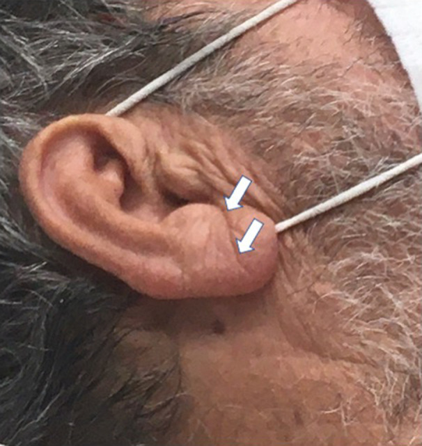
Frank's sign
3. DISCUSSION
The pathophysiological basis for the relationship between FS and cardiovascular disease is unclear. The earlobe crease has been associated with the aging process, as suggested by excessive telomere loss. 7 In addition, a recent histopathologic study showed a significant correlation between morphological changes of the myocardium and the presence of the ear lobe creases, with arterial myoelastofibrosis, Wallerian‐like degeneration in peripheral nerves, and deep‐tissue fibrosis found in the base of the crease. 8
FS has been associated with the severity of coronary artery disease, as demonstrated by the SYNTAX score. 5 Specifically, crossing creases not originating from the ear hole had the highest positive predictive value, and vertical creases on the face side had the highest negative predictive value for detecting intermediate and high SYNTAX scores. 5 FS has been associated not only with the presence and severity of CAD but also with long‐term prognosis. 9 Specifically, in patients with myocardial infarction, the presence of FS was associated with an increased risk of death. 9 The association of ear creases on the helix with coronary artery disease has also been proposed but needs more data to be confirmed. 10
By contrast, an observational study found no significant association between FS and abnormal ankle‐branchial index. 11 Furthermore, a recent systematic review showed that the diagnostic accuracy of diagonal earlobe crease for detecting chronic coronary syndromes is insufficient. 12 Additionally, the Fremantle Diabetes Study assessed the clinical usefulness of FS as a sign of coronary artery disease or retinopathy in type 2 diabetes. 13 This study found that FS was not a significant independent predictor of either coronary artery disease or retinopathy. 13
In the stroke setting, FS was more prevalent in patients with ischemic stroke than hemorrhagic stroke. 14 Moreover, in stroke patients, atherosclerotic plaques were observed more frequently in patients with FS. 14 Another study proposed that FS could predict ischemic cerebrovascular events while patients with classical cardiovascular risk factors had FS at a higher frequency. 15
4. CONCLUSIONS
Classical risk factors for coronary artery disease should be assessed in every patient. The presence of one or multiple risk factors should raise the suspicion of coronary artery disease, especially in symptomatic patients. We propose that the assessment of classical risk factors and the presence of FS should be part of the routine cardiovascular clinical examination.
AUTHOR CONTRIBUTIONS
George Bazoukis: Management of the patient, wrote the first draft, major revision, approval of the final manuscript. Stamatis S Papadatos: literature review, major revision, approval of the final manuscript. Dimitrios Varrias: literature review, major revision, approval of the final manuscript. Gary Tse: literature review, major revision, approval of the final manuscript.
CONFLICT OF INTEREST
The authors declare no conflicts of interest.
CONSENT
The authors have obtained written informed consent from the patient.
ACKNOWLEDGMENTS
None.
Bazoukis G, Papadatos SS, Varrias D, Tse G. The importance of inspection in clinical cardiology: Frank's sign. Clin Case Rep. 2022;10:e06150. doi: 10.1002/ccr3.6150
DATA AVAILABILITY STATEMENT
None.
REFERENCES
- 1. Frank ST. Ear‐crease sign of coronary disease. N Engl J Med. 1977;297:282. [DOI] [PubMed] [Google Scholar]
- 2. Galassi FM, Borghi C, Ballestriero R, Habicht ME, Henneberg M, Ruhli FJ. Palaeopathology of the earlobe crease (Frank's sign): new insights from renaissance art. Int J Cardiol. 2017;236:82‐84. [DOI] [PubMed] [Google Scholar]
- 3. Gutiu IA, Galetescu E, Gutiu LI, Raducu L. Diagonal earlobe crease: a coronary risk factor, a genetic marker of coronary heart disease, or a mere wrinkle. ancient greco‐roman evidence. Rom J Intern Med. 1996;34:271‐278. [PubMed] [Google Scholar]
- 4. Ono R, Iwahana T, Kobayashi Y. Frank's sign in recurrent triple‐vessel disease. BMJ Case Rep. 2020;13(9):e239173. [DOI] [PMC free article] [PubMed] [Google Scholar]
- 5. Liu Z, Qiu C, Xu J, et al. Ear crease features are associated with complexity of coronary lesions. Med Sci Monit. 2020;26:e923343. [DOI] [PMC free article] [PubMed] [Google Scholar]
- 6. Ogan N, Gunay E, Ekici B, et al. Morphological overview of cardiovascular comorbidities in chronic obstructive pulmonary disease: Frank's sign. Heart Lung. 2020;49:331‐335. [DOI] [PubMed] [Google Scholar]
- 7. Higuchi Y, Maeda T, Guan JZ, Oyama J, Sugano M, Makino N. Diagonal earlobe crease are associated with shorter telomere in male Japanese patients with metabolic syndrome. Circ J. 2009;73:274‐279. [DOI] [PubMed] [Google Scholar]
- 8. Stoyanov GS, Dzhenkov D, Petkova L, Sapundzhiev N, Georgiev S. The histological basis of Frank's sign. Head Neck Pathol. 2021;15:402‐407. [DOI] [PMC free article] [PubMed] [Google Scholar]
- 9. Thilo C, Meisinger C, Heier M, von Scheidt W, Kirchberger I. Diagonal earlobe crease and long‐term survival after myocardial infarction. BMC Cardiovasc Disord. 2021;21:597. [DOI] [PMC free article] [PubMed] [Google Scholar]
- 10. Pathmarajah P, Rowland PC. Paired ear creases of the helix (PECH): a possible physical sign. Cureus. 2017;9:e1884. [DOI] [PMC free article] [PubMed] [Google Scholar]
- 11. Del Brutto OH, Mera RM, Costa AF, Zambrano M, Sedler MJ. The association between earlobe crease (Frank's sign) and abnormal ankle‐brachial index determination is related to age: a population‐based study. Int J Vasc Med. 2018;2018:4735731‐4735734. [DOI] [PMC free article] [PubMed] [Google Scholar]
- 12. Wieckowski K, Gallina T, Surdacki A, Chyrchel B. Diagonal earlobe crease (Frank's sign) for diagnosis of coronary artery disease: a systematic review of diagnostic test accuracy studies. J Clin Med. 2021;10(13):2799. [DOI] [PMC free article] [PubMed] [Google Scholar]
- 13. Davis TM, Balme M, Jackson D, Stuccio G, Bruce DG. The diagonal ear lobe crease (Frank's sign) is not associated with coronary artery disease or retinopathy in type 2 diabetes: the Fremantle diabetes study. Aust N Z J Med. 2000;30:573‐577. [DOI] [PubMed] [Google Scholar]
- 14. Sanchez‐Cirera L, Bashir S, Ciscar A, et al. Prevalence of the Frank's sign by aetiopathogenic stroke subtype: a prospective analysis. PLoS One. 2021;16:e0261080. [DOI] [PMC free article] [PubMed] [Google Scholar]
- 15. Nazzal S, Hijazi B, Khalila L, Blum A. Diagonal earlobe crease (Frank's sign): a predictor of cerebral vascular events. Am J Med. 2017;130:1324 e1‐1324 e5. [DOI] [PubMed] [Google Scholar]
Associated Data
This section collects any data citations, data availability statements, or supplementary materials included in this article.
Data Availability Statement
None.