Abstract
Embryo implantation is one of the hottest topics during female reproduction since it is the first dialogue between maternal uterus and developing embryo whose disruption will contribute to adverse pregnancy outcome. Numerous achievements have been made to decipher the underlying mechanism of embryo implantation by genetic and molecular approaches accompanied with emerging technological advances. In recent decades, raising concepts incite insightful understanding on the mechanism of reciprocal communication between implantation competent embryos and receptive uterus. Enlightened by these gratifying evolvements, we aim to summarize and revisit current progress on the critical determinants of mutual communication between maternal uterus and embryonic signaling on the perspective of embryo implantation to alleviate infertility, enhance fetal health, and improve contraceptive design.
Keywords: Embryo implantation, diapause, activation, embryonic signal
We revisit the progress on the critical determinants of mutual communication between maternal uterus and embryonic signaling on the perspective of embryo implantation and aim to advance the mechanism study of embryo implantation and ultimately alleviate infertility, enhance fetal health, and improve contraceptive design.
Introduction
Embryo implantation involves the adequate mutual conversations between implantation competent embryo and receptive uterus under the precise orchestration of molecular interactions directed by steroid hormones estrogen and progesterone. Embryo implantation is the one of the critical rate-limiting steps of pregnancy success, whose quality is sophisticatedly determined by both embryonic and maternal signaling [1]. Accompanied with technological advance, the elaboration of embryo implantation is updated encompassing earlier genetic evidence, current epigenetic modification and further to endometrium and embryo cell heterogeneity. Even with these abundant emergent evidences, the detailed and ingenious mechanism governing the orderly transitions of implantation events is still far to fully decrypt.
The hormone responsive uterus is derived from the intermediate mesoderm and Mullerian ducts with a monolayer of luminal epithelium surrounded by undifferentiated mesenchyme. The epithelium subsequently buds and invaginates into endometrium to form glands at the developmental window at postnatal day (PND) 6–9 [2, 3]. The undifferentiated mesenchyme develops into stroma under the guiding of subepithelial Amhr2 positive cells [4]. The formation of functional endometrium is accompanied with the development of myometrium from mesenchyme which is not fully illustrated as well as the infiltration of distinct immune cells [3]. In adult mouse uterus, the mesenchyme derived stromal cells are further classified as five distinct cell types, including vascular smooth muscle cells located around large basal blood vessels, pericytes positioned around smaller vessels throughout the tissue, and three divergent fibroblast cells [5]. The deciphering of cell-type specific functions of these cells will open new avenues to decrypt the regulatory apparatus of transient embryo implantation window and embryo–uterus communications.
Embryo implantation
Although the strategies of embryo implantation are species dependent, the ultimate goal is to establish adequate communications between fetus and mother. For mice, this process is a little tortuous since the embryo needs to settle down in the uterine implantation chamber by the adhesion of lateral trophectoderm to uterine wall and subsequently to remove the surrounding epithelium by a nonapoptotic process termed entosis and apoptotic process mediated by embryonic Tumor necrosis factor (TNF) and epithelial TNFRI to establish the embryo–stroma communication and induce stromal cells decidualization at anti-mesometrial (AM) site, then deeply invade into maternal uterus at mesometrial (M) site, the entry site of blood vessels into the uterus [6, 7]. For humans, this process is more straightforward by orienting the polar trophectoderm (TE) with inner cell mass (ICM) toward the epithelium to penetrate into stroma to initial intimate dialogue between blastocyst and uterus [1].
Newly developed 3D staining and tissue clearing provides spatial cues for this enigmatic process and cast new opinions for this black box. Although the even distribution of implantation site in uterus is described for many decades [8], it remains disputable about how the implantation chamber forms which have two prevailing conjectures: formed before embryo enters into the uterus and formed by the implanting embryo. The advancement of 3D staining provides innovative perspective that there are regular wrinkles-like epithelial evaginations evenly distributed at both sides of luminal epithelium [9]. Whether these folds herald future implantation sites warrant further attempts. VANGL2, a critical member of planar cell polarity (PCP) whose lose remarkably disrupts the epithelial architecture and contributes to inferior embryo implantation, is supposed to participate the appropriate implantation chamber formation by regulating the arrangement of epithelium [9]. After the embryos enter into the lumen, they gradually gather in the middle of uterus and travel back and forth to be evenly distributed in the uterus [10]. Additionally, the appropriate embryo distribution is also regulated by LPA3 and myometrial β-AR (β2-adrenoreceptor) signaling [11, 12]. It is of potential interest whether the pre-distributed embryos will home to these wrinkles-like pockets. It is well recognized that the mouse embryos need to invade into decidualized stromal cells at AM site accompanied with thinner decidua with the progress of pregnancy based on the observation of 2D histological section, while 3D staining and the fact of rare stromal cells proliferation after day 8 provide an alternative explanation that the thinner decidual would be primarily ascribed to the rapid growing embryo similar to inflated balloon. The revisit of embryo implantation at another dimension will remarkably advance our understanding on embryo implantation.
The stability of the receptor of pregnancy hormone: progesterone receptor
Receptivity preparation involves intricate reciprocal interaction between stromal and epithelial cells under the instruction of steroid hormones estrogen and progesterone (P4). During early pregnancy, influenced by preovulatory estrogen (E2), the epithelium proliferation is almost ceased on D1 (the first day post-coitum) then undergoes massive apoptosis on day 2 owing to long exposure to ovulation estrogen [13]. The uterus then gradually switches from an estrogen dominate to progesterone dominate milieu on day 3 characterized with incited stromal cell proliferation in stromal PR-dependent manner which account for synthesized progesterone from newly formed corpus luteum [14]. Synchronized with small estrogen surge on day 4, P4 prepares the hostile uterus to implantation favorable environment by stimulating intensive stromal cells proliferation, epithelium cell cycle quiescence, and differentiation [15]. Although the genomic regulation of PR after binding with P4 has been unraveled in both mouse and human uterus, the stability of PR protein is not fully explored. Utilizing genetic, biochemical, and pathophysiological approaches, P4-PR responsiveness is evidenced by post-translational modification via Bmi1 polycomb ring finger oncogene (BMI1) and ubiquitin protein ligase E3A (UBE3A) mediated PR ubiquitination in a polycomb complex independent manner [16]. Normally, as a critical component of the PRC1, BMI1 directs the ubiquitination of lysine 119 of histone H2A through provoking the complex’s E3 ligase activity to restrain gene expression [17, 18]. The potentially novel regulatory mechanism of BMI1 in governing endometrial P4-PR responsiveness through ubiquitination provides an alternative regulation of PR transcriptional activation in embryo implantation. Our recent work also provides evidence that SOX4 (SRY-box 4), the highest expressed SOX member in mouse and human uterus, modulates PGR stability by repressing E3 ubiquitin ligase HERC4-mediated degradation [19]. In a word, emerging evidence imply that PR stability is regulated by ubiquitination modification through divergence mechanisms (Figure 1). Since PR can also be modulated by phosphorylation [20] and acetylation [21], whether there are other covalent modifications of PR and how these modifications affect PR function in pregnancy deserve further efforts.
Figure 1.
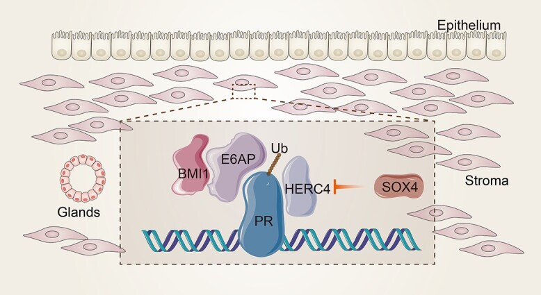
PR stability and transcriptional activity regulation in uterus.
The landmark of transcription regulated by ERα in embryo implantation
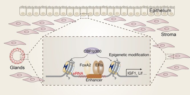
Epigenetic landmarks dynamically and reversibly regulate gene expression without changing DNA sequence. As an important sensor of external environment, epigenetic medication also contributes to inherent diversity by programmed DNA packaging [22]. Accumulated data have been emerged for those modifications in reproduction. As an essential transcription factor, ERα plays vital roles in uterine development and embryo implantation [23–25]. The direct target genes of ERα are identified in uterus by integrating genomic-wide mRNA and binding site assay, such as insulin-like growth factor 1 (IGF1) and PR [26, 27]. The poised promoter-proximal phosphor-Ser5 polymerase II (p-Ser5-Pol II) enrichment in some rapidly induced genes after estrogen treatment supports the underlying mechanism for acute estrogen response. Noticeably, the binding of ERα and Pol II at enhancer regions insinuates the transcription of potential enhancer RNA (eRNA) [28, 29]. Recently, the deletion of enhancer located in the upstream of IGF1 remarkably ablates IGF1 expression, which further corroborates the physiological function of these enhancer in uterus [30]. It is interesting to notice that there is a significant enrichment of ERα and Pol II at 20 kb downstream of LIF [27], whether this region behaves as an eRNA responsible for LIF expression needs to be further determined.
FoxA2 is a pivotal factor for implantation by regulating LIF expression in glands [31, 32]. As a multiple face factor, the function of FoxA2 is overtly different in neonatal and adult mice [33, 34]. The FoxA family members interact with ERα to influence epigenetic signature establishment in the presence of E2 in MCF7 cells [35]. In vivo evidence also shows that the FoxA1/A2 deficient female mice predispose to develop liver cancer, but not in male, ascribing to the mostly overlapping peaks of Foxa2 and ERα in female liver [36]. Whether FoxA2 would regulate estrogen targets through affecting ERα activity in glands warrants further efforts.
With respect to PR, genomic evidence reveals that there is an array of binding sites in both promoter and enhancer [37]. The widely overlapping of ERα and PR binding sites indicates that ERα and PR would potentially function together to alter histone modification to guide gene expression. Meanwhile, PR also directs gene expression by cooperating with other transcription factors. For instance, PR and SOX17 regulate Indian hedgehog (IHH), expression in the epithelium at distal enhancer to modify epithelium–stroma communication [38]. GATA2 is a recently identified essential gene for embryo implantation and decidualization by not only directly regulating PR expression but also modulating genome-wide PR transcriptional activity as a coregulator of PR [39].
Spatial–temporal expression of embryo implantation associated genes is a complex process involving transcription factor binding, chromatin modification mediated by histone alteration, and subsequent chromatin architecture configuration (Figure 2). Until now, the genome-wide binding of transcription factors in the uterus is very limited, further efforts are encouraged to untangle previously unappreciated uterus specific pioneer factors and their interactions with histone landmark and chromosome conformation [40, 41].
Figure 2.
The genomic transcriptional activation of ER in uterus.
Maternal epigenetic modification regulates embryo implantation
FOXAs and GATAs, representative pioneer factors possessing nucleosome-binding properties that distinguish from other DNA-binding factors, actively facilitate the assembly of regulatory factors on the DNA to modify opening the chromatin locally to recruit other chromatin modifiers and coregulators, including hormone receptor ERa [42]. Among them, Gata2, one of critical PR downstream target genes, is highly expressed in uterus modulating a key regulatory network of gene expression for progesterone signaling in uterus [39, 43, 44]. While how these uterine pioneer factors direct local epigenetic alteration remain intangible. Epigenetic modification signature participates in many fundamental biological processes by influencing gene expression and some of these modifications inherit to the next generation through germ cells without changing genome sequence. There are two important complexes: polycomb repressive complex 1 (PRC1) and polycomb repressive complex 2 (PRC2) which contain different components. The core components of PRC1 encompass Ring1A and Ring1B, which are E3 protein ligases responsible for ubiquitylation of histone H2A and gene repression [45], chromoboxs (CBXs) family, polycomb group RING finger protein (PCGF) family, polyhomeotic-like (PHC) family, and RING1 and YY1-binding protein/YY1 associated factor 2 (Rybp/Yaf2) [46].
Most of the components of PRC1 are differentially and spatiotemporally expressed in the peri-implantation uteri with H2AK119ub1 (mono ubiquitination of histone-H2A at lysine-119) and H3K27me3 colocalized with Cbx4/2 and Ring1B in the polyploid decidual cells. And inhibition of PRC1 activity by Ring1A/B inhibitor compromises decidualization and polyploidy development during early pregnancy. Meanwhile, interfering CBX4 expression in stroma cells also shows defective stromal cell decidualization and polyploidy development in vitro [47]. While the in vivo function of these components of PRC1 remains ambiguous.
PRC2 is an important H3K27 methyltransferase regulating gene repression in the presence of enhancer of zeste 1 PRC2 subunit (EZH1) or enhancer of zeste 2 PRC2 subunit (EZH2), embryonic ectoderm development (EED), SUZ12 PRC2 subunit (SUZ12), and RB binding protein 7 (RBAP46) or RB binding protein 4 (RBAP48) [48]. The increased H3K27me3 enrichment at the promoters of stromal chemokine (C–C motif) ligand 8 (CCL8) and chemokine (C–C motif) ligand 9 (CCL9) contributes to T cell migration from stroma to myometrium to confer a local immune privilege region for embryo developing, indicating the importance of this epigenetic hallmark in pregnancy [49]. EZH2 reported dynamic change in human uterus and obviously depressed in decidualized stromal cells, emphasizing the essential role of EZH2-PRC2 mediated chromatin remodeling in human endometrium [50, 51].
The prevailing hypothesis is the hierarchical model that PRC2 is recruited to target locus to catalyze H3K27me3 then recognized by PRC1 to modify H2AK119ub1 [52]. While emerging evidence also show that PRC2 functions as downstream of PRC1 by reading H2AK119ub1 [53]. The physiological significance of PRC2 complex and its components in embryo implantation still remains to be elucidated.
Currently studies primarily focus on H2 and H3 modification, emerging evidence concerning H4 modification is also been well recognized recently. Histone H4 Lys 20 methylation (H4K20me1) and the methyl-transferase activity of SET8 (PR-Set7/KMT5a) are involved in regulating distinct processes ranging from the DNA damage response, chromatin condensation, and DNA replication to gene regulation [54]. Loss of PR-Set7 is catastrophic for the mouse embryonic development due to embryonic lethality and arrest between the four- and eight-cell stages [55]. These findings have placed PR-Set7 and H4K20me as central nodes of many important pathways. Genome-wide study shows that H4K20me1 is enriched at the coding region of active genes and associates with chromatin compaction [56, 57]. PR-Set7 is extensively expressed in the postnatal uteri, whose conditional deletion resulted in a complete lack of endometrial glands in postnatal uterus which attributed to abolishment of the dynamic endometrial epithelial population growth during the short window of gland formation from PNDs 3–9 [58]. This study also raises a novel hypothesis of ‘epithelial population growth threshold’ for adenogenesis governing uterine gland formation.
Maternal signaling is vital for embryo activation
Embryo implantation necessitates the intricated communication between both uterus and embryo. Embryo implantation would be postponed for several days in ovariectomized mice on pregnancy Day 4 before the presumed estrogen surge in the presence of P4 which can be terminated by E2 and P4 administration on Day 7, undoubtedly implying the indispensable role of maternal uterus on embryo activation. While little to rare is known about the maternal signaling curbing embryo activation, there is an ingenious experiment by transferring dormant embryos into estrogen pre-injected recipients for different time points. The results show that the most effective activation of dormant embryo is observed in 1 h estrogen pre-injected recipients and then gradually decreased and totally failed 4 h later (Figure 3) [59]. These results denote that the rapid activation of both embryos and uterus is critical for embryo implantation. There are evidence that E2 activates uterus and embryo separately. For uterus, E2 is documented to induce receptivity opening in delayed uterus for up to 24 h [23]. Meanwhile, the E2 metabolite 4-hydroxy-E2 (4-OH-E2) is proven to be one of the critical estromedins to activate embryo by maternal CYP1B1 [60]. In summary, the above proofs signify that embryo is mainly activated by E2 and occurs at a very transient time, probably through 4-OH-E2 in ERα independent manner. While it is notable that CYP1b1 only expresses in stromal cells but not in epithelium. To depict, the detailed maternal signaling contributing to embryo activation remains a formidable challenge.
Figure 3.
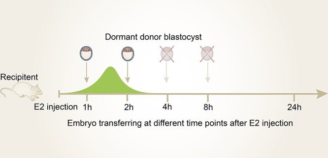
Embryo activation by estrogen at short window. Green color represents serum estrogen concentration after estrogen injection.
Currently, there are several factors that have been reported related to implantation clock disruption. Msh homeobox 1 (MSX1) and Msh homeobox 2 (MSX2) are mainly expressed on Days 3 and 4 uterine epithelium, whose loss contributes to deferred embryo implantation [61]. Moreover, mechanism study reveals that MSX1/2 regulates LIF expression, which is a critical maternal factor for receptivity regulation by activating STAT3 phosphorylation via LIFR and GP130 [61–64]. Additionally, MSX1/2 ablation also leads to uterine PCP aberrant in epithelium by modulating WNT5a/RORs/VANGL2/SCRIB signaling pathway [64–66]. Furthermore, the observation of sustained uterine expression of MSX1/2 in multiple diapause species designates its conserved role in divergent mammalian [67, 68]. Leukemia inhibitory factor (LIF), an essential implantation factor derived from glands of Day 4 uterus, is supposed to be critical for receptivity opening [69]. It is interesting that there is complicated entanglement between LIF and MSX1/2. On the one hand, MSX1/2 deficiency compromises LIF expression on Day 4 glands. On the contrary, E2-induced epithelial MSX1 disappearance and dormant embryo implantation in delayed uteri are dependent on maternal LIF [61]. The casual relationship between these two factors deserves further efforts.
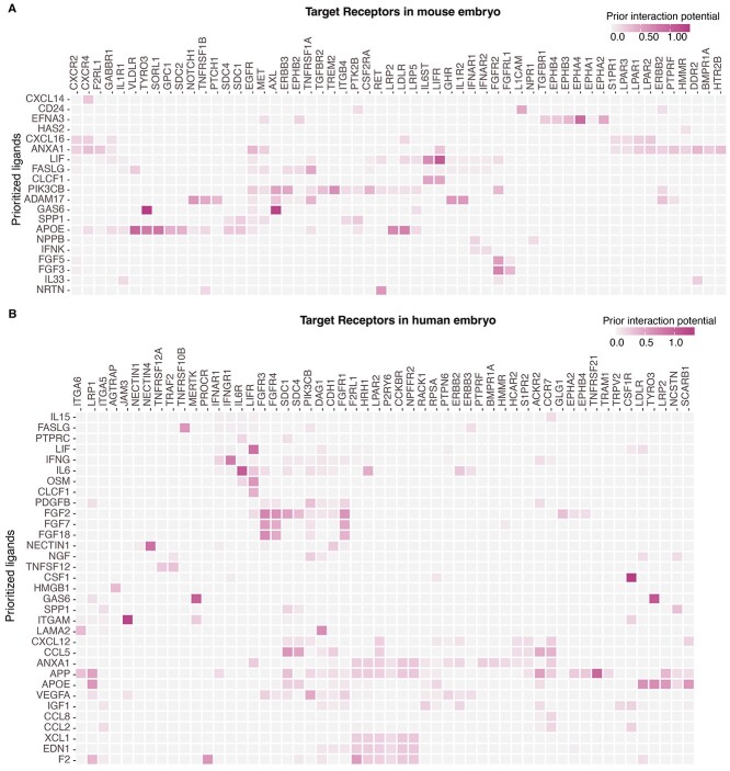
Since LIF is critical for stem cell stemness, it is imaginable that maternal factors also play a vital role in embryo development and activation. Similar scenario is also observed for HB-EGF. HB-EGF is expressed in maternal epithelial cells with its receptor expression in blastocyst as well as its binding on the surface of blastocyst [70, 71]. While the detailed regulatory regiment of these maternal growth factors on embryo activation remains obscure, there is an explicit experiment proven that 4-OH-E2, PGE2, and its downstream secondary messenger cAMP activate dormant embryo efficiently to implant into delayed uteri treated by 2-fluoroestradiol (2-FL-E2), an E2 derivation which cannot be metabolized to 4-OH-E2 and only activate maternal uterus but not embryos [60]. It is possible that uterine epithelial COX2-PGE2 activates diapausing embryos by eliciting cAMP level after binding to its receptor. Additionally, our previous work shows that RBPJ instructs embryonic-uterine orientation to ensure decidual patterning in a stage-specific manner corroborating the concept that embryonic-uterine orientation requires appropriate guidance from developmentally controlled uterine signaling [72], while the underlying mechanism underpinning maternal uterus directs embryo remains largely uncertain. To decipher the latent crosstalk between embryo and epithelial cells in human and mouse, we integrate published data in receptive epithelium and implantation competent embryo data and probe the prioritized ligands and corresponding receptors [73–75]. Intriguingly, apart from LIF, it appears that there are some previously unappreciated ligand–receptor pairs deserving further attention, such as CSF1-CSF1R in human (Figure 4).
Figure 4.
Potential interactions between receptive epithelium and implantation competent embryos. The prioritized ligands in receptive epithelium and their target signaling pathways in competent embryos in mouse (A) and human (B).
In conclusion, until now, marginal progress has been made on maternal factors responding to embryo activation. There is another possibility that some small molecules derived from epithelial cells via extracellular vesicles endow the embryo diapause, such as Let-7 [76]. How progesterone maintains high level of Let-7 in epithelial cells remains elusive.
Embryo diapause and activation
In wild animals, nutrition is considered to be a critical factor for embryos diapause based on the observation of a delay of parturition to ensure sufficient nutrition and survival of the infant, which bring out the conjecture that metabolism is one of the critical determinants for embryo diapause [77]. Especially, evidence support that the inhibition of polyamine synthesis largely causes embryo diapause in both mouse and mink [78, 79]. A recent study also shows that the content of amino acids changes significantly in delayed and activated uterine fluid in roe deer, which is supposed to be relevant with mTOR signaling activation [80].
Especially, a recent study comparing the transcriptomic and proteomic changes in diapause and activated embryos reveals downregulated mTOR signaling with decreased glycolysis in diapause embryo [81–83]. Moreover, arginine, leucine, and glutamine are proven effective to stimulate porcine trophectoderm cells’ proliferation [84]. Since leucine and glutamine are reported to promote mTORC1 translocation to the lysosome to incite downstream signaling pathway [85], it is very possible that similar scenario is also applicable for embryo development. The observation that mTOR inhibitors targeting both mTORC1 and mTORC2 induce reversible pausing of mouse blastocyst strongly corroborates this hypothesis. The mechanism is partly due to global profound suppression of gene transcription [86]. The fact that targeting mTORC1 only marginally extend blastocyst survival indicates the essential role of mTORC2 in delayed blastocyst, while convincing evidence warrants further efforts. Another interesting study provides evidence that diapause embryo is characterized by increased lipolysis which might be due to reduced fatty acid β-oxidation. The metabolite analysis in diapause and pre-implantation blastocyst offers insightful information for the mechanism of embryo delay and reactivation. The increased leucine degradation associated genes and leucine degradation metabolites in diapause blastocyst in consistent with the conception that leucine-activated mTOR signal is critical for embryo activation [87]. While how to reconcile the observation of increased glutamine demand and diminished mTOR requires further experiments to define the enigmatic mechanisms.
A fascinating feature of dormant blastocysts is the activation of autophagy to prolong its survival and the disruption of autophagy is associated with reduced blastocyst survival [88]. Since mTOR signaling is downregulated in diapause embryos, how activated autophagy is regulated in diapause embryo remains unclear.
Embryonic signaling guides embryo implantation
The mechanism of embryo diapause and activation is widely discussed, while how implantation competent blastocyst educates receptive uterus to facilitate implantation is largely ambiguous. The evidences originate from growth factors soaped beads transferring into receptivity uterus support that embryonic HB-EGF and IGF1 are effective to initiate implantation [89]. These embryonic signals are first supposed to direct the formation of tight junctional permeability barrier in the decidualizing stroma [90]. To globally depict the critical embryonic factors essential for embryo implantation, microarray was first applied to determine the potential molecule. HB-EGF pathway as well as metabolism, transcriptional regulation, and cell cycle genes are differentially expressed in delayed and activated blastocyst [83]. To profoundly illustrate the potential embryonic determinants, high-throughput RNA-Seq was utilized to compare the transcriptome of delayed, activating (6 h after E2 injection), and activated (12 h after E2 injection) embryos. We notice that the proinflammatory factors, including TNFα and S100A9, are obviously increased in the activated embryos [82]. Our previous work shows that embryonic TNF is critical for epithelium removal through epithelial RAC1-Pak1-ERM pathway via TNFR1 and p38 [7]. Furthermore, our lab also notices that S100A9, which is highly expressed in activated embryos, significantly promotes embryo implantation [82].
IGF1 is assumed to be a critical embryo derived factor to promote embryo implantation [89]. We first detected the expression of its receptor in peri-implantation uterus. Intriguingly, IGF1R is specifically localized in epithelium. The compromised embryo implantation by abrogating IGF1R in whole uterus or only epithelium strongly underscores the vital role of IGF1R. Our result also surprisingly observed that embryonic IGF2 is essential for embryo implantation initiation [91]. Collectively, we have very limited evidence of how embryo interplay with maternal uterus to facilitate embryo implantation. To comprehensively interrogate, the communication between embryo and uterus still remains a huge challenge and requires further investigation (Figure 5).
Figure 5.
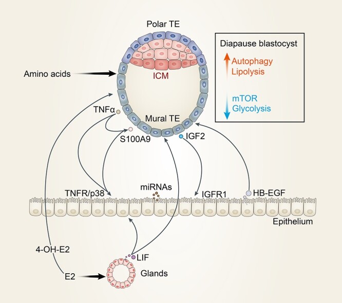
The communications between embryo and maternal uterus.
Concluding remarks and future perspectives
Accumulating evidences show that there is adequate entanglement between implantation competent blastocyst and receptive uterus. While this process is largely constrained due to the rare material accessibility of embryos. Advances in chromatin and chromosome research using sequencing-based genomic approaches with limited number of cells will largely pave the way for the epigenetic landmarks of the diapause and delayed embryos [92]. Since blastocyst encompasses epiblast, ICM, polar TE, and mural TE, the contribution of each cell type in embryo implantation gradually attracts scientist attention, the multi-omic single-cell signatures of heterogenic embryo will also definitely shed new light on the mechanism study of embryonic signal on embryo implantation [93]. Due to the ethical limitation, it is difficult to investigate the process of human implantation in vivo. The development of suitable in vitro embryo implantation model resembles human is imperative. The successfully established various assembled endometrium in vitro will ensure the study of the cross talk between embryo and endometrium [94–99]. Meanwhile, there are also diverse trials to construct blastoids or blastocyst-like cysts in vitro to effectively and faithfully mimic cellular determination and morphogenesis according to the in vivo developmental pace [100–106]. Future functional studies leveraging genetic modification in these in vitro models will greatly advance the mechanism study of embryo implantation and ultimately improve pregnancy outcome.
Acknowledgments
The authors regret that, because of page limitations, the contributions of many investigators to the study of cell chatting between embryo and uterus could not be credited in this article.
Contributor Information
Wenbo Deng, Fujian Provincial Key Laboratory of Reproductive Health Research, Department of Obstetrics and Gynecology, The First Affiliated Hospital of Xiamen University, School of Medicine, Xiamen University, Xiamen, Fujian, China.
Haibin Wang, Fujian Provincial Key Laboratory of Reproductive Health Research, Department of Obstetrics and Gynecology, The First Affiliated Hospital of Xiamen University, School of Medicine, Xiamen University, Xiamen, Fujian, China.
Conflict of interest
The authors declare that they have no competing interests.
Authors’ contributions
H.W. conceptualized this manuscript and W.D. and H.W. discussed and wrote the manuscript. All authors read and approved the manuscript for publication.
References
- 1. Cha J, Sun X, Dey SK. Mechanisms of implantation: strategies for successful pregnancy. Nat Med 2012; 18:1754–1767. [DOI] [PMC free article] [PubMed] [Google Scholar]
- 2. Branham WS, Sheehan DM, Zehr DR, Medlock KL, Nelson CJ, Ridlon E. Inhibition of rat uterine gland genesis by tamoxifen. Endocrinology 1985; 117:2238–2248. [DOI] [PubMed] [Google Scholar]
- 3. Brody JR, Cunha GR. Histologic, morphometric, and immunocytochemical analysis of myometrial development in rats and mice: I. Normal development. Am J Anat 1989; 186:1–20. [DOI] [PubMed] [Google Scholar]
- 4. Saatcioglu HD, Kano M, Horn H, Zhang L, Samore W, Nagykery N, Meinsohn MC, Hyun M, Suliman R, Poulo J, Hsu J, Sacha Cet al. Single-cell sequencing of neonatal uterus reveals an Misr2+ endometrial progenitor indispensable for fertility. Elife 2019; 8:e46349. [DOI] [PMC free article] [PubMed] [Google Scholar]
- 5. Kirkwood PM, Gibson DA, Smith JR, Wilson-Kanamori JR, Kelepouri O, Esnal-Zufiaurre A, Dobie R, Henderson NC, Saunders PTK. Single-cell RNA sequencing redefines the mesenchymal cell landscape of mouse endometrium. FASEB J 2021; 35:e21285. [DOI] [PMC free article] [PubMed] [Google Scholar]
- 6. Li Y, Sun X, Dey SK. Entosis allows timely elimination of the luminal epithelial barrier for embryo implantation. Cell Rep 2015; 11:358–365. [DOI] [PMC free article] [PubMed] [Google Scholar]
- 7. Tu Z, Wang Q, Cui T, Wang J, Ran H, Bao H, Lu J, Wang B, Lydon JP, DeMayo F, Zhang S, Kong Set al. Uterine RAC1 via Pak1-ERM signaling directs normal luminal epithelial integrity conducive to on-time embryo implantation in mice. Cell Death Differ 2016; 23:169–181. [DOI] [PMC free article] [PubMed] [Google Scholar]
- 8. O'Grady JE, Heald PJ. The position and spacing of implantation sites in the uterus of the rat during early pregnancy. J Reprod Fertil 1969; 20:407–412. [DOI] [PubMed] [Google Scholar]
- 9. Yuan J, Deng W, Cha J, Sun X, Borg JP, Dey SK. Tridimensional visualization reveals direct communication between the embryo and glands critical for implantation. Nat Commun 2018; 9:603. [DOI] [PMC free article] [PubMed] [Google Scholar]
- 10. Flores D, Madhavan M, Wright S, Arora R. Mechanical and signaling mechanisms that guide pre-implantation embryo movement. Development 2020; 147:dev193490. [DOI] [PubMed] [Google Scholar]
- 11. Chen Q, Zhang Y, Peng H, Lei L, Kuang H, Zhang L, Ning L, Cao Y, Duan E. Transient {beta}2-adrenoceptor activation confers pregnancy loss by disrupting embryo spacing at implantation. J Biol Chem 2011; 286:4349–4356. [DOI] [PMC free article] [PubMed] [Google Scholar]
- 12. Ye X, Hama K, Contos JJ, Anliker B, Inoue A, Skinner MK, Suzuki H, Amano T, Kennedy G, Arai H, Aoki J, Chun J. LPA3-mediated lysophosphatidic acid signalling in embryo implantation and spacing. Nature 2005; 435:104–108. [DOI] [PMC free article] [PubMed] [Google Scholar]
- 13. Winuthayanon W, Hewitt SC, Orvis GD, Behringer RR, Korach KS. Uterine epithelial estrogen receptor alpha is dispensable for proliferation but essential for complete biological and biochemical responses. Proc Natl Acad Sci U S A 2010; 107:19272–19277. [DOI] [PMC free article] [PubMed] [Google Scholar]
- 14. Franco HL, Rubel CA, Large MJ, Wetendorf M, Fernandez-Valdivia R, Jeong JW, Spencer TE, Behringer RR, Lydon JP, Demayo FJ. Epithelial progesterone receptor exhibits pleiotropic roles in uterine development and function. FASEB J 2012; 26:1218–1227. [DOI] [PMC free article] [PubMed] [Google Scholar]
- 15. Wang H, Dey SK. Roadmap to embryo implantation: clues from mouse models. Nat Rev Genet 2006; 7:185–199. [DOI] [PubMed] [Google Scholar]
- 16. Xin Q, Kong S, Yan J, Qiu J, He B, Zhou C, Ni Z, Bao H, Huang L, Lu J, Xia G, Liu Xet al. Polycomb subunit BMI1 determines uterine progesterone responsiveness essential for normal embryo implantation. J Clin Invest 2018; 128:175–189. [DOI] [PMC free article] [PubMed] [Google Scholar]
- 17. Wang H, Wang L, Erdjument-Bromage H, Vidal M, Tempst P, Jones RS, Zhang Y. Role of histone H2A ubiquitination in Polycomb silencing. Nature 2004; 431:873–878. [DOI] [PubMed] [Google Scholar]
- 18. Cao R, Tsukada Y, Zhang Y. Role of Bmi-1 and Ring1A in H2A ubiquitylation and Hox gene silencing. Mol Cell 2005; 20:845–854. [DOI] [PubMed] [Google Scholar]
- 19. Huang P, Deng W, Bao H, Lin Z, Liu M, Wu J, Zhou X, Qiao M, Yang Y, Cai H, Rao F, Chen Jet al. SOX4 facilitates PGR protein stability and FOXO1 expression conducive for human endometrial decidualization. Elife 2022; 11:e72073. [DOI] [PMC free article] [PubMed] [Google Scholar]
- 20. Denner LA, Weigel NL, Maxwell BL, Schrader WT, O'Malley BW. Regulation of progesterone receptor-mediated transcription by phosphorylation. Science 1990; 250:1740–1743. [DOI] [PubMed] [Google Scholar]
- 21. Chung HH, Sze SK, Tay AS, Lin VC. Acetylation at lysine 183 of progesterone receptor by p300 accelerates DNA binding kinetics and transactivation of direct target genes. J Biol Chem 2014; 289:2180–2194. [DOI] [PMC free article] [PubMed] [Google Scholar]
- 22. Arrowsmith CH, Bountra C, Fish PV, Lee K, Schapira M. Epigenetic protein families: a new frontier for drug discovery. Nat Rev Drug Discov 2012; 11:384–400. [DOI] [PubMed] [Google Scholar]
- 23. Ma WG, Song H, Das SK, Paria BC, Dey SK. Estrogen is a critical determinant that specifies the duration of the window of uterine receptivity for implantation. Proc Natl Acad Sci U S A 2003; 100:2963–2968. [DOI] [PMC free article] [PubMed] [Google Scholar]
- 24. Hewitt SC, Kissling GE, Fieselman KE, Jayes FL, Gerrish KE, Korach KS. Biological and biochemical consequences of global deletion of exon 3 from the ER alpha gene. FASEB J 2010; 24:4660–4667. [DOI] [PMC free article] [PubMed] [Google Scholar]
- 25. Lubahn DB, Moyer JS, Golding TS, Couse JF, Korach KS, Smithies O. Alteration of reproductive function but not prenatal sexual development after insertional disruption of the mouse estrogen receptor gene. Proc Natl Acad Sci U S A 1993; 90:11162–11166. [DOI] [PMC free article] [PubMed] [Google Scholar]
- 26. Mehta FF, Son J, Hewitt SC, Jang E, Lydon JP, Korach KS, Chung SH. Distinct functions and regulation of epithelial progesterone receptor in the mouse cervix, vagina, and uterus. Oncotarget 2016; 7:17455–17467. [DOI] [PMC free article] [PubMed] [Google Scholar]
- 27. Hewitt SC, Li L, Grimm SA, Chen Y, Liu L, Li Y, Bushel PR, Fargo D, Korach KS. Research resource: whole-genome estrogen receptor alpha binding in mouse uterine tissue revealed by ChIP-seq. Mol Endocrinol 2012; 26:887–898. [DOI] [PMC free article] [PubMed] [Google Scholar]
- 28. Kim TK, Hemberg M, Gray JM, Costa AM, Bear DM, Wu J, Harmin DA, Laptewicz M, Barbara-Haley K, Kuersten S, Markenscoff-Papadimitriou E, Kuhl Det al. Widespread transcription at neuronal activity-regulated enhancers. Nature 2010; 465:182–187. [DOI] [PMC free article] [PubMed] [Google Scholar]
- 29. Core LJ, Waterfall JJ, Lis JT. Nascent RNA sequencing reveals widespread pausing and divergent initiation at human promoters. Science 2008; 322:1845–1848. [DOI] [PMC free article] [PubMed] [Google Scholar]
- 30. Hewitt SC, Lierz SL, Garcia M, Hamilton KJ, Gruzdev A, Grimm SA, Lydon JP, Demayo FJ, Korach KS. A distal super enhancer mediates estrogen-dependent mouse uterine-specific gene transcription of Igf1 (insulin-like growth factor 1). J Biol Chem 2019; 294:9746–9759. [DOI] [PMC free article] [PubMed] [Google Scholar]
- 31. Jeong JW, Kwak I, Lee KY, Kim TH, Large MJ, Stewart CL, Kaestner KH, Lydon JP, DeMayo FJ. Foxa2 is essential for mouse endometrial gland development and fertility. Biol Reprod 2010; 83:396–403. [DOI] [PMC free article] [PubMed] [Google Scholar]
- 32. Kelleher AM, Peng W, Pru JK, Pru CA, DeMayo FJ, Spencer TE. Forkhead box a2 (FOXA2) is essential for uterine function and fertility. Proc Natl Acad Sci U S A 2017; 114:E1018–E1026. [DOI] [PMC free article] [PubMed] [Google Scholar]
- 33. Filant J, Lydon JP, Spencer TE. Integrated chromatin immunoprecipitation sequencing and microarray analysis identifies FOXA2 target genes in the glands of the mouse uterus. FASEB J 2014; 28:230–243. [DOI] [PMC free article] [PubMed] [Google Scholar]
- 34. Cha J, Dey SK. Hunting for Fox(A2): dual roles in female fertility. Proc Natl Acad Sci U S A 2017; 114:1226–1228. [DOI] [PMC free article] [PubMed] [Google Scholar]
- 35. Lupien M, Eeckhoute J, Meyer CA, Wang Q, Zhang Y, Li W, Carroll JS, Liu XS, Brown M. FoxA1 translates epigenetic signatures into enhancer-driven lineage-specific transcription. Cell 2008; 132:958–970. [DOI] [PMC free article] [PubMed] [Google Scholar]
- 36. Li Z, Tuteja G, Schug J, Kaestner KH. Foxa1 and Foxa2 are essential for sexual dimorphism in liver cancer. Cell 2012; 148:72–83. [DOI] [PMC free article] [PubMed] [Google Scholar]
- 37. Rubel CA, Lanz RB, Kommagani R, Franco HL, Lydon JP, DeMayo FJ. Research resource: genome-wide profiling of progesterone receptor binding in the mouse uterus. Mol Endocrinol 2012; 26:1428–1442. [DOI] [PMC free article] [PubMed] [Google Scholar]
- 38. Wang X, Li X, Wang T, Wu SP, Jeong JW, Kim TH, Young SL, Lessey BA, Lanz RB, Lydon JP, DeMayo FJ. SOX17 regulates uterine epithelial-stromal cross-talk acting via a distal enhancer upstream of IHH. Nat Commun 2018; 9:4421. [DOI] [PMC free article] [PubMed] [Google Scholar]
- 39. Rubel CA, Wu SP, Lin L, Wang T, Lanz RB, Li X, Kommagani R, Franco HL, Camper SA, Tong Q, Jeong JW, Lydon JPet al. A Gata2-dependent transcription network regulates uterine progesterone responsiveness and endometrial function. Cell Rep 2016; 17:1414–1425. [DOI] [PMC free article] [PubMed] [Google Scholar]
- 40. Pott S, Lieb JD. What are super-enhancers? Nat Genet 2015; 47:8–12. [DOI] [PubMed] [Google Scholar]
- 41. Dekker J, Mirny L. The 3D genome as moderator of chromosomal communication. Cell 2016; 164:1110–1121. [DOI] [PMC free article] [PubMed] [Google Scholar]
- 42. Zaret KS, Carroll JS. Pioneer transcription factors: establishing competence for gene expression. Genes Dev 2011; 25:2227–2241. [DOI] [PMC free article] [PubMed] [Google Scholar]
- 43. Jeong JW, Lee KY, Kwak I, White LD, Hilsenbeck SG, Lydon JP, DeMayo FJ. Identification of murine uterine genes regulated in a ligand-dependent manner by the progesterone receptor. Endocrinology 2005; 146:3490–3505. [DOI] [PubMed] [Google Scholar]
- 44. Rubel CA, Franco HL, Jeong JW, Lydon JP, DeMayo FJ. GATA2 is expressed at critical times in the mouse uterus during pregnancy. Gene Expr Patterns 2012; 12:196–203. [DOI] [PubMed] [Google Scholar]
- 45. Margueron R, Reinberg D. The Polycomb complex PRC2 and its mark in life. Nature 2011; 469:343–349. [DOI] [PMC free article] [PubMed] [Google Scholar]
- 46. Schwartz YB, Pirrotta V. A new world of Polycombs: unexpected partnerships and emerging functions. Nat Rev Genet 2013; 14:853–864. [DOI] [PubMed] [Google Scholar]
- 47. Bian F, Gao F, Kartashov AV, Jegga AG, Barski A, Das SK. Polycomb repressive complex 1 controls uterine decidualization. Sci Rep 2016; 6:26061. [DOI] [PMC free article] [PubMed] [Google Scholar]
- 48. Blackledge NP, Rose NR, Klose RJ. Targeting Polycomb systems to regulate gene expression: modifications to a complex story. Nat Rev Mol Cell Biol 2015; 16:643–649. [DOI] [PMC free article] [PubMed] [Google Scholar]
- 49. Nancy P, Tagliani E, Tay CS, Asp P, Levy DE, Erlebacher A. Chemokine gene silencing in decidual stromal cells limits T cell access to the maternal-fetal interface. Science 2012; 336:1317–1321. [DOI] [PMC free article] [PubMed] [Google Scholar]
- 50. Grimaldi G, Christian M, Steel JH, Henriet P, Poutanen M, Brosens JJ. Down-regulation of the histone methyltransferase EZH2 contributes to the epigenetic programming of decidualizing human endometrial stromal cells. Mol Endocrinol 2011; 25:1892–1903. [DOI] [PMC free article] [PubMed] [Google Scholar]
- 51. Grimaldi G, Christian M, Quenby S, Brosens JJ. Expression of epigenetic effectors in decidualizing human endometrial stromal cells. Mol Hum Reprod 2012; 18:451–458. [DOI] [PubMed] [Google Scholar]
- 52. Wang L, Brown JL, Cao R, Zhang Y, Kassis JA, Jones RS. Hierarchical recruitment of polycomb group silencing complexes. Mol Cell 2004; 14:637–646. [DOI] [PubMed] [Google Scholar]
- 53. Blackledge NP, Farcas AM, Kondo T, King HW, McGouran JF, Hanssen LL, Ito S, Cooper S, Kondo K, Koseki Y, Ishikura T, Long HKet al. Variant PRC1 complex-dependent H2A ubiquitylation drives PRC2 recruitment and polycomb domain formation. Cell 2014; 157:1445–1459. [DOI] [PMC free article] [PubMed] [Google Scholar]
- 54. Beck DB, Oda H, Shen SS, Reinberg D. PR-Set7 and H4K20me1: at the crossroads of genome integrity, cell cycle, chromosome condensation, and transcription. Genes Dev 2012; 26:325–337. [DOI] [PMC free article] [PubMed] [Google Scholar]
- 55. Oda H, Okamoto I, Murphy N, Chu J, Price SM, Shen MM, Torres-Padilla ME, Heard E, Reinberg D. Monomethylation of histone H4-lysine 20 is involved in chromosome structure and stability and is essential for mouse development. Mol Cell Biol 2009; 29:2278–2295. [DOI] [PMC free article] [PubMed] [Google Scholar]
- 56. Jorgensen S, Schotta G, Sorensen CS. Histone H4 lysine 20 methylation: key player in epigenetic regulation of genomic integrity. Nucleic Acids Res 2013; 41:2797–2806. [DOI] [PMC free article] [PubMed] [Google Scholar]
- 57. Li Z, Nie F, Wang S, Li L. Histone H4 Lys 20 monomethylation by histone methylase SET8 mediates Wnt target gene activation. Proc Natl Acad Sci U S A 2011; 108:3116–3123. [DOI] [PMC free article] [PubMed] [Google Scholar]
- 58. Cui T, He B, Kong S, Zhou C, Zhang H, Ni Z, Bao H, Qiu J, Xin Q, Reinberg D, Lydon JP, Lu Jet al. PR-Set7 deficiency limits uterine epithelial population growth hampering postnatal gland formation in mice. Cell Death Differ 2017; 24:2013–2021. [DOI] [PMC free article] [PubMed] [Google Scholar]
- 59. Paria BC, Huet-Hudson YM, Dey SK. Blastocyst's state of activity determines the "window" of implantation in the receptive mouse uterus. Proc Natl Acad Sci U S A 1993; 90:10159–10162. [DOI] [PMC free article] [PubMed] [Google Scholar]
- 60. Paria BC, Lim H, Wang XN, Liehr J, Das SK, Dey SK. Coordination of differential effects of primary estrogen and catecholestrogen on two distinct targets mediates embryo implantation in the mouse. Endocrinology 1998; 139:5235–5246. [DOI] [PubMed] [Google Scholar]
- 61. Daikoku T, Cha J, Sun X, Tranguch S, Xie H, Fujita T, Hirota Y, Lydon J, DeMayo F, Maxson R, Dey SK. Conditional deletion of Msx homeobox genes in the uterus inhibits blastocyst implantation by altering uterine receptivity. Dev Cell 2011; 21:1014–1025. [DOI] [PMC free article] [PubMed] [Google Scholar]
- 62. Sun X, Bartos A, Whitsett JA, Dey SK. Uterine deletion of Gp130 or Stat3 shows implantation failure with increased estrogenic responses. Mol Endocrinol 2013; 27:1492–1501. [DOI] [PMC free article] [PubMed] [Google Scholar]
- 63. Lee JH, Kim TH, Oh SJ, Yoo JY, Akira S, Ku BJ, Lydon JP, Jeong JW. Signal transducer and activator of transcription-3 (Stat3) plays a critical role in implantation via progesterone receptor in uterus. FASEB J 2013; 27:2553–2563. [DOI] [PMC free article] [PubMed] [Google Scholar]
- 64. Cha J, Bartos A, Park C, Sun X, Li Y, Cha SW, Ajima R, Ho HY, Yamaguchi TP, Dey SK. Appropriate crypt formation in the uterus for embryo homing and implantation requires Wnt5a-ROR signaling. Cell Rep 2014; 8:382–392. [DOI] [PMC free article] [PubMed] [Google Scholar]
- 65. Yuan J, Cha J, Deng W, Bartos A, Sun X, Ho HH, Borg JP, Yamaguchi TP, Yang Y, Dey SK. Planar cell polarity signaling in the uterus directs appropriate positioning of the crypt for embryo implantation. Proc Natl Acad Sci U S A 2016; 113:E8079–E8088. [DOI] [PMC free article] [PubMed] [Google Scholar]
- 66. Yuan J, Aikawa S, Deng W, Bartos A, Walz G, Grahammer F, Huber TB, Sun X, Dey SK. Primary decidual zone formation requires scribble for pregnancy success in mice. Nat Commun 2019; 10:5425. [DOI] [PMC free article] [PubMed] [Google Scholar]
- 67. Cha J, Sun X, Bartos A, Fenelon J, Lefevre P, Daikoku T, Shaw G, Maxson R, Murphy BD, Renfree MB, Dey SK. A new role for muscle segment homeobox genes in mammalian embryonic diapause. Open Biol 2013; 3:130035. [DOI] [PMC free article] [PubMed] [Google Scholar]
- 68. Cha J, Fenelon JC, Murphy BD, Shaw G, Renfree MB, Dey SK. A role for Msx genes in mammalian embryonic diapause. Biosci Proc 2020; 10:44–51. [DOI] [PMC free article] [PubMed] [Google Scholar]
- 69. Stewart CL, Kaspar P, Brunet LJ, Bhatt H, Gadi I, Kontgen F, Abbondanzo SJ. Blastocyst implantation depends on maternal expression of leukaemia inhibitory factor. Nature 1992; 359:76–79. [DOI] [PubMed] [Google Scholar]
- 70. Paria BC, Elenius K, Klagsbrun M, Dey SK. Heparin-binding EGF-like growth factor interacts with mouse blastocysts independently of ErbB1: a possible role for heparan sulfate proteoglycans and ErbB4 in blastocyst implantation. Development 1999; 126:1997–2005. [DOI] [PubMed] [Google Scholar]
- 71. Raab G, Kover K, Paria BC, Dey SK, Ezzell RM, Klagsbrun M. Mouse preimplantation blastocysts adhere to cells expressing the transmembrane form of heparin-binding EGF-like growth factor. Development 1996; 122:637–645. [DOI] [PubMed] [Google Scholar]
- 72. Zhang S, Kong S, Wang B, Cheng X, Chen Y, Wu W, Wang Q, Shi J, Zhang Y, Wang S, Lu J, Lydon JPet al. Uterine Rbpj is required for embryonic-uterine orientation and decidual remodeling via notch pathway-independent and -dependent mechanisms. Cell Res 2014; 24:925–942. [DOI] [PMC free article] [PubMed] [Google Scholar]
- 73. Aikawa S, Deng W, Liang X, Yuan J, Bartos A, Sun X, Dey SK. Uterine deficiency of high-mobility group box-1 (HMGB1) protein causes implantation defects and adverse pregnancy outcomes. Cell Death Differ 2020; 27:1489–1504. [DOI] [PMC free article] [PubMed] [Google Scholar]
- 74. Dang Y, Yan L, Hu B, Fan X, Ren Y, Li R, Lian Y, Yan J, Li Q, Zhang Y, Li M, Ren Xet al. Tracing the expression of circular RNAs in human pre-implantation embryos. Genome Biol 2016; 17:130. [DOI] [PMC free article] [PubMed] [Google Scholar]
- 75. Chi RA, Wang T, Adams N, Wu SP, Young SL, Spencer TE, DeMayo F. Human endometrial transcriptome and progesterone receptor cistrome reveal important pathways and epithelial regulators. J Clin Endocrinol Metab 2020; 105:e1419–e1439. [DOI] [PMC free article] [PubMed] [Google Scholar]
- 76. Liu WM, Cheng RR, Niu ZR, Chen AC, Ma MY, Li T, Chiu PC, Pang RT, Lee YL, Ou JP, Yao YQ, Yeung WSB. Let-7 derived from endometrial extracellular vesicles is an important inducer of embryonic diapause in mice. Sci Adv 2020; 6:eaaz7070. [DOI] [PMC free article] [PubMed] [Google Scholar]
- 77. Renfree MB, Shaw G. Embryo-endometrial interactions during early development after embryonic diapause in the marsupial tammar wallaby. Int J Dev Biol 2014; 58:175–181. [DOI] [PubMed] [Google Scholar]
- 78. Fenelon JC, Murphy BD. Inhibition of polyamine synthesis causes entry of the mouse blastocyst into embryonic diapause. Biol Reprod 2017; 97:119–132. [DOI] [PubMed] [Google Scholar]
- 79. Lefevre PL, Palin MF, Chen G, Turecki G, Murphy BD. Polyamines are implicated in the emergence of the embryo from obligate diapause. Endocrinology 2011; 152:1627–1639. [DOI] [PubMed] [Google Scholar]
- 80. Weijden VA, Bick JT, Bauersachs S, Ruegg AB, Hildebrandt TB, Goeritz F, Jewgenow K, Giesbertz P, Daniel H, Derisoud E, Chavatte-Palmer P, Bruckmaier RMet al. Amino acids activate mTORC1 to release roe deer embryos from decelerated proliferation during diapause. Proc Natl Acad Sci U S A 2021; 118:e2100500118. [DOI] [PMC free article] [PubMed] [Google Scholar]
- 81. Fu Z, Wang B, Wang S, Wu W, Wang Q, Chen Y, Kong S, Lu J, Tang Z, Ran H, Tu Z, He Bet al. Integral proteomic analysis of blastocysts reveals key molecular machinery governing embryonic diapause and reactivation for implantation in mice. Biol Reprod 2014; 90:52. [DOI] [PubMed] [Google Scholar]
- 82. He B, Zhang H, Wang J, Liu M, Sun Y, Guo C, Lu J, Wang H, Kong S. Blastocyst activation engenders transcriptome reprogram affecting X-chromosome reactivation and inflammatory trigger of implantation. Proc Natl Acad Sci U S A 2019; 116:16621–16630. [DOI] [PMC free article] [PubMed] [Google Scholar]
- 83. Hamatani T, Daikoku T, Wang H, Matsumoto H, Carter MG, Ko MS, Dey SK. Global gene expression analysis identifies molecular pathways distinguishing blastocyst dormancy and activation. Proc Natl Acad Sci U S A 2004; 101:10326–10331. [DOI] [PMC free article] [PubMed] [Google Scholar]
- 84. Kim J, Song G, Wu G, Gao H, Johnson GA, Bazer FW. Arginine, leucine, and glutamine stimulate proliferation of porcine trophectoderm cells through the MTOR-RPS6K-RPS6-EIF4EBP1 signal transduction pathway. Biol Reprod 2013; 88:113. [DOI] [PubMed] [Google Scholar]
- 85. Jewell JL, Kim YC, Russell RC, Yu FX, Park HW, Plouffe SW, Tagliabracci VS, Guan KL. Metabolism. Differential regulation of mTORC1 by leucine and glutamine. Science 2015; 347:194–198. [DOI] [PMC free article] [PubMed] [Google Scholar]
- 86. Bulut-Karslioglu A, Biechele S, Jin H, Macrae TA, Hejna M, Gertsenstein M, Song JS, Ramalho-Santos M. Inhibition of mTOR induces a paused pluripotent state. Nature 2016; 540:119–123. [DOI] [PMC free article] [PubMed] [Google Scholar]
- 87. Hussein AM, Wang Y, Mathieu J, Margaretha L, Song C, Jones DC, Cavanaugh C, Miklas JW, Mahen E, Showalter MR, Ruzzo WL, Fiehn Oet al. Metabolic control over mTOR-dependent diapause-like state. Dev Cell 2020; 52:236–250.e7. [DOI] [PMC free article] [PubMed] [Google Scholar]
- 88. Lee JE, Oh HA, Song H, Jun JH, Roh CR, Xie H, Dey SK, Lim HJ. Autophagy regulates embryonic survival during delayed implantation. Endocrinology 2011; 152:2067–2075. [DOI] [PubMed] [Google Scholar]
- 89. Paria BC, Ma W, Tan J, Raja S, Das SK, Dey SK, Hogan BL. Cellular and molecular responses of the uterus to embryo implantation can be elicited by locally applied growth factors. Proc Natl Acad Sci U S A 2001; 98:1047–1052. [DOI] [PMC free article] [PubMed] [Google Scholar]
- 90. Wang X, Matsumoto H, Zhao X, Das SK, Paria BC. Embryonic signals direct the formation of tight junctional permeability barrier in the decidualizing stroma during embryo implantation. J Cell Sci 2004; 117:53–62. [DOI] [PubMed] [Google Scholar]
- 91. Zhou C, Lv M, Wang P, Guo C, Ni Z, Bao H, Tang Y, Cai H, Lu J, Deng W, Yang X, Xia Get al. Sequential activation of uterine epithelial IGF1R by stromal IGF1 and embryonic IGF2 directs normal uterine preparation for embryo implantation. J Mol Cell Biol 2021; 13:646–661. [DOI] [PMC free article] [PubMed] [Google Scholar]
- 92. Agbleke AA, Amitai A, Buenrostro JD, Chakrabarti A, Chu L, Hansen AS, Koenig KM, Labade AS, Liu S, Nozaki T, Ovchinnikov S, Seeber Aet al. Advances in chromatin and chromosome research: perspectives from multiple fields. Mol Cell 2020; 79:881–901. [DOI] [PMC free article] [PubMed] [Google Scholar]
- 93. Zhang K, Hocker JD, Miller M, Hou X, Chiou J, Poirion OB, Qiu Y, Li YE, Gaulton KJ, Wang A, Preissl S, Ren B. A single-cell atlas of chromatin accessibility in the human genome. Cell 2021; 184:5985–6001.e19. [DOI] [PMC free article] [PubMed] [Google Scholar]
- 94. Boretto M, Cox B, Noben M, Hendriks N, Fassbender A, Roose H, Amant F, Timmerman D, Tomassetti C, Vanhie A, Meuleman C, Ferrante Met al. Development of organoids from mouse and human endometrium showing endometrial epithelium physiology and long-term expandability. Development 2017; 144:1775–1786. [DOI] [PubMed] [Google Scholar]
- 95. Turco MY, Gardner L, Hughes J, Cindrova-Davies T, Gomez MJ, Farrell L, Hollinshead M, Marsh SGE, Brosens JJ, Critchley HO, Simons BD, Hemberger Met al. Long-term, hormone-responsive organoid cultures of human endometrium in a chemically defined medium. Nat Cell Biol 2017; 19:568–577. [DOI] [PMC free article] [PubMed] [Google Scholar]
- 96. Al-Juboori AAA, Ghosh A, Jamaluddin MFB, Kumar M, Sahoo SS, Syed SM, Nahar P, Tanwar PS. Proteomic analysis of stromal and epithelial cell Communications in Human Endometrial Cancer Using a unique 3D co-culture model. Proteomics 2019; 19:e1800448. [DOI] [PubMed] [Google Scholar]
- 97. Heidari-Khoei H, Esfandiari F, Hajari MA, Ghorbaninejad Z, Piryaei A, Baharvand H. Organoid technology in female reproductive biomedicine. Reprod Biol Endocrinol 2020; 18:64. [DOI] [PMC free article] [PubMed] [Google Scholar]
- 98. Alzamil L, Nikolakopoulou K, Turco MY. Organoid systems to study the human female reproductive tract and pregnancy. Cell Death Differ 2021; 28:35–51. [DOI] [PMC free article] [PubMed] [Google Scholar]
- 99. Fitzgerald HC, Dhakal P, Behura SK, Schust DJ, Spencer TE. Self-renewing endometrial epithelial organoids of the human uterus. Proc Natl Acad Sci U S A 2019; 116:23132–23142. [DOI] [PMC free article] [PubMed] [Google Scholar]
- 100. Kagawa H, Javali A, Khoei HH, Sommer TM, Sestini G, Novatchkova M, Scholte Op Reimer Y, Castel G, Bruneau A, Maenhoudt N, Lammers J, Loubersac Set al. Human blastoids model blastocyst development and implantation. Nature 2022; 601:600–605. [DOI] [PMC free article] [PubMed] [Google Scholar]
- 101. Rivron NC, Frias-Aldeguer J, Vrij EJ, Boisset JC, Korving J, Vivie J, Truckenmuller RK, Oudenaarden A, Blitterswijk CA, Geijsen N. Blastocyst-like structures generated solely from stem cells. Nature 2018; 557:106–111. [DOI] [PubMed] [Google Scholar]
- 102. Sozen B, Amadei G, Cox A, Wang R, Na E, Czukiewska S, Chappell L, Voet T, Michel G, Jing N, Glover DM, Zernicka-Goetz M. Self-assembly of embryonic and two extra-embryonic stem cell types into gastrulating embryo-like structures. Nat Cell Biol 2018; 20:979–989. [DOI] [PubMed] [Google Scholar]
- 103. Kime C, Kiyonari H, Ohtsuka S, Kohbayashi E, Asahi M, Yamanaka S, Takahashi M, Tomoda K. Induced 2C expression and implantation-competent blastocyst-like cysts from primed pluripotent stem cells. Stem Cell Reports 2019; 13:485–498. [DOI] [PMC free article] [PubMed] [Google Scholar]
- 104. Li R, Zhong C, Yu Y, Liu H, Sakurai M, Yu L, Min Z, Shi L, Wei Y, Takahashi Y, Liao HK, Qiao Jet al. Generation of blastocyst-like structures from mouse embryonic and adult cell cultures. Cell 2019; 179:687–702.e18. [DOI] [PMC free article] [PubMed] [Google Scholar]
- 105. Fan Y, Min Z, Alsolami S, Ma Z, Zhang E, Chen W, Zhong K, Pei W, Kang X, Zhang P, Wang Y, Zhang Yet al. Generation of human blastocyst-like structures from pluripotent stem cells. Cell Discov 2021; 7:81. [DOI] [PMC free article] [PubMed] [Google Scholar]
- 106. Zhang S, Chen T, Chen N, Gao D, Shi B, Kong S, West RC, Yuan Y, Zhi M, Wei Q, Xiang J, Mu Het al. Implantation initiation of self-assembled embryo-like structures generated using three types of mouse blastocyst-derived stem cells. Nat Commun 2019; 10:496. [DOI] [PMC free article] [PubMed] [Google Scholar]


