Abstract
Little information is available on the systemic effects of Bacillus thuringiensis toxins in the hemocoel of insects. In order to test whether B. thuringiensis-activated toxins elicit a toxic response in the hemocoel, we measured the effect of intrahemocoelic injections of several Cry1 toxins on the food intake, growth, and survival of Lymantria dispar (Lepidoptera) and Neobellieria bullata (Diptera) larvae. Injection of Cry1C was highly toxic to the Lymantria larvae and resulted in the complete inhibition of food intake, growth arrest, and death in a dose-dependent manner. Cry1Aa and Cry1Ab (5 μg/0.2 g [fresh weight] [g fresh wt]) also affected growth and food intake but were less toxic than Cry1C (0.5 μg/0.2 g fresh wt). Cry1E and Cry1Ac (5 μg/0.2 g fresh wt) had no toxic effect upon injection. Cry1C was also highly toxic to N. bullata larvae upon injection. Injection of 5 μg/0.2 g fresh wt resulted in rapid paralysis, followed by hemocytic melanization and death. Lower concentrations delayed pupariation or gave rise to malformation of the puparium. Finally, Cry1C was toxic to brain cells of Lymantria in vitro. The addition of Cry1C (20 μg/ml) to primary cultures of Lymantria brain cells resulted in rapid lysis of the cultured neurons.
Bacillus thuringiensis is a gram-positive, spore-forming bacterium which, during sporulation, produces protein crystals. It is characterized as a widespread insect pathogen, and its insecticidal activity is attributed to the parasporal crystals. A variety of strains have been isolated from different habitats and, to date, more than 100 crystal protein genes have been sequenced (for a review, see reference (10). A classification with respect to amino acid sequence homology of the full-length crystal proteins is generally employed (2). The toxicity of these crystal proteins against certain insects and their high specificity led to the development of bioinsecticides for the control of pest insect species among the orders Lepidoptera, Diptera, and Coleoptera (10).
In general, most lepidopteran-specific B. thuringiensis toxins are known to be synthesized as protein crystals composed of protoxin molecules of ca. 130 to 140 kDa which, upon ingestion by larvae of a susceptible species, are dissolved by alkaline midgut fluid and proteolytically processed to an active toxin of ca. 60 kDa. Subsequently, the active toxin binds to specific receptors on the surface of midgut epithelial cells, followed by the insertion of the hydrophobic region of the toxin molecule into the cell membrane and formation of a transmembrane pore, which eventually results in cell lysis. Disruption of the gut epithelium leads to starvation, septicemia, and ultimately to death of the intoxicated larvae (5, 9).
To date, most in vivo studies have focused on this general mode of action. In the present study, we investigated some other aspects of the toxicity of Cry1 B. thuringiensis toxins both upon injection in life insects (Lymantria dispar [Lepidoptera: Lymantriidae] and Neobellieria bullata [Diptera: Sarcophagidae]) and in vitro on primary cultures of Lymantria brain cells. Cry1C, which was considered to be a lepidopteran-specific toxin for a long time, has recently been shown to also possess mosquitocidal activity (11). We demonstrated that, upon injection, additional targets for the Cry1C toxin reside in the insects body cavity of both the gipsy moth, L. dispar and the gray fleshfly, N. bullata. The evidence that we provide strongly suggests that the nervous system might be one of them.
MATERIALS AND METHODS
Insect rearing.
Eggs of the European gypsy moth, L. dispar, were obtained from the Biologische Bundesanstalt für Land und Forstwirtschaft (Darmstadt, Germany). L. dispar larvae were reared at room temperature on an artificial medium (100 ml contains 12 g of germ wheat, 12.5 g of milk powder, 0.8 g of Wesson salt mixture [ICN], 1 g of ICN vitamin diet fortification mixture, 0.2 g of benzoic acid, 0.1 g of nipagin, 1.5 g of Agar powder, and 80 ml of distilled water). Neonate larvae were grown in petri dishes (20 per dish).
N. bullata was reared as described by Huybrechts and De Loof (3).
Toxin preparation and quantification.
The activated toxins were kindly provided by Aventis CropScience (Ghent, Belgium). Cry1Aa, Cry1Ab, Cry1Ac, Cry1C, and Cry1E toxins were prepared by proteolytic activation of protoxins with trypsin and subsequently purified by high-pressure liquid chromatography and quantified as described previously (14). Stock solutions were prepared in 20 mM Tris-HCl–150 mM NaCl (pH 8.6) and kept at −70°C. Dilutions were made to appropriate concentrations immediately prior to use in a saline buffer solution (NaCl, 154 mM; KCl, 2.68 mM; CaCl2, 1.8 mM; NaHCO3, 0.7 mM; d-glucose, 11.1 mM; pH 7).
Primary culture of Lymantria brain cells.
L. dispar larvae were cold anesthetized, surface sterilized with 70% ethanol in water, and pinned down on a dissection cuvette with the dorsal side up. In a laminar flow cabinet and by using sterile dissection techniques, under direct observation through a stereomicroscope, the heads were cut open dorsally. The cerebral ganglia were carefully excised and transferred to a saline buffer solution (described above) containing 25 μl of gentamicin (Gibco)/ml. The brain was then transferred to a Nunc plastic culture dish (25 mm in diameter) in 100 μl of saline buffer solution. The perineural sheath was removed, and the cells were mechanically dissociated with the help of sterile microneedles. Each culture dish was left undisturbed for 15 min to allow the cells in suspension to settle and adhere to the bottom of the dish. After three rinses to remove unattached cells and cellular debris, the dishes were filled with 1 ml of a serum-free culture medium which consisted of equal parts of Eagle basal medium with Hanks' salts (Gibco) and Grace's insect medium (Sigma). All cultures were kept at 26°C in a humidified atmosphere. After isolation of the Lymantria neurons, the cultures were examined under an inverted phase-contrast microscope.
Assessment of neuronal viability.
Cell viability in the presence or absence of B. thuringiensis toxins was determined by trypan blue (0.01 mg/ml, 2-min incubation) exclusion. The percentage of cell death in a culture dish was estimated by calculating the number of dead cells (Nd) divided by the total number of cells (Nt) times 100 (Nd*100/Nt).
Injection assay on L. dispar
For the insect bioassays, freshly molted fourth- or fifth-instar larvae were placed individually in plastic vials (with perforated covers for aeration) together with 1 g of artificial food. The larvae received lateral injections at halfway the body length with a Hamilton syringe (20 μl). During the injection the needle was kept parallel to the cuticle to prevent any physical damage of the internal tissues. The different toxins to be tested were dissolved in saline solution to appropriate concentrations and administered in volumes of 2 μl of a given dose per 0.2 g (fresh weight) (g fresh wt) of the larvae. As a result, each larva was given the same dose in relation to its body weight. Control animals were injected with corresponding volumes of the saline buffer solution. To evaluate the effect of the injected toxins on inhibition of food intake in Lymantria, the individual larvae were weighed before (W0) and 48 h after (W48) injection. The growth of the larvae was expressed as the relative increase in fresh body weight as a percentage: [(W48 − W0) × 100]/W0. Correspondingly, the amount of food in each vial was weighed before (F0) and 48 h after (F48) injection. Weight loss due to evaporation of water was never >0.7% and therefore was not factored into the calculations. The relative food intake of each animal was calculated as the total fresh weight of food consumed over a 48-h period relative to W0, i.e., [(F0 − F48) × 100]/W0. Data were analyzed with the Student's t test comparing the mean relative growth or relative food intake of the experimental group with a control group. As a benchmark measure for the toxic effect, we determined the effective dose that induced a toxic effect in 50% of the injected animals (ED50). Estimation of ED50 values and statistical analysis was done by probit analysis using the EPA Probit Analysis Program, version 1.5 (source: http://www.its.uidaho.edu/etox/resources.htm).
Pupariation assay.
The N. bullata pupariation assay as described elsewhere (15) was used to evaluate the toxic effect of Cry1C. Zd'árek et al. (16) proved this assay to be a valuable and informative means for monitoring effects of drugs, venoms, and other neurotoxic compounds. These authors screened 62 neuroactive compounds and compared their pharmacological actions to their morphogenetic effects on pupation, which led to the following generalizations. Agents that paralyze neuromuscular systems at the peripheral level or suppress or modify basic motor patterns centrally cause the retention of larval morphologic characters in the pupae. Compounds that stimulate convulsive contractions of segmental musculature cause retention of larval segmentation on longitudinally contracted pupae.
RESULTS
Effect of Cry1 B. thuringiensis toxins on food intake and relative growth of L. dispar larvae upon injection.
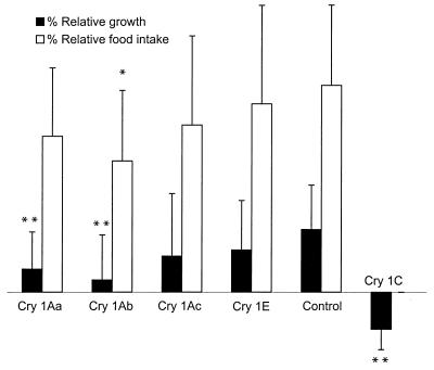
A single dose of 5 μg of the activated Cry1C toxin/0.2 g fresh wt resulted in an irreversible inhibition of food intake in all of the injected animals. The animals also failed to defecate after injection. No lethal effect was observed during the first 48 h. After this period the treated animals gradually died, 60% within the first week after injection. Some of the remaining animals survived up to 2 weeks without resuming food intake. Whether the animals died from a direct toxic effect or starvation was not determined. In contrast, injection of 5 μg of Cry1Ac or Cry1E per 0.2 g fresh wt resulted in no effect after 48 h. An injection of 5 μg of Cry1Aa or Cry1Ab per 0.2 g fresh wt resulted in an inhibitory effect on the feeding behavior of L. dispar larvae but to a much smaller extent then Cry1C (Fig. 1).
FIG. 1.
Effect of B. thuringiensis toxins on the relative food intake and relative growth of L. dispar fourth- and fifth-instar larvae. Larvae were injected with a dose of 5 μg/0.2 g fresh wt. The relative growth was calculated as the relative percent increase in the fresh weight of the larvae at 48 h postinjection. The relative food intake is the food intake as a percentage relative to the fresh weight of the larvae (n = 30). Values are means ± the standard deviations (∗∗, P ≪ 0.01; ∗, P < 0.05) .
The body weight increased with 21.7% over 48 h in control animals, whereas animals injected with Cry1Aa and Cry1Ab gained only 8.0 and 4.2%, respectively (Fig. 1). The relative food intake (individual food intake per gram of body weight) of the animals injected with 5 μg/0.2 g fresh wt Cry1Ab was significantly (P < 0.05) lower than in the control animals, i.e., 45% of the body weight versus 72% for the controls. Injection of lower doses (<5 μg/0.2 g fresh wt) of Cry1Aa, Cry1Ab, Cry1Ac, and Cry1E resulted in no observable effect in the larvae.
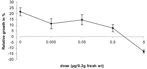
Cry1C proved to be the most potent delta-endotoxin in the bioassay (Fig. 2). A dose as small as 0.5 μg could inhibit the food intake of the injected animals significantly (P < 0.05) (45% ± 18% of the fresh body weight versus 72% ± 25% in the control animals) and had a growth-inhibiting effect.
FIG. 2.
Dose-response curve of the Cry1C toxin on the relative growth of L. dispar larvae. Values are means ± the standard error of the mean (n ≥ 20).
Comparison of the toxic effect of Cry1 toxins upon injection and upon feeding.
The toxicity data from force-feeding assays do not reflect the intrahemocoelic activity of the Cry1 toxins. When the animals were force-fed, the toxicity of Cry1C was much less pronounced (Table 1). Cry1Ab and Cry1Aa, which are particularly active on Lymantria in force-feeding assays, display only a moderate effect upon injection. Finally, the larvae showed very low sensitivity toward Cry1Ac and Cry1E both in force-feeding and upon injection.
TABLE 1.
Comparison of the effect of injecting or force-feeding B. thuringiensis toxins in L. dispar larvae
Cry1C is toxic to brain cells of L. dispar in vitro
The potent toxic effect of Cry1C, when injected into the hemolymph of Lymantria, suggests that other targets are present and accessible for this toxin after injection. We examined the effect of Cry1C on the viability of primary cultures of Lymantria brain cells (Table 2).
TABLE 2.
In vitro cytotoxicity of B. thuringiensis toxins (20 μg/ml) on L. dispar brain cells in primary culture
| Toxin | % Mortalitya ± SD | nb |
|---|---|---|
| Control | 26.4 ± 6.6 | 6 |
| Cry1Ab | 35.3 ± 10.3 | 5 |
| Cry1Ac | 73.2 ± 4.6 | 5 |
| Cry1C | 100 ± 0 | 5 |
| Cry1E | 24.9 ± 3.1 | 6 |
% Mortality = [number of lysed cells (Nl)/total number of cells (Nt)] × 100.
n = number of cell cultures tested.
The activated toxins were diluted with the culture medium to appropriate concentrations. One-day-old cultures of the neurons were incubated with the toxins for 24 h at 26°C. A dose as low as 20 μg of Cry1C/ml was sufficient to induce 100% lysis of the cultured neurons. With the exception of Cry1Ac, other toxins had no effect. The data show that Cry1C is extremely toxic to the brain cells of Lymantria larvae.
Cry1C toxicity to N. bullata larvae.
The strong toxicity to Lymantria upon injection led us to perform the same experiment in a dipteran species. Injection of 5 μg of Cry1C/larva into the last larval instars of N. bullata was extremely lethal. It resulted in rapid death, within seconds. In addition, an immediate, extensive, local hemocytic melanization occurred. As a negative control, 5 μg of Cry1E/larva was injected; this had no observable effect on the maggots over a period of 24 h.
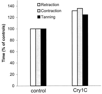
The effect of a sublethal dose of 0.05 μg/larva was monitored in wandering larvae. After synchronization (i.e., selection of red spiracle larvae), the maggots (n = 30) were injected with 0.05 μg of Cry1C. Three criteria (retraction, contraction, and tanning) were monitored for the following 6 h. The toxin markedly affected pupariation (Fig. 3). In 75% of the larvae, retraction and contraction were slowed down compared to the controls. A total of 25% of the treated larvae completely failed to retract the anterior segments and contract longitudinally and also retained larval morphology even after tanning of the cuticle. A total of 50% of the treated larvae did not survive metamorphosis and died during pupation.
FIG. 3.
Effect on pupariation of Cry1C (0.05 μg/larva) injected into red spiracle larvae of N. bullata compared to controls. A total of 25% of the treated animals were not able to form a puparium and were therefore not taken into account.
DISCUSSION
The present study for the first time reports that some B. thuringiensis toxins are lethal to primary cultures of neuronal cells of an insect species. Previous reports have shown that Cry1C is toxic to various cell lines derived from Spodoptera frugiperda, Manduca sexta, Plodia interpunctuella (4), Aedes aegypti, and Anopheles gambiae (11). Spodoptera exigua cell lines show intermediate sensitivity, whereas the sensitivity of Mamestra brassicae and Drosophila melanogaster cell lines is low (6). Cells derived from Choristoneura fumiferana (4) and Culex quinquefasciatus (11) were insensitive.
The doses of Cry1C, used in this study on primary neuronal cultures, which provoke a toxic response of 100%, are considerably smaller (20 μg ml−1) than those reported in toxicity studies on cell lines of A. aegypti and A. gambiae (250 μg ml−1) (11) or S. frugiperda (50 μg ml−1) (6).
It has been known for a long time now that B. thuringiensis toxins, after oral administration, are species specific as well as target specific (the apical membrane of the midgut epithelium cells of susceptible insects). To date, most in vivo studies have focused on this general mode of action. A toxic effect of intrahemocoelic injections of crystals of B. thuringiensis was noted in Pieris brassicae as early as 1967 (7), but since then all attention has been paid to the toxicity of B. thuringiensis toxins after oral intake. Little is known about other putative target organs and cells in insects that are possibly accessible upon injection of the toxin. The potent effect of Cry1C upon injection in the hemolymph strongly suggests that a target other than the midgut epithelial cells must be present for this delta-endotoxin in the larvae of L. dispar. Peyronnet et al. (8) showed that neither Cry1Aa nor Cry1C had any depolarizing effect when applied on the basolateral side of the midgut membrane of L. dispar. On the other hand, Butko et al. (1) demonstrated a membrane-perturbing activity of Cry1C in receptor-free phospholipid vesicles. In vitro, and at low pH, Cry1C undergoes a conformational change that leads to membrane interaction and promotes flux of ions across the lipid bilayer (1). It is conceivable that, in vivo, a transition to a similar conformational state could be triggered by means of another, as-yet-unknown, physiological condition.
The intrahemocoelic activity, however, is not a general property of Cry1 toxins since Lymantria was only weakly sensitive to Cry1Aa and Cry1Ab and not sensitive at all toward Cry1Ac and Cry1E. Comparison of the toxicity upon injection of these toxins with data obtained in force-feeding assays (12, 13) shows quite the opposite pharmacologic profile for Cry1C, Cry1Aa, and Cry1Ab. Cry1Ac and Cry1E, on the other hand, are inactive in vivo both when force-fed and when injected. Moreover, a potent toxic effect was observed after injection of Cry1C into the fleshfly, N. bullata. In sublethal doses, the toxin affected pupation and gave rise to deformed pupae. The morphologic deformation (retaining of larval characteristics in the pupae) strongly resembles the one described earlier in flies treated with bee venom (16) and suggests a suppression of the peripheral neuromuscular system.
These data demonstrate that Cry1C inhibits food intake, and concomitantly growth of the insect, when injected into the hemolymph of Lymantria. Evidently, the mode of action after injection is quite different from the one that occurs after oral intake of the toxins. In vitro toxicity to Lymantria neurons suggests that the insect nervous system is a target for the toxin. In vivo experiments in the fleshfly support this view. According to Kwa et al. (6), sensitivity toward Cry1C of cell lines of S. frugiperda and S. exigua are correlated to the binding of the toxin to a 40-kDa protein. Whether the toxin-specific effects that we observed involve an aspecific mechanism or binding to a distinct receptor remains, however, inconclusive and needs further study. Its putative mode of action upon injection in the hemolymph will require more fundamental research. The question arises if the observed specificity of Cry1C toxicity after oral intake coincides with a specificity after injection.
The model proposed for Cry1C is that injected toxins may reach a neural target, as demonstrated by the good correlation between cytotoxicity and toxicity upon injection. On the other hand, Cry1Ac is nontoxic upon injection but quite cytotoxic to the neural cells in vitro. Apparently, the active Cry1Ac toxin does not reach the brain cells in vivo or the toxin is no longer active when it reaches the cells. Perhaps the toxin is not able to cross the blood-brain barrier or, in the hemolymph, it may be degraded, denatured, or aggregated in a nonfunctional form that would prevent binding or pore formation.
In conclusion, the results of this study suggest that B. thuringiensis toxin targets are not only present in the gut. This is an important result of biological relevance at the level of B. thuringiensis mode of action. Second, insects or other organisms that are susceptible to “injection” (for example, insects or parasitoids that may have ingested the toxin might inject it into their hosts or preys) may be affected. Therefore, the toxic effect described here may be relevant to the more general field of biocontrol, including trophic interactions. The benefit of, or the damage caused by, the phenomenon at the ecological level remains to be investigated. Also, the health protection of workers involved in B. thuringiensis spraying, etc., and the nonspecific effects on nontarget species need to be carefully scrutinized.
ACKNOWLEDGMENTS
This research was supported by a bilateral project (BIL97/61) between the Catholic University of Leuven, the Limburgs Universitair Centrum, and the University of Montreal. A.C. is a postdoctoral researcher of the Research Council of the K. U. Leuven. P.V. is a recipient of a grant from the Flemish Science Foundation (FWO).
We thank Aventis Cropscience for kindly providing the toxins.
REFERENCES
- 1.Butko P, Cournoyer M, Pusztai-Carey M, Surewicz W K. Membrane interactions and surface hydrofobicity of Bacillus thuringiensis δ-endotoxin Cry1C. FEBS Lett. 1994;340:89–92. doi: 10.1016/0014-5793(94)80178-9. [DOI] [PubMed] [Google Scholar]
- 2.Crickmore N, Zeigler D R, Feitelson J, Schneph E, Van Rie J, Lereclus D, Baum J, Dean D H. Revision of the nomenclature for the Bacillus thuringiensis pesticidal crystal proteins. Microbiol Mol Biol Rev. 1998;62:807–813. doi: 10.1128/mmbr.62.3.807-813.1998. [DOI] [PMC free article] [PubMed] [Google Scholar]
- 3.Huybrechts R, De Loof A. Induction of vitellogenin synthesis in male Sarcophaga bullata by ecdysterone. J Insect Physiol. 1977;23:1359–1362. [Google Scholar]
- 4.Johnson D E. Cellular toxicity's and membrane binding characteristics of insecticidal crystal proteins from Bacillus thuringiensis toward cultured insect cells. J Invertebr Pathol. 1994;63:123–129. doi: 10.1006/jipa.1994.1024. [DOI] [PubMed] [Google Scholar]
- 5.Knowles B H. Mechanism of action of Bacillus insecticidal δ-endotoxins. Adv Insect Physiol. 1994;24:275–308. [Google Scholar]
- 6.Kwa M S, de Maagd R A, Stiekema W J, Vlak J M, Bosch D. Toxicity and binding properties of the Bacillus thuringiensis delta-endotoxin Cry1C to cultured insect cells. J Invertebr Pathol. 1998;71:121–127. doi: 10.1006/jipa.1997.4723. [DOI] [PubMed] [Google Scholar]
- 7.Lecadet M M, Martouret D. Enzymatic hydrolysis of the crystals of Bacillus thuringiensis by the proteases of Pieris brassicae. II. Toxicity of the different fractions of the hydrolysate for larvae of Pieris brassicae. J Invertebr Pathol. 1967;9:322–330. [Google Scholar]
- 8.Peyronnet O, Vachon V, Brousseau R, Baines D, Schwartz J-L, Laprade R. Effect of Bacillus thuringiensis toxins on the membrane potential of lepidopteran insect midgut cells. Appl Environ Microbiol. 1997;63:1679–1684. doi: 10.1128/aem.63.5.1679-1684.1997. [DOI] [PMC free article] [PubMed] [Google Scholar]
- 9.Rajamohan F, Lee M K, Dean D H. Bacillus thuringiensis insecticidal proteins: molecular mode of action. Prog Nucleic Acid Res Mol Biol. 1998;60:1–27. doi: 10.1016/s0079-6603(08)60887-9. [DOI] [PubMed] [Google Scholar]
- 10.Schneph E, Crickmore N, Van Rie J, Lereclus D, Baum J, Feitelson J, Zeigler D R, Dean D H. Bacillus thuringiensis and its pesticidal crystal proteins. Microbiol Mol Biol Rev. 1998;62:775–806. doi: 10.1128/mmbr.62.3.775-806.1998. [DOI] [PMC free article] [PubMed] [Google Scholar]
- 11.Smith G P, Merrick J D, Bone E J, Ellar D J. Mosquitocidal activity of the Cry1C δ-endotoxin from Bacillus thuringiensis subsp. aizawai. Appl Environm Microbiol. 1996;62:680–684. doi: 10.1128/aem.62.2.680-684.1996. [DOI] [PMC free article] [PubMed] [Google Scholar]
- 12.van Frankenhuyzen K, Gringorten J L, Gauthier D, Milne R E, Masson L, Peferoen M. Toxicity of activated Cry1 proteins from Bacillus thuringiensis to six forest Lepidoptera and Bombyx mori. J Invertebr Pathol. 1993;62:295–301. [Google Scholar]
- 13.van Frankenhuyzen K, Gringorten J L, Milne R E, Gauthier D, Pusztai M, Brousseau R, Masson L. Specificity of activated Cry1A proteins from Bacillus thuringiensis subsp. kurstaki HD-1 for defoliating forest Lepidoptera. Appl Environ Microbiol. 1991;57:1650–1655. doi: 10.1128/aem.57.6.1650-1655.1991. [DOI] [PMC free article] [PubMed] [Google Scholar]
- 14.Van Rie J, Jansens S, Höfte H, Degheele D, Van Mellaert H. Receptors on the brush border membrane of the insect midgut as determinants of the specificity of Bacillus thuringiensis delta-endotoxins. Appl Environ Microbiol. 1990;56:1378–1385. doi: 10.1128/aem.56.5.1378-1385.1990. [DOI] [PMC free article] [PubMed] [Google Scholar]
- 15.Zd'árek J, Fraenkel G. Pupariation in flies: a tool for monitoring effects of drugs, venoms, and other neurotoxic compounds. Arch Insect Biochem Physiol. 1987;4:29–46. [Google Scholar]
- 16.Zd'árek J, Slama K, Fraenkel G. Changes in internal pressure during puparium formation in flies. J Exp Biol. 1979;207:187–195. [Google Scholar]


