
Fig. 8.
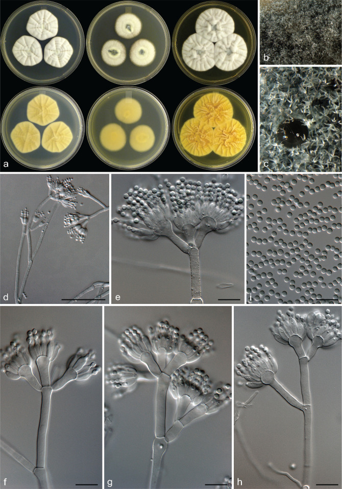
Penicillium eickeri. a. Colonies (top row, left to right: CYA, MEA, YES; bottom row, left to right: CYA reverse, MEA reverse, YES reverse); b, c. colony texture on MEA; d–h. conidiophores; i. conidia. — Scale bars: d = 25 μm, e–i = 10 μm.
Official websites use .gov
A
.gov website belongs to an official
government organization in the United States.
Secure .gov websites use HTTPS
A lock (
) or https:// means you've safely
connected to the .gov website. Share sensitive
information only on official, secure websites.
Penicillium eickeri. a. Colonies (top row, left to right: CYA, MEA, YES; bottom row, left to right: CYA reverse, MEA reverse, YES reverse); b, c. colony texture on MEA; d–h. conidiophores; i. conidia. — Scale bars: d = 25 μm, e–i = 10 μm.