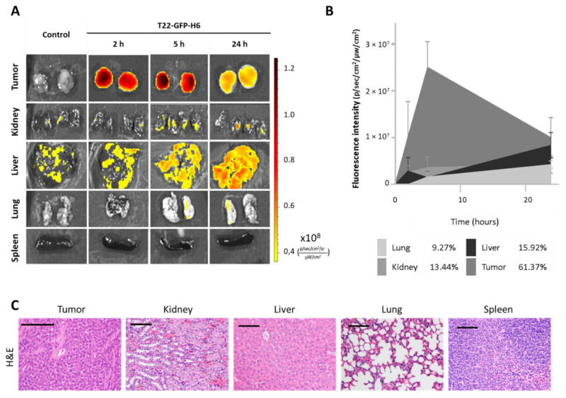
Figure 5.
In vivo biodistribution and toxicity assessment of nanocarrier T22-GFP-H6 in a subcutaneous mouse model derived from CXCR4+ AN3CA cells. (A) Representative images of fluorescence emitted by the nanocarrier after 2, 5 and 24 h after intravenous injection of 200 µg of T22-GFP-H6. (B) Area under the curve representation of fluorescence emitted over time by tumor and non-tumor tissues, and their respective percentage of nanocarrier uptake out of the total fluorescence emitted by T22-GFP-H6 in all tissues (n = 4/group; mean ± s.e.m). (C) Hematoxylin-eosin staining of tumor and non-tumor organs 48 h after administration of the nanocarrier. Bar: 100 µm.

