Abstract
Septo-optic dysplasia (SOD) is a developmental phenotype characterized by midline neuroradiological anomalies, optic nerve hypoplasia, and pituitary anomalies, with a high degree of variability and additional systemic anomalies present in some cases. While disruption of several transcription factors has been identified in SOD cohorts, most cases lack a genetic diagnosis, with multifactorial risk factors being thought to play a role. Exome sequencing in a cohort of families with a clinical diagnosis of SOD identified a genetic diagnosis in 3/6 families, de novo variants in SOX2, SHH, and ARID1A, and explored variants of uncertain significance in the remaining three. The outcome of this study suggests that investigation for a genetic etiology is warranted in individuals with SOD, particularly in the presence of additional syndromic anomalies and when born to older, multigravida mothers. The identification of causative variants in SHH and ARID1A further expands the phenotypic spectra associated with these genes and reveals novel pathways to explore in septo-optic dysplasia.
Keywords: septo-optic dysplasia, SHH, ARID1A, SOX2
1. Introduction
Septo-optic dysplasia (SOD) is a rare, heterogenous phenotype reported in 1 out of 10,000 live births and is characterized by any combination of midline neuroradiological anomalies, including absence of the septum pellucidum or agenesis of the corpus callosum, hypoplasia of the optic chiasma/nerves, and hypothalamic–pituitary dysfunction [1]. The phenotype is highly variable, with additional anomalies present in many cases. Several transcription factors were shown to play a role in SOD, with the first factor, HESX1, identified in 1998 [2] and variants in SOX3, SOX2, and OTX2 being more recently implicated [1,3]. However, variants in these genes explain only a small number of cases, highlighting the need to identify additional novel players. In the majority of cases, SOD is sporadic with a low recurrence risk; young maternal age, primigravida status, viral infections, and other environmental causes have also been implicated, suggesting that the phenotype is likely to be multifactorial in many cases [1,3].
2. Materials and Methods
This human study was conducted according to the guidelines of the Declaration of Helsinki and approved by the Institutional Review Board of Children’s Wisconsin. Exome sequencing was undertaken through Psomagen (previously Axeq; Rockville, MD, USA), the University of Washington Center for Mendelian Genomics, or via clinical testing and then analyzed, as previously described, utilizing the SNP &Variation Suite or VarSeq software (Golden Helix, Bozeman, MT, USA), including annotations for gnomAD v2.1.1, OMIM genes, CADD Scores 1.4, and REVEL Functional Predictions [4]. The data were reviewed for rare variants in known SOD genes (HESX1, SOX3, SOX2, OTX2) and OMIM genes, along with a standard trio analysis (when available) and review of ultra-rare (≤5 alleles in gnomAD) and damaging (Loss of Function (LOF) or REVEL > 0.4 and CADD > 20) coding variants. Causative variants were confirmed by Sanger sequencing using region-specific primers. Six families with a clinical diagnosis of SOD in the proband underwent exome sequencing, including four trios (affected proband plus unaffected parents), one quad (affected siblings plus unaffected parents), and one singleton. Tissue-expression patterns for novel genes were investigated in The Human Protein Atlas (proteinatlas.org).
3. Results
Causative variants were identified in three SOD families, including one variant in SOX2. No rare variants were identified in other SOD genes.
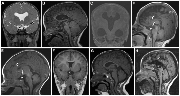
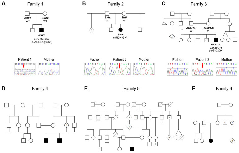
In Family 1, the affected individual is a 5-year-old White male with mild optic nerve hypoplasia, absent septum pellucidum, hypoplasia of corpus callosum, and dilated lateral ventricles identified by Brain MRI; the individual was born as the second child to a 30-year-old mother (Figure 1). He has global developmental delay, hypotonia with unsteady gait requiring the use of supports (forearm crutches), complex seizures, and hyperopia with mild optic disc pallor by clinical exam. Trio exome sequencing identified a de novo variant in SOX2, NM_003106.4:c.70_89del20 p.(Asn24Argfs*65); Sanger sequencing confirmed the presence of the variant in the child and its absence in the mother (Table 1; Figure 2).
Figure 1.
Magnetic resonance imaging (MRI) data for affected individuals. Family 1 (A,B) Coronal T2 image (A) from the proband showing absent septum pellucidum; Sagittal T1 image (B) showing hypoplasia of the corpus callosum. Family 3 (C) Coronal T1 image of the proband showing absent septum pellucidum, absent corpus callosum, and ventriculomegaly. Family 4 (D,E) Sagittal T1 image of proband (D) showing hypoplastic corpus callosum (curved white arrow), ectopic posterior pituitary (straight white arrow), and hypoplastic optic chiasm (arrowhead); Sagittal T1 image of affected brother (E) demonstrating a slightly hypoplastic corpus callosum (curved white arrow), ectopic posterior pituitary (white arrow), and hypoplastic optic chiasm (arrowhead). Family 5 (F,G) Coronal T1 image (F) with absent septum pellucidum and a small hypoplastic optic chiasm (arrowhead) (F); Sagittal T1 image (G) showing hypoplastic small pituitary (arrow). Family 6 (H) Sagittal T1 image showing marked hypoplasia of corpus callosum (curved white arrow) and hypoplastic optic chiasm (arrowhead).
Table 1.
Pathogenic variants identified in individuals with a clinical diagnosis of SOD.
| Family | Gene | Nucleotide Change | Predicted Effect | MAF 1 | ACMG/AMP Classification | Family History |
|---|---|---|---|---|---|---|
| 1 | SOX2 | NM_003106.4: c.70_89del20 | p.(Asn24Argfs*65) | NP | Pathogenic (PVS1, PS2, PM2, PP5) |
De Novo |
| 2 | SHH | NM_000193.2: c.562+1G>A | Abnormal splicing | NP | Pathogenic (PVS1, PS2, PM2, PP5) |
De Novo |
| 3 | ARID1A | NM_006015.6: c.6625C>T | p.(Gln2209*) | NP | Pathogenic (PVS1, PS2, PM2) |
De Novo |
1 Frequency in gnomAD v2.1.1; NP, not present.
Figure 2.
Pedigrees of Families 1–6 with a clinical diagnosis of SOD. (A–C) Pedigrees of genetically explained families with genotypes indicated for corresponding variants in SOX2 (Family 1), SHH (Family 2), and ARID1A (Family 3) and Sanger traces showing variants in each affected individual as well as absence in unaffected parent(s). (D–F) Pedigrees of genetically unexplained families. Filled symbols represent affected individuals; empty symbols represent unaffected individuals.
In Family 2, the affected individual is an 18-month-old Hispanic female with absent septum pellucidum, partially absent corpus callosum (anterior absent), left optic nerve hypoplasia, and right retinal coloboma with mild asymmetry of the orbits noted by clinical exam, with the right eye appearing somewhat smaller than the left, born as the third child of a 35-year-old mother. She also has a bilateral complete cleft palate and a right complete cleft lip, a single central incisor, microcephaly, mandibular hypoplasia, alveolar gap, global developmental delay, failure to thrive (1st centile), asymmetric thigh creases, coxa valga, dysphagia requiring g-tube feeding, and diabetes insipidus due to pituitary dysfunction. At 13 months of age, measurements confirmed normal placement of the eyes with an inner canthal distance of 2 cm (50th centile) and an outer canthal distance of 7 cm (25th–50th centile). Trio exome sequencing identified a de novo variant in SHH, NM_000193.2:c.562+1G>A, which was confirmed to be present in the child and absent in both parents by Sanger sequencing (Figure 2).
In Family 3, the affected individual is a 6-week-old White male with absent septum pellucidum, absent corpus callosum, ventriculomegaly, aqueductal stenosis, and intraventricular hemorrhage identified on Brain MRI (Figure 1), born at 33 weeks of gestation as the 5th child of a 32-year-old mother. Eye exam at 5 weeks of age identified bilateral optic disc pallor, asymmetric optic discs with the right larger than the left, and a peripapillary halo also more notable on the right. Additional systemic anomalies included a ventricular septal defect and a patent foramen ovale, 13 pairs of ribs, bilateral clinodactyly, single palmar crease, broad large toe with hypoplastic nail, cleft palate, choanal atresia, seizures, apnea, and dysmorphic facial features, including down-slanting palpebral fissures, long columella, low-set and posteriorly rotated ears, depressed nasal bridge, scant hair due to premature birth; he died at 6 weeks of age. Trio exome sequencing identified a de novo variant in ARID1A, NM_006015.6:c.6625C>T p.(Gln2209*), which was confirmed to be present in the child and absent in both parents by Sanger sequencing (Figure 2). The variant appeared to be mosaic: it was present in 59/179 (33%) exome reads and had a lower peak as determined by Sanger sequencing (Figure 2).
Family 4 consists of a 4-year-old White/Native Hawaiian male with hypoplastic corpus callosum and genu, bilateral optic nerve hypoplasia, and panhypopituitarism with an ectopic posterior and a severely hypoplastic anterior pituitary gland on Brain MRI (Figure 1) along with global delay and seizure-like activity, gastroschisis with jejunal atresia, and a ventricular septal defect, born to a 23-year-old primigravida mother. He has a 2-year-old affected brother with bilateral optic nerve hypoplasia, a mildly hypoplastic corpus callosum, and panhypopituitarism with posterior pituitary ectopia with small sella and hypoplasia of the anterior pituitary gland on Brain MRI (Figure 1) along with global delay, depressed nasal bridge, and simple, cupped ears. Quad exome sequencing did not identify a causative variant and no rare variants were identified in known SOD genes; variants of uncertain significance included compound heterozygous variants in MIB2 and a hemizygous missense variant in AKAP4 (Table 2). A review of the shared, ultra-rare, damaging variants identified seven inherited variants of uncertain significance shared by both brothers (Supplemental Table S1), including a heterozygous missense variant in CHD5.
Table 2.
Select variants of uncertain significance discovered in individuals with a clinical diagnosis of SOD.
| Family | Gene | Zygosity | Nucleotide Change | Predicted Effect |
MAF 1 | CADD/ REVEL3 |
Segregation | |
|---|---|---|---|---|---|---|---|---|
| 4 | MIB2 | Compound heterozygous |
NM_080875.3:c.-48C>T NM_001170688.1:c.124C>T |
? p.(Arg42*) |
4/185998 | 33 | N/A | Paternal (het) |
| 4 | MIB2 | Compound heterozygous | NM_080875.3:c.1766A>G | p.(Gln589Arg | 270/ 259488 |
10.19 | 0.041 | Maternal (het) |
| 4 | AKAP4 | Hemizygous | NM_003886.3:c.1835G>A | p.(Cys612Tyr) | 1/183267, 0 hemi | 14.58 | 0.089 | Maternal (het) |
| 4 | CHD5 | Heterozygous | NM_015557.3:c.5809G>A | p.(Gly1937Arg) | NP | 24.3 | 0.443 | Paternal (het) |
| 5 | FAT3 | Compound heterozygous | NM_001008781.3:c.9772G>A | p.(Val3258Ile) | 3/271900 | 24 | 0.131 | Paternal (het) |
| 5 | FAT3 | Compound heterozygous | NM_001008781.3:c.11546G>A | p.(Arg3849Gln) | 33/ 249054 |
24.2 | 0.41 | Maternal (het) |
| 5 | RPTN | Homozygous | NM_001122965.1:c.489delA | p.(Lys163Asnfs*48) | NP | 14.88 | N/A | Biparental (het) |
| 5 | TRPM3 | Heterozygous | NM_001366145.2:c.871C>T | p.(His291Tyr) | 1/31412 | 23.2 | 0.419 | Paternal (het) |
| 6 | DMXL1 | Homozygous | NM_005509.6:c.9002G>A | p.(Gly3001Glu) | 1/251182 | 31 | 0.614 | Unknown |
| 6 | CCDC13 | Heterozygous | NM_144719.4:c.631C>T | p.(Gln211*) | NP | 40 | N/A | Unknown |
1 Frequency in gnomAD v2.1.1; 3 CADDphredhg19 and REVEL scores (from dbNSFP v4.1a, accessed through Varseq). N/A, not applicable; NP, not present.
Family 5 consists of a 5-month-old White male with absent septum pellucidum, hypoplastic corpus collosum, and bilateral optic nerve hypoplasia on Brain MRI (Figure 1) along with growth hormone deficiency, borderline micropenis, and mild global developmental delay, born to a 29-year-old primigravida mother. Trio exome sequencing did not identify a causative variant; variants of uncertain significance include compound heterozygous missense variants in FAT3 and a homozygous frameshift variant in RPTN (Table 2). Review of ultra-rare damaging variants identified seven inherited variants of uncertain significance (Supplemental Table S1), including a missense variant in TRPM3.
Family 6 consists of a 7-year-old Hispanic female with a hypoplastic corpus callosum and midline anomalies as well as bilateral optic nerve hypoplasia on Brain MRI (Figure 2) along with highly arched palate, spastic hemiplegia, dysphagia, enamel hypoplasia, high-arched palate, hemolytic anemia, and global delay, born to a 20-year-old primigravida mother. Singleton exome sequencing did not identify a causative variant. A review of ultra-rare damaging variants identified 17 variants of uncertain significance (Supplemental Table S1), including a homozygous variant in DMXL1 and a heterozygous variant in CCDC13 (Table 2).
4. Discussion
While septo-optic dysplasia (SOD) typically has a low rate of genetic diagnosis, the genetic analysis of this small cohort of individuals with SOD identified a genetic etiology in 50% of the families. Cases with a genetic diagnosis were more likely to have atypical optic nerve findings, but all had been given a clinical diagnosis of septo-optic dysplasia prior to genetic testing, highlighting the variability of this phenotype. Interestingly, cases without a genetic diagnosis were more likely to be born to younger, primigravida mothers, consistent with previous associations [1,3], although potentially contributing variants were identified in novel genes in each of these families.
SOX2 intragenic variants and deletions are the most common cause of anophthalmia/microphthalmia, typically syndromic with commonly seen esophageal, genitourinary, and neurological anomalies [5]. A connection to SOD was noted in a mouse model, and subsequent screening in a cohort of individuals with SOX2 variants identified pituitary hypoplasia and hypogonadotropic hypogonadism along with anomalies of the corpus callosum and medial temporal structures [6]. As expected, all of the individuals with loss-of-function variants also had anophthalmia/microphthalmia, with variable additional syndromic features. Absent septum pellucidum was only noted in two individuals with missense variants (p.(Gly130Ala) and p.(Ala191Thr)) and an isolated SOD phenotype with normal eye size; both of these variants were inherited from phenotypically normal parents and are now known to be present in the general population, with population max frequencies of 0.02% and 0.05% in gnomAD, higher than the frequency of SOD, suggesting that these are likely to be benign, population-specific variants. Individual 1 of this study is the first case of SOD with normal eye size and a lack of additional birth defects to have a loss-of-function variant in SOX2. Interestingly, the identified SOX2 variant, c.70_89del, is a recurrent variant now reported in 20 individuals; while the majority of cases presented with a severe phenotype of syndromic anophthalmia/microphthalmia, phenotypic variability has been reported in some cases [5]. An abnormal gait, often described as ataxic and requiring the use of a walker or other assistive devices, is typical for SOX2 disruption [5].
In humans, SHH variants are associated with holoprosencephaly (HPE), another developmental anomaly affecting the brain [7], and explain up to 37% of dominant HPE [8]. HPE is characterized by incomplete separation of the forebrain into right and left hemispheres, typically associated with craniofacial anomalies including microcephaly, hypotelorism, single central incisor, and cleft lip/palate [8]. Phenotypic variability is well-recognized for SHH, with family members often presenting with only subtle midline craniofacial features or developmental delays/ADHD [9,10]. While the conditional knockout of Shh in the hypothalamus of mice resulted in an SOD phenotype [11], this is the first association of variants of this gene with an SOD diagnosis in humans. The specific variant identified in Family 2 was previously reported in two cases with holoprosencephaly [12,13]. The presence of a cleft lip/palate along with SOD may suggest SHH disruption. Interestingly, Sox2 and Shh act in the same pathway, with Sox2/3 expression being required for Shh expression in the hypothalamus (via direct action of a long-range Shh enhancer) and disruption of this pathway in the hypothalamus was found to result in SOD in mice [11]. Another major SOD gene, the paired homeodomain transcription factor HESX1, is also implicated in the Sox2 pathway, with SOX2 binding to the Hesx1 promoter in vitro; observations of decreased expression of Hesx1 in Sox2-deficient mice indicate likely direct regulation of Hesx1 by Sox2 [6].
ARID1A encodes a member of the SWItch/Sucrose NonFermenting (SWI/SNF) complex; variants in genes encoding the components of the SWI/SNF complex result in Coffin-Siris syndrome, a syndromic form of intellectual disability frequently associated with agenesis or hypoplasia of the corpus callosum [14,15]. While SOD has not been reported, other syndromic features seen in this individual show a strong overlap and early lethality has been seen with ARID1A variants in particular [16]. The variant identified here, c.6625C>T p.(Gln2209*), is the most C-terminal variant identified to date, but five other premature termination alleles within this final exon have been reported (HGMD [17]). The presence of multiple additional syndromic anomalies—particularly a hypoplastic big toenail, sparse hair, and heart defects—in an individual with SOD may indicate the presence of Coffin–Siris syndrome. The identification of a role for ARID1A in SOD proposes the involvement of a novel pathway in this disorder. Examination of ARID1A and related factors in SOD is warranted.
With regard to the latter finding, it is interesting to note the identification of compound heterozygous variants of uncertain significance in MIB2 shared by the two affected siblings in Family 4. MIB2 (skeletrophin) is a RING finger-dependent ubiquitin ligase first identified in a screen for genes that were upregulated by truncated ARID1A (SWI1) in neuroblastoma cells displaying increased cell–cell adhesions and aggregations. MIB2 was also found to bind JAG2, a ligand in the Notch family [18,19]; Notch and Hedgehog are major signaling pathways that regulate the early steps of pituitary organogenesis and eye development, with interplay between these pathways including the restriction of Jag2 expression by Shh [20]. A mouse model of Mib2 deficiency showed variable neural tube closure defects [21], and an abnormal eye morphology was reported in the Mouse Genome Informatics database (http://www.informatics.jax.org/, accessed on 18 April 2022). Since the nonsense variant affects only a single transcript (out of several known isoforms) and the missense variant has weak predictions, the significance of these variants is unclear. The brothers also shared a heterozygous missense variant in CDH5, whose knockdown has been associated with reduced head and eye size in zebrafish [22]; however, this variant was inherited from the unaffected father.
The compound heterozygous missense variants in atypical cadherin 3, FAT3, that were identified in Family 5 both fall within identified domains, the Cadherin 30 and Laminin G-like domains [23]. Fat3 has been shown to be strongly expressed in the embryonic but not adult brain in mice and rats [24], making it an interesting candidate for SOD. Variants in other members of this family, FAT1 and FAT2, were found to be associated with recessive syndromic ocular coloboma [25] and dominant spinocerebellar ataxia-45 [26]. The heterozygous missense variant in TRPM3, with links to ocular development and intellectual disability [27,28], is also notable, but it was inherited from the unaffected father.
In Family 6, the most interesting candidate variant is a homozygous missense variant in DMXL1, which encodes a WD-repeat protein. This gene was identified as a candidate gene within the 5q22.3q23.3 deletion region in a patient with iris coloboma and Chiari I malformation [29]; a homozygous frameshift variant was reported in an individual with global delay, seizures, hypotonia, and optic disc edema, along with heart and kidney defects [30]. Abnormal development was also noted in a Drosophila mutant [31]. A heterozygous nonsense variant in the Coiled-Coil domain containing 13 gene, CCDC13, is also notable for its high CADD score (40) and absence in gnomAD. While little is known about the gene beyond a possible role in ciliogenesis [32], its RNA expression was found to be enriched in human brain and eye tissues [33].
The outcome of this study suggests that investigation for a genetic etiology is warranted in individuals with a clinical diagnosis of SOD, particularly in the presence of additional syndromic anomalies and when born to older, multigravida mothers. The identification of causative variants in SHH and ARID1A further expands the phenotypic spectra associated with these genes and identifies novel pathways to explore in septo-optic dysplasia.
Acknowledgments
We are grateful to the patients and their families for their participation in this study and to Samuel Thompson for his assistance with the Sanger sequencing.
Supplementary Materials
The following supporting information can be downloaded at: https://www.mdpi.com/article/10.3390/genes13071165/s1, Supplemental Table S1: Variants of uncertain significance identified in Families 4–6.
Author Contributions
Conceptualization, E.V.S.; methodology, E.V.S. and L.M.R.; formal analysis, L.M.R., M.M. and S.S.; resources, D.B., L.W. and J.M.; writing—original draft preparation, L.M.R. and E.V.S.; writing—review and editing, M.M., S.S., D.B., L.W. and J.M.; funding acquisition, E.V.S. All authors have read and agreed to the published version of the manuscript.
Institutional Review Board Statement
The study was conducted according to the guidelines of the Declaration of Helsinki and approved by the Institutional Review Board of Children’s Wisconsin (protocol 124172-76, with initial approval in 2003).
Informed Consent Statement
Informed consent was obtained from all subjects (and/or legal guardians) involved in the study, including permission to publish.
Data Availability Statement
There are no other data associated with this manuscript.
Conflicts of Interest
The authors declare no conflict of interest.
Funding Statement
This work was supported by NIH grants R01EY015518 and R01EY025718 as well as by funds provided by the Children’s Research Institute Foundation at Children’s Wisconsin (EVS) and Grant No. 1UL1RR031973 from the Clinical and Translational Science Award (CTSA) program. The University of Washington Center for Mendelian Genomics (UW-CMG) was funded by NHGRI and NHLBI Grant Nos. UM1HG006493 and U24HG008956. The content is the sole responsibility of the authors and does not necessarily represent the official views of the National Institutes of Health.
Footnotes
Publisher’s Note: MDPI stays neutral with regard to jurisdictional claims in published maps and institutional affiliations.
References
- 1.McCabe M.J., Alatzoglou K.S., Dattani M.T. Septo-optic dysplasia and other midline defects: The role of transcription factors: HESX1 and beyond. Best Pract. Res. Clin. Endocrinol. Metab. 2011;25:115–124. doi: 10.1016/j.beem.2010.06.008. [DOI] [PubMed] [Google Scholar]
- 2.Dattani M.T., Martinez-Barbera J.P., Thomas P.Q., Brickman J.M., Gupta R., Martensson I.L., Toresson H., Fox M., Wales J.K., Hindmarsh P.C., et al. Mutations in the homeobox gene HESX1/Hesx1 associated with septo-optic dysplasia in human and mouse. Nat. Genet. 1998;19:125–133. doi: 10.1038/477. [DOI] [PubMed] [Google Scholar]
- 3.Kelberman D., Dattani M.T. Genetics of septo-optic dysplasia. Pituitary. 2007;10:393–407. doi: 10.1007/s11102-007-0055-5. [DOI] [PubMed] [Google Scholar]
- 4.Reis L.M., Sorokina E.A., Thompson S., Muheisen S., Velinov M., Zamora C., Aylsworth A.S., Semina E.V. De Novo Missense Variants in WDR37 Cause a Severe Multisystemic Syndrome. Am. J. Hum. Genet. 2019;105:425–433. doi: 10.1016/j.ajhg.2019.06.015. [DOI] [PMC free article] [PubMed] [Google Scholar]
- 5.Amlie-Wolf L., Bardakjian T., Kopinsky S.M., Reis L.M., Semina E.V., Schneider A. Review of 37 patients with SOX2 pathogenic variants collected by the Anophthalmia/Microphthalmia Clinical Registry and DNA Research Study. Am. J. Med. Genet. Part A. 2021. accepted . [DOI] [PMC free article] [PubMed]
- 6.Kelberman D., Rizzoti K., Avilion A., Bitner-Glindzicz M., Cianfarani S., Collins J., Chong W.K., Kirk J.M., Achermann J.C., Ross R., et al. Mutations within Sox2/SOX2 are associated with abnormalities in the hypothalamo-pituitary-gonadal axis in mice and humans. J. Clin. Investig. 2006;116:2442–2455. doi: 10.1172/JCI28658. [DOI] [PMC free article] [PubMed] [Google Scholar]
- 7.Roessler E., Belloni E., Gaudenz K., Jay P., Berta P., Scherer S.W., Tsui L.C., Muenke M. Mutations in the human Sonic Hedgehog gene cause holoprosencephaly. Nat. Genet. 1996;14:357–360. doi: 10.1038/ng1196-357. [DOI] [PubMed] [Google Scholar]
- 8.Nanni L., Ming J.E., Bocian M., Steinhaus K., Bianchi D.W., Die-Smulders C., Giannotti A., Imaizumi K., Jones K.L., Campo M.D., et al. The mutational spectrum of the sonic hedgehog gene in holoprosencephaly: SHH mutations cause a significant proportion of autosomal dominant holoprosencephaly. Hum. Mol. Genet. 1999;8:2479–2488. doi: 10.1093/hmg/8.13.2479. [DOI] [PubMed] [Google Scholar]
- 9.Hehr U., Gross C., Diebold U., Wahl D., Beudt U., Heidemann P., Hehr A., Mueller D. Wide phenotypic variability in families with holoprosencephaly and a sonic hedgehog mutation. Eur. J. Pediatr. 2004;163:347–352. doi: 10.1007/s00431-004-1459-0. [DOI] [PubMed] [Google Scholar]
- 10.Heussler H.S., Suri M., Young I.D., Muenke M. Extreme variability of expression of a Sonic Hedgehog mutation: Attention difficulties and holoprosencephaly. Arch. Dis. Child. 2002;86:293–296. doi: 10.1136/adc.86.4.293. [DOI] [PMC free article] [PubMed] [Google Scholar]
- 11.Zhao L., Zevallos S.E., Rizzoti K., Jeong Y., Lovell-Badge R., Epstein D.J. Disruption of SoxB1-dependent Sonic hedgehog expression in the hypothalamus causes septo-optic dysplasia. Dev. Cell. 2012;22:585–596. doi: 10.1016/j.devcel.2011.12.023. [DOI] [PMC free article] [PubMed] [Google Scholar]
- 12.Roessler E., El-Jaick K.B., Dubourg C., Velez J.I., Solomon B.D., Pineda-Alvarez D.E., Lacbawan F., Zhou N., Ouspenskaia M., Paulussen A., et al. The mutational spectrum of holoprosencephaly-associated changes within the SHH gene in humans predicts loss-of-function through either key structural alterations of the ligand or its altered synthesis. Hum. Mutat. 2009;30:E921–E935. doi: 10.1002/humu.21090. [DOI] [PMC free article] [PubMed] [Google Scholar]
- 13.Xiong H.Y., Alipanahi B., Lee L.J., Bretschneider H., Merico D., Yuen R.K., Hua Y., Gueroussov S., Najafabadi H.S., Hughes T.R., et al. RNA splicing. The human splicing code reveals new insights into the genetic determinants of disease. Science. 2015;347:1254806. doi: 10.1126/science.1254806. [DOI] [PMC free article] [PubMed] [Google Scholar]
- 14.Tsurusaki Y., Okamoto N., Ohashi H., Kosho T., Imai Y., Hibi-Ko Y., Kaname T., Naritomi K., Kawame H., Wakui K., et al. Mutations affecting components of the SWI/SNF complex cause Coffin-Siris syndrome. Nat. Genet. 2012;44:376–378. doi: 10.1038/ng.2219. [DOI] [PubMed] [Google Scholar]
- 15.Kosho T., Okamoto N., Coffin-Siris Syndrome International C. Genotype-phenotype correlation of Coffin-Siris syndrome caused by mutations in SMARCB1, SMARCA4, SMARCE1, and ARID1A. Am. J. Med. Genet. C Semin. Med. Genet. 2014;166:262–275. doi: 10.1002/ajmg.c.31407. [DOI] [PubMed] [Google Scholar]
- 16.Kosho T., Okamoto N., Ohashi H., Tsurusaki Y., Imai Y., Hibi-Ko Y., Kawame H., Homma T., Tanabe S., Kato M., et al. Clinical correlations of mutations affecting six components of the SWI/SNF complex: Detailed description of 21 patients and a review of the literature. Am. J. Med. Genet. A. 2013;161:1221–1237. doi: 10.1002/ajmg.a.35933. [DOI] [PubMed] [Google Scholar]
- 17.Stenson P.D., Mort M., Ball E.V., Chapman M., Evans K., Azevedo L., Hayden M., Heywood S., Millar D.S., Phillips A.D., et al. The Human Gene Mutation Database (HGMD((R))): Optimizing its use in a clinical diagnostic or research setting. Hum. Genet. 2020;139:1197–1207. doi: 10.1007/s00439-020-02199-3. [DOI] [PMC free article] [PubMed] [Google Scholar]
- 18.Takeuchi T., Adachi Y., Ohtsuki Y. Skeletrophin, a novel ubiquitin ligase to the intracellular region of Jagged-2, is aberrantly expressed in multiple myeloma. Am. J. Pathol. 2005;166:1817–1826. doi: 10.1016/S0002-9440(10)62491-1. [DOI] [PMC free article] [PubMed] [Google Scholar]
- 19.Takeuchi T., Heng H.H., Ye C.J., Liang S.B., Iwata J., Sonobe H., Ohtsuki Y. Down-regulation of a novel actin-binding molecule, skeletrophin, in malignant melanoma. Am. J. Pathol. 2003;163:1395–1404. doi: 10.1016/S0002-9440(10)63497-9. [DOI] [PMC free article] [PubMed] [Google Scholar]
- 20.Traiffort E., Zakaria M., Laouarem Y., Ferent J. Hedgehog: A Key Signaling in the Development of the Oligodendrocyte Lineage. J. Dev. Biol. 2016;4:28. doi: 10.3390/jdb4030028. [DOI] [PMC free article] [PubMed] [Google Scholar]
- 21.Wu J.I., Rajendra R., Barsi J.C., Durfee L., Benito E., Gao G., Kuruvilla M., Hrdlickova R., Liss A.S., Artzt K. Targeted disruption of Mib2 causes exencephaly with a variable penetrance. Genesis. 2007;45:722–727. doi: 10.1002/dvg.20349. [DOI] [PubMed] [Google Scholar]
- 22.Bishop B., Ho K.K., Tyler K., Smith A., Bonilla S., Leung Y.F., Ogas J. The chromatin remodeler chd5 is necessary for proper head development during embryogenesis of Danio rerio. Biochim. Biophys. Acta. 2015;1849:1040–1050. doi: 10.1016/j.bbagrm.2015.06.006. [DOI] [PMC free article] [PubMed] [Google Scholar]
- 23.UniProt C. UniProt: The universal protein knowledgebase in 2021. Nucleic Acids. Res. 2021;49:D480–D489. doi: 10.1093/nar/gkaa1100. [DOI] [PMC free article] [PubMed] [Google Scholar]
- 24.Sadeqzadeh E., de Bock C.E., Thorne R.F. Sleeping giants: Emerging roles for the fat cadherins in health and disease. Med. Res. Rev. 2014;34:190–221. doi: 10.1002/med.21286. [DOI] [PubMed] [Google Scholar]
- 25.Lahrouchi N., George A., Ratbi I., Schneider R., Elalaoui S.C., Moosa S., Bharti S., Sharma R., Abu-Asab M., Onojafe F., et al. Homozygous frameshift mutations in FAT1 cause a syndrome characterized by colobomatous-microphthalmia, ptosis, nephropathy and syndactyly. Nat. Commun. 2019;10:1180. doi: 10.1038/s41467-019-08547-w. [DOI] [PMC free article] [PubMed] [Google Scholar]
- 26.Nibbeling E.A.R., Duarri A., Verschuuren-Bemelmans C.C., Fokkens M.R., Karjalainen J.M., Smeets C., de Boer-Bergsma J.J., van der Vries G., Dooijes D., Bampi G.B., et al. Exome sequencing and network analysis identifies shared mechanisms underlying spinocerebellar ataxia. Brain. 2017;140:2860–2878. doi: 10.1093/brain/awx251. [DOI] [PubMed] [Google Scholar]
- 27.Shiels A. TRPM3_miR-204: A complex locus for eye development and disease. Hum. Genom. 2020;14:7. doi: 10.1186/s40246-020-00258-4. [DOI] [PMC free article] [PubMed] [Google Scholar]
- 28.Dyment D.A., Terhal P.A., Rustad C.F., Tveten K., Griffith C., Jayakar P., Shinawi M., Ellingwood S., Smith R., van Gassen K., et al. De Novo substitutions of TRPM3 cause intellectual disability and epilepsy. Eur. J. Hum. Genet. 2019;27:1611–1618. doi: 10.1038/s41431-019-0462-x. [DOI] [PMC free article] [PubMed] [Google Scholar]
- 29.Garcia-Minaur S., Ramsay J., Grace E., Minns R.A., Myles L.M., FitzPatrick D.R. Interstitial deletion of the long arm of chromosome 5 in a boy with multiple congenital anomalies and mental retardation: Molecular characterization of the deleted region to 5q22.3q23.3. Am. J. Med. Genet. A. 2005;132:402–410. doi: 10.1002/ajmg.a.30421. [DOI] [PubMed] [Google Scholar]
- 30.Monies D., Abouelhoda M., AlSayed M., Alhassnan Z., Alotaibi M., Kayyali H., Al-Owain M., Shah A., Rahbeeni Z., Al-Muhaizea M.A., et al. The landscape of genetic diseases in Saudi Arabia based on the first 1000 diagnostic panels and exomes. Hum. Genet. 2017;136:921–939. doi: 10.1007/s00439-017-1821-8. [DOI] [PMC free article] [PubMed] [Google Scholar]
- 31.Yamamoto S., Jaiswal M., Charng W.L., Gambin T., Karaca E., Mirzaa G., Wiszniewski W., Sandoval H., Haelterman N.A., Xiong B., et al. A drosophila genetic resource of mutants to study mechanisms underlying human genetic diseases. Cell. 2014;159:200–214. doi: 10.1016/j.cell.2014.09.002. [DOI] [PMC free article] [PubMed] [Google Scholar]
- 32.Staples C.J., Myers K.N., Beveridge R.D., Patil A.A., Howard A.E., Barone G., Lee A.J., Swanton C., Howell M., Maslen S., et al. Ccdc13 is a novel human centriolar satellite protein required for ciliogenesis and genome stability. J. Cell Sci. 2014;127:2910–2919. doi: 10.1242/jcs.147785. [DOI] [PubMed] [Google Scholar]
- 33.Uhlen M., Fagerberg L., Hallstrom B.M., Lindskog C., Oksvold P., Mardinoglu A., Sivertsson A., Kampf C., Sjostedt E., Asplund A., et al. Proteomics. Tissue-based map of the human proteome. Science. 2015;347:1260419. doi: 10.1126/science.1260419. [DOI] [PubMed] [Google Scholar]
Associated Data
This section collects any data citations, data availability statements, or supplementary materials included in this article.
Supplementary Materials
Data Availability Statement
There are no other data associated with this manuscript.


