Abstract
Background
Ubiquitous and diverse marine microorganisms utilise the abundant organosulfur molecule dimethylsulfoniopropionate (DMSP), the main precursor of the climate-active gas dimethylsulfide (DMS), as a source of carbon, sulfur and/or signalling molecules. However, it is currently difficult to discern which microbes actively catabolise DMSP in the environment, why they do so and the pathways used.
Results
Here, a novel DNA-stable isotope probing (SIP) approach, where only the propionate and not the DMS moiety of DMSP was 13C-labelled, was strategically applied to identify key microorganisms actively using DMSP and also likely DMS as a carbon source, and their catabolic enzymes, in North Sea water. Metagenomic analysis of natural seawater suggested that Rhodobacterales (Roseobacter group) and SAR11 bacteria were the major microorganisms degrading DMSP via demethylation and, to a lesser extent, DddP-driven DMSP lysis pathways. However, neither Rhodobacterales and SAR11 bacteria nor their DMSP catabolic genes were prominently labelled in DNA-SIP experiments, suggesting they use DMSP as a sulfur source and/or in signalling pathways, and not primarily for carbon requirements. Instead, DNA-SIP identified gammaproteobacterial Oceanospirillales, e.g. Amphritea, and their DMSP lyase DddD as the dominant microorganisms/enzymes using DMSP as a carbon source. Supporting this, most gammaproteobacterial (with DddD) but few alphaproteobacterial seawater isolates grew on DMSP as sole carbon source and produced DMS. Furthermore, our DNA-SIP strategy also identified Methylophaga and other Piscirickettsiaceae as key bacteria likely using the DMS, generated from DMSP lysis, as a carbon source.
Conclusions
This is the first study to use DNA-SIP with 13C-labelled DMSP and, in a novel way, it identifies the dominant microbes utilising DMSP and DMS as carbon sources. It highlights that whilst metagenomic analyses of marine environments can predict microorganisms/genes that degrade DMSP and DMS based on their abundance, it cannot disentangle those using these important organosulfur compounds for their carbon requirements. Note, the most abundant DMSP degraders, e.g. Rhodobacterales with DmdA, are not always the key microorganisms using DMSP for carbon and releasing DMS, which in this coastal system were Oceanospirillales containing DddD.
Video abstract.
Supplementary Information
The online version contains supplementary material available at 10.1186/s40168-022-01304-0.
Keywords: Dimethylsulfoniopropionate (DMSP), Dimethylsulfide (DMS), DNA-stable isotope probing (DNA-SIP), Oceanospirillales, DddD DMSP lyase, Seawater, Biogeochemical sulfur cycling
Background
Petagrams of the sulfonium compound dimethylsulfoniopropionate (DMSP) are produced in the Earth’s oceans and marine sediment annually [1–4]. Organisms produce DMSP for its anti-stress functions, e.g. as an osmoprotectant [5], grazing deterrent [6, 7], antioxidant [8] and protectant against hydrostatic pressure [9]. In the environment, DMSP is imported by diverse bacteria and algae [10, 11] and used for its anti-stress properties, in signalling [12] or as a major source of carbon, sulfur and/or energy via DMSP catabolic pathways [13, 14]. Microbial DMSP catabolism is an important source of climate-active gases, e.g. methanethiol (MeSH) via DMSP demethylation [15] and dimethylsulfide (DMS) via DMSP lysis [16].
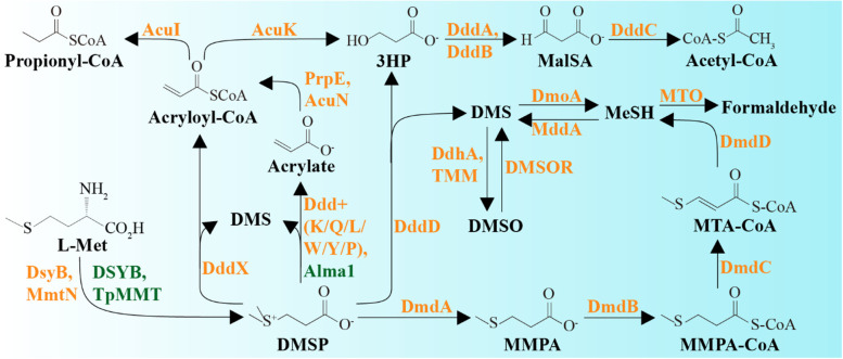
DMSP demethylation is initiated by the bacterial DmdA enzyme (EC 2.1.1.269, Fig. 1) that generates methylmercaptopropionate (MMPA) [17–19]. dmdA is widespread in marine Alphaproteobacteria, notably Rhodobacterales (also known as the Roseobacter group) and SAR11, and some Gammaproteobacteria [19]. MMPA can be further catabolised to MeSH and used as a source of carbon and/or sulfur via dmdBCD gene products that are common in marine and terrestrial bacteria [20]. DMSP demethylation is thought to dominate in marine systems accounting for ~75% of DMSP catabolism [21].
Fig. 1.
Pathways of DMSP synthesis and degradation. DMSP can be synthesised by both phytoplankton and bacterioplankton from methionine (L-Met). The SAM-dependent S-methyltransferase of the transamination pathway has been identified from phytoplankton (DSYB and TpMMT), and bacterioplankton (DsyB). Bacteria also can synthesise DMSP through a methylation pathway mediated by a L-Met-S-methylating enzyme MmtN. DMSP can be degraded through two competing pathways. The demethylation pathway involves DmdABCD and leads to the formation of methylmercaptopropionate (MMPA), methylthioacryloyl-CoA (MTA-CoA) and methanethiol (MeSH). MeSH can be oxidised to formaldehyde by MeSH oxidase (MTO). The cleavage pathway catalysed by Ddd enzymes in some bacteria, fungi and viruses, or Alma1 in algae, liberates DMS and acrylate, acryloyl-CoA or 3-hydroxypropionate (3HP). DMS can be further oxidised by marine microbes through trimethylamine monooxygenase (TMM) or dimethylsulfide dehydrogenase (DdhA) to generate dimethyl sulfoxide or by dimethylsulfide monooxgenase (DmoA) to generate MeSH. Methanethiol S-methyltransferase (MddA) and dimethyl sulfoxide reductase (DMSOR) mediate the production of DMS from MeSH and DMSO, respectively. Potentially toxic acrylate is detoxified by bacteria through several enzymes (PrpE, AcuI, AcuN and AcuK). Bacterial catabolism of 3HP involves DddABC proteins and generates malonate semi-aldehyde (MalSA) and acetyl-CoA. Enzymes from phytoplankton and bacteria are shown in green and orange, respectively
DMSP lyase enzymes cleave DMSP to generate DMS and either acrylate (EC 4.4.1.3), 3-hydroxypropionate (3HP; EC 3.1.2), or acryloyl-CoA as co-products [20, 22]. Eight DMSP lyases have been discovered in bacteria, fungi and viruses: DddD [22], DddL [23], DddQ [24], DddW [25], DddY [26], DddK [27], DddP [28] and DddX [29], and currently only Alma1 [30] in algae. Organisms using DMSP as a carbon source require ancillary (ddd, acu and prp) genes to incorporate acrylate, 3HP, or acryloyl-CoA into central metabolism (Fig. 1) [31–33]. Many bacteria, particularly the Rhodobacterales, possess both DMSP demethylation and cleavage pathways [34, 35].
Many metagenomics and metatranscriptomics studies examine the abundance and taxonomy of DMSP catabolic genes and their transcripts to infer the activity of DMSP-degrading microorganisms in marine environments [3, 19, 36]. However, such studies cannot elucidate with certainty the microbes and pathways used to catabolise DMSP for carbon requirements because many microbes, particularly Rhodobacterales with dmdA and/or ddd genes, cannot grow on DMSP as a carbon source [37] and may degrade DMSP for signalling and/or reduced sulfur requirements [12, 38]. Thus, there is a clear need to establish which microbes in marine samples use DMSP as a carbon source and what DMSP catabolic pathways they contain.
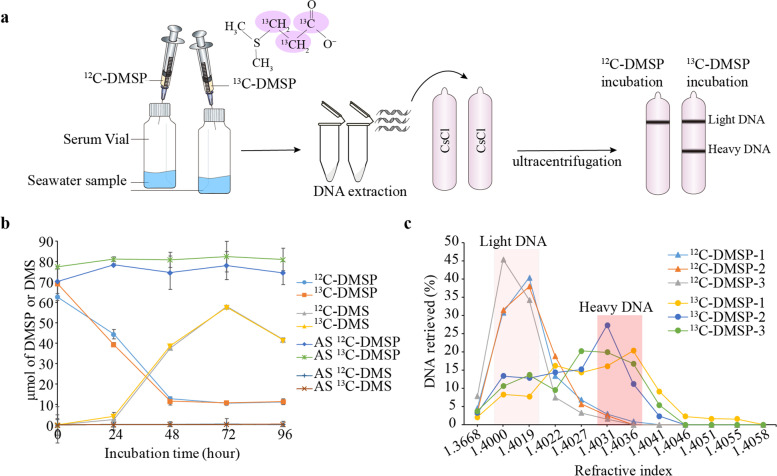
Here we use 13C-labelled DMSP (with only the propionate moiety 13C-labelled; Fig. 2a) and DNA-stable isotope probing (DNA-SIP) [39, 40] combined with metagenomics and 16S rRNA gene amplicon sequencing to identify organisms in coastal seawater that utilise DMSP (enriched in 13C-heavy fractions) and DMS (enriched in 13C-light fractions) as a carbon source (Fig. 2a). Culture-dependent methods were also used to examine bacteria using DMSP as a carbon source. Thus, this study uses DNA-SIP in a novel way to provide key insights into microorganisms utilising DMSP and DMS as carbon sources in natural seawater samples and to distinguish them from microbes that might catabolise these compounds for other uses.
Fig. 2.
DNA-SIP experiments with 13C-labelled DMSP and subsequent separation of heavy and light DNA. a Schematic diagram of SIP experiments with 13C-DMSP and 12C-DMSP (control). b DMSP and DMS levels in seawater samples incubated with 13C- and 12C-DMSP. Autoclaved seawater (AS) was used as abiotic control. Values show the average of three biological replicates. c DNA retrieved as function of refractive index of each fraction recovered after isopycnic ultracentrifugation. Samples in shaded backgrounds were used for downstream analysis. Triangles: seawater samples incubated with 12C-DMSP (control). Circles: seawater samples incubated with 13C-labelled DMSP
Results and discussion
Characterisation of DMSP cycling in coastal seawater
North Sea coastal water, sampled in Great Yarmouth, UK, in January 2018, contained 3.7 ± 0.4 nM DMSP and 0.9 ± 0.4 nM DMS. After addition of exogenous 100 μM DMSP, significant initial rates of DMSP removal (1.1 ± 0.1 μmol h−1) and DMS production (0.8 ± 0.02 μmol h−1) were detected over the first 48 h of incubation (Fig. 2b). Interestingly, no MeSH was detected in the incubation experiments, suggesting either that DMSP lysis dominated over the demethylation pathway in these samples or that the MeSH consumption rates were equal to the synthesis rates.
Identification of candidate microbes cycling DMSP in natural (T0) coastal seawater
16S rRNA gene amplicon (16S) and metagenomic (MG) sequencing was used to identify microorganisms in the natural (T0) seawater samples with the potential to cycle DMSP. Analysis of both 16S and MG data revealed that the T0 microbial community was dominated by bacteria, especially Proteobacteria (relative abundance, RA 82.6 ± 7.3% of the total 16S reads), with archaea and eukaryotes present at very low RA (<1%; Fig. S1). Although algae, thought to be the major DMSP producers in photic seawater, were not abundant, potential DMSP-producing cryptophytes Teleaulax [41] and Guillardia, and the chlorophyte Bathycoccus [42] were identified in these T0 seawater samples (Fig. S2a). Furthermore, algal DMSP synthesis genes encoding proteins 100% and 86.6% identical to Prymnesium parvum DSYB and Thalassiosira pseudonana TpMMT [43, 44] were not abundant in the T0 seawater metagenomes (Fig. S2b) and were present at similar levels to bacterial dsyB (Table S1). This is not surprising given the DMSP synthesis genes in chlorophyte or cryptophyte algae are not known. Furthermore, few algal DMSP lyase (Alma1) sequences were detected compared to bacterial DMSP lysis genes suggesting that bacteria are likely the major drivers of DMSP cycling in these seawater samples; see below (Table S1; Fig. S2b−c).
The most abundant bacteria in T0 seawater were from gammaproteobacterial (mostly Alteromonadales) and alphaproteobacterial (mostly Rhodobacterales) classes, which comprised respectively 50.2 ± 19.1% and 25.5 ± 10.9% of the 16S and 30.6 ± 13.6% and 25.1 ± 9.5% of MG data (Fig. S1 and Fig. 3). Less than 1% of the T0 microbial community was of genera predicted to produce DMSP [45, 46] and/or have DMSP synthesis genes (dsyB or mmtN), according to the 16S (Fig. S3a) and MG analysis (Fig. S3b), which was consistent with the relatively low DMSP concentrations observed in these T0 coastal waters.
Fig. 3.
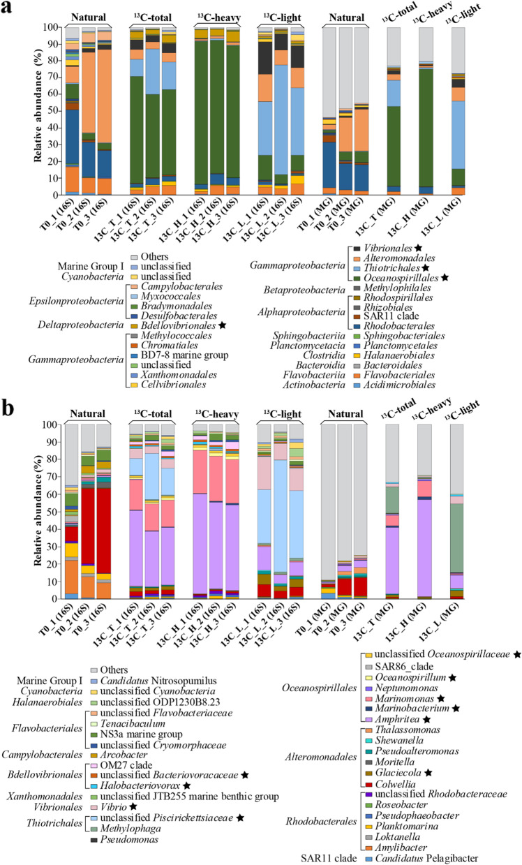
Microbial community profile of coastal seawater samples at order (a) and genus (b) levels. Bacterial diversity of the natural (T0) and labelled (heavy; H) and unlabelled (light; L) fractions of 13C-DMSP seawater incubations was analysed by 16S rRNA gene amplicon (16S) and metagenomics (MG) sequencing. “_1”, “_2” and “_3” after the sample name represent biological replicates. Biological replicates from 13C-heavy and 13C-light fractions were respectively combined before MG sequencing due to their highly similar 16S rRNA gene community profile shown by DGGE (Fig. S7). Only classes and genera with RA >0.5% in at least one of the conditions are represented. Statistically enriched genera in the incubations with 13C-labelled DMSP (13C_T) compared the natural (T0) samples based on 16S data are labelled with an asterisk. Classes and genera with RA <0.5% are grouped into “others”. For 16S and MG data of samples incubated with 12C-DMSP (controls), see Fig. S8
Regarding DMSP catabolism, Colwellia strains (RA of 33.2 ± 21.9%) from the Alteromonadales order together with the Rhodobacterales Amylibacter (13.4 ± 5.1%) and Planktomarina (5.3 ± 2.4%) were predicted to be the major DMSP degraders in these T0 seawater samples (Fig. 3, Table S2). Indeed, marine Colwellia spp. isolates have previously been suggested to be important DMSP catabolisers in polar samples [47], and like several Amylibacter spp., they contain dmdA [48]. Furthermore, many Amylibacter spp. and Planktomarina spp., abundant in other coastal samples [48], contain proteins homologous to ratified DddP DMSP lyases (Table S3). However, it was surprising that Oceanospirillales and SAR11 clade bacteria, often abundant in surface seawater [49] and known DMSP degraders via lysis and/or demethylation pathways [17, 27], only comprised 5.3 ± 3.0% and 1.9 ± 1.8% of the T0 microbial community, respectively, according to the 16S data (Fig. 3; Table S4).
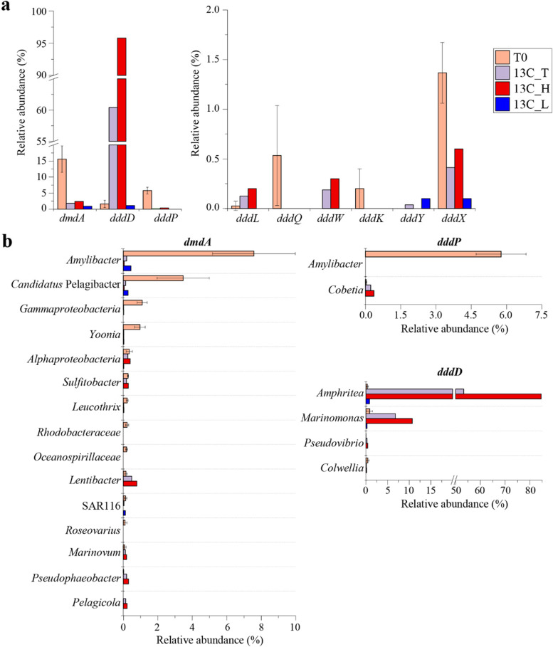
MG analysis revealed that dmdA and dddP were the most abundant DMSP catabolic genes in the T0 samples, with 15.6 ± 4.1% and 5.8 ± 1% of bacteria predicted to contain them, respectively (Fig. 4a; Table S1). The majority of dmdA sequences (48.3%) were from Rhodobacterales, mainly homologous to Amylibacter dmdA, and 21.7% were from Candidatus Pelagibacter of the SAR11 clade (Fig. 4b and Fig. S4). Nearly all detected dddP sequences in T0 metagenomes (>98.8%) were also affiliated to Amylibacter (Fig. 4b and Fig. S5). Bacteria containing dddD (1.6 ± 1.1%; mainly related to gammaproteobacterial Amphritea, Marinomonas and Colwellia strains), dddX (1.4 ± 0.3%) and dddQ (0.5 ± 0.5%) were also relatively abundant in the T0 seawater microbial community (Fig. 4a, Fig. S6 and Table S1). Other known DMSP lyases (dddL, dddW, dddK and dddY) were predicted to be in only <0.5% of T0 bacteria (Fig. 4a, Fig. S6 and Table S1). These data suggest a typical coastal system where Rhodobacterales and SAR11 bacterial DMSP demethylation dominates over DMSP lysis through DddP and, to a lesser extent, DddD, DddX and DddQ [17, 50, 51]. However, the abundance and taxonomic profile of DMSP catabolic genes cannot predict their corresponding levels of transcription, enzyme abundance and/or activity. Furthermore, it is impossible for such an ‘omics study alone to elucidate why these microbes catabolise DMSP, e.g. to provide carbon, sulfur or signalling needs. Therefore, we conducted a DNA-SIP experiment with 13C-labelled DMSP to determine which microbes use DMSP as carbon source in these seawater samples.
Fig. 4.
Relative abundance and taxonomic affiliation of DMSP cycling genes retrieved from coastal seawater metagenomes. a Relative abundance (RA) of DMSP demethylation (dmdA) and lysis (ddd) genes in seawater metagenomes. b Taxonomic affiliation of key genes involved in DMSP catabolism in seawater samples retrieved from metagenomic data. T0: metagenomes from natural samples; 13C_T: total microbial community from samples incubated with 13C-DMSP; 13C_H: metagenomes from 13C-heavy fractions; 13C_L: metagenomes from 13C-light fractions. T0 values represent the average of three biological replicates. Biological replicates from 13C-heavy and 13C-light fractions were respectively combined prior to metagenomic sequencing (see the “Methods” section)
Identification of microorganisms degrading DMSP for use as a carbon source by DNA-SIP
Known DMSP demethylation and lysis pathways in model organisms that use DMSP as a carbon source release MeSH and DMS, respectively, as gaseous products and utilise the propionate component for carbon (Fig. 1). Thus, to identify microbes using DMSP as a carbon source, we synthesised DMSP from 13C3-acrylic acid and 12C-DMS (see the “Methods” section), the product hereafter being referred to as 13C-DMSP, which was used in our DNA-SIP experiments with 12C-DMSP as a control (Fig. 2a). This strategy was chosen to also enable identification of microorganisms utilising the DMS component of DMSP, i.e. those microbes enriched in incubations with DMSP but not necessarily being represented in the heavy fraction of 13C-DMSP incubations.
The T0 seawater samples collected above were incubated with 13C-DMSP or 12C-DMSP (control) as substrate (100 μM) under 12-h light/dark cycling conditions to consider both phototrophic and heterotrophic DMSP catabolism. No MeSH production was detected in any of the incubations. DMSP degradation, DMS production and subsequent DMS removal processes were similar between the samples incubated with 13C-DMSP and 12C-DMSP (Fig. 2b). DNA was extracted after 96 h when 77 μmol DMSP L−1 (231 μmol C L−1) was assimilated and DMS levels were decreasing (by 0.7 ± 0.006 μmol h−1 during the last 24 h of incubation), suggesting that DMS degraders were also active at this timepoint. DNA was then separated into heavy (13C-labelled) and light (12C-labelled) fractions by isopycnic centrifugation (see the “Methods” section; Fig. 2c), which were analysed by denaturing gradient gel electrophoresis (DGGE). This technique showed clear differences in the 16S rRNA gene profiles from 13C-heavy and 13C-light fractions, whereas fractions from 12C-DMSP (control) incubations had similar profiles (Fig. S7), indicating that DNA from microorganisms using 13C-DMSP for carbon had been successfully labelled.
Subsequently, the heavy and light DNA fractions from 13C-DMSP and 12C-DMSP (control) incubations were subjected to MG and 16S sequencing to identify those microorganisms that used DMSP as carbon source. Sequence analysis revealed that Gammaproteobacteria dominated in both the heavy (13C_H) and light (13C_L) fractions from 13C-DMSP incubations (Fig. 3a), but there were major differences at lower taxonomic levels (i.e. order and genus; Fig. 3b). Oceanospirillales, with a RA of 70.2% in MG and 81.2 ± 3.5% in 16S data, was the most abundant order in the 13C-heavy fractions (13C_H), but only represented <10% of the bacterial community from 13C-light fractions (13C_L; Fig. 3b, Table S4). Importantly, there was no such increase in Oceanospirillales RA between heavy (12C_H) and light fractions (12C_L) when the T0 samples were incubated with control 12C-DMSP (Fig. S8a), indicating that these Gammaproteobacteria were the major microorganisms assimilating carbon from the propionate moiety of DMSP in the DNA-SIP experiments.
At the genus level, Amphritea dominated the heavy fractions from the 13C-labelled microbial community (13C_H), comprising 55.9% and 52 ± 4.3% of the MG and 16S reads, respectively, followed by Marinomonas (9.1% RA in MG and 25.2 ± 0.5% in 16S data; Fig. 3b, Table S2). Other less abundant Oceanospirillales genera significantly enriched (P <0.05) in the 13C-heavy fractions compared to the T0 samples, were Marinobacterium (to 1.3% RA in MG and 0.6 ± 0.1% in 16S data) and Oceanospirillum (to 0.5% RA in MG and 1.8 ± 0.1% in 16S; Table S2). These four Oceanospirillales genera were 6- to 60-fold more abundant (P <0.05) in 13C-heavy (13C_H) than in the 13C-light (13C_L) fractions and constituted 0.4−36% of the total microbial community of the samples incubated with 13C-DMSP (13C_T, 16S data; Fig. 3b, Table S2). As expected, these genera were also highly abundant in both the 12C-light and 12C-heavy fractions (12C_L and 12C_H; Fig. S8b), further supporting them being key bacteria that used DMSP as a carbon source in the DMSP incubations.
In addition, two genera of the Bdellovibrionales order, Halobacteriovorax and an unclassified Bacteriovoracaceae, were also enriched during the 13C-DMSP incubations, with their RA increasing to 0.7 ± 0.2% and 1 ± 0.1% of 16S reads in the 13C_T samples from <0.1% at T0 (Fig. 3b, Table S2). These Bdellovibrionales bacteria were ~10-fold more abundant in the heavy (13C_H) than in the light (13C_L) fractions from incubations with 13C-DMSP (Table S2). No sequenced Halobacteriovorax strains contain known DMSP catabolic genes, so if they are true DMSP degraders they may utilise novel pathways and/or enzymes.
As only one DNA-SIP timepoint was analysed here, it is possible that some microorganisms enriched in 13C-heavy fractions were labelled due to cross-feeding, i.e. they assimilated 13C-DMSP catabolites released from primary degraders. This is unlikely for the Oceanospirillales given their dominance in the 13C_H fractions and that several previously studied Oceanospirillales strains, e.g. Marinomonas [22], Oceanimonas [52] and Halomonas [31], and bacterial isolates from this work, used DMSP as a sole carbon source and contained DddD (Table 1 and see below). However, it is plausible that the predatory Bdellovibrionales may have consumed primary DMSP degraders and become labelled due to cross-feeding [53].
Table 1.
Characteristics of DMSP-degrading bacterial strains isolated from seawater samples incubated with DMSP
| Strain | Top-hit taxon identified by 16S rRNA gene sequences | Reference straina | Accession number of genomes from sequenced isolates | Homologues in reference strain or genome from sequenced isolateb | DMS production ratec | MeSH productiond | Growth on DMSPe | Class |
|---|---|---|---|---|---|---|---|---|
| GY12 | Litoreibacter albidus | DSM 26922 | dddA, dddC, prpE, acuI, acuK, dmdA, dmdB, dmdC | ND | Y | N | Alphaproteobacteria | |
| MB12-2 | Neptunicoccus sediminis | CY02 | dddP, prpE, acuI, acuK, dmdA, dmdB, dmdC | 6 ± 2 | ND | N | Alphaproteobacteria | |
| GY7 | Pseudophaeobacter arcticus | DSM 23566 | dddW, dddA, dddC, prpE, acuI, acuN, acuK, dmdA, dmdB, dmdC, dmdD | 558 ± 260 | ND | N | Alphaproteobacteria | |
| MB12-4 | Sulfitobacter pontiacus | DSM 10014 | dddL, dmdB, dmdC, dmdD | 648 ± 153 | ND | N | Alphaproteobacteria | |
| GY16 | Sulfitobacter pseudonitzschiae | H3 | dddL, dddA, dddC, prpE, acuI, acuK, dmdB, dmdC, dmdD | 201 ± 40 | ND | N | Alphaproteobacteria | |
| D12-10 | Alteromonas stellipolaris | LMG 21861 | dmdC | 60 ± 6 | ND | Y | Gammaproteobacteria | |
| GY8 | Marinobacter sediminum | JAGTWY000000000 | dddL, dddA, dddC, prpE, dmdB, dmdC | 4560 ± 785 | ND | Y | Gammaproteobacteria | |
| MC12-9 | Marinobacter similis | A3d10 | dddL, dddA, prpE, acuN, acuK, dmdC | 86 ± 3 | ND | Y | Gammaproteobacteria | |
| GY20 | Pseudoalteromonas hodoensis | H7 | ND | Y | N | Gammaproteobacteria | ||
| D13-2 | Cobetia amphilecti | KMM 296 | dddD, dddA, dddC, dddT, prpE, acuI, acuK, dmdC, tmm | 1342 ± 101 | ND | N | Gammaproteobacteria | |
| MC13-5 | Cobetia litoralis | CP073342 | dddD, dddA, dddC, dddT, prpE, acuI, dmdC, tmm | 1316 ± 94 | ND | Y | Gammaproteobacteria | |
| GY6 | Amphritea atlantica | CP073344, CP073345 | dddD, prpE (2), acuI, acuK, dddB, dddC, dddT, dmdB (4), dmdC, tmm | 1173 ± 208 | ND | Y | Gammaproteobacteria | |
| D13-1 | Marinobacterium rhizophilum | CP073347 | dddD, dddP, prpE (2), acuI, acuN, acuK, dddA, dddB, dddC, dddT, dmdB (2), dmdC | 1039 ± 21 | ND | Y | Gammaproteobacteria | |
| MC13-7 | Marinobacterium profundum | PAMC 27536 | dddD, dddP, dddA, dddB, dddC, dddT, prpE (2), acuI, dmdB, dmdC | 1132 ± 31 | ND | Y | Gammaproteobacteria | |
| GY1 | Marinomonas atlantica | Cmf 18.22 | dddD, dddB, dddC, prpE, acuI, dmdC | 59 ± 30 | ND | Y | Gammaproteobacteria | |
| MB12-3 | Marinomonas foliarum | CECT 7731 | dddC, acuI, dmdC | 11 ± 0.4 | ND | N | Gammaproteobacteria | |
| MB12-11 | Marinomonas rhizomae | CP073343 | dddD, dddB, dddC, dddT, acuI, dmdC | 500 ± 36 | ND | Y | Gammaproteobacteria | |
| D13-4 | Pseudomonas benzenivorans | CP073346 | dddD, dddP, acuK, dddB, dddC, dddT, dmdB, dmdC | 2462 ± 123 | ND | Y | Gammaproteobacteria | |
| GY22 | Pseudomonas leptonychotis | CCM 8849 | dddD, dddB, dddC, dddT, dmdB, dmdC | 2350 ± 343 | ND | Y | Gammaproteobacteria | |
| GY17 | Pseudomonas taeanensis | MS-3 | dddD, dddP, dddB, dddC, dddT, acuI, dmdB, dmdC, tmm | 829 ± 186 | ND | Y | Gammaproteobacteria | |
| GY15 | Vibrio splendidus | 10N.286.45 | prpE, acuI, DMSOR | ND | Y | Y | Gammaproteobacteria |
aReference strain: most closely related strain with publicly available genome
bNumber of prpE and dmdB genes in genomes with multiple copies are indicated in brackets
cRate of DMSP-dependent DMS production expressed in nmol DMS mg protein−1 h−1
dY, detectable MeSH production from DMSP. ND, not detected
eY, growth on DMSP as sole carbon source (Student’s t-test, P < 0.05); N, no growth on DMSP as sole carbon source (P > 0.05)
Interestingly, Gammaproteobacteria, including members of the orders Alteromonadales (Glaciecola), Vibrionales (Vibrio) and, most notably, Thiotrichales (unclassified Piscirickettsiaceae), were also enriched during the incubations with 13C-labelled DMSP (RA 0.003−0.2% in T0 to 1.8−17.3% in 13C_T samples according to 16S data; Table S2). These Gammaproteobacteria were more abundant in the 13C-light than in the 13C-heavy fractions (RA >15-fold higher in 13C_L than in 13C_H fractions; Fig. 3b, Table S2), suggesting that they likely assimilated the 12C-DMS generated from the lysis of 13C-DMSP during the incubations with 13C-DMSP (see below).
Strikingly, Rhodobacterales and SAR11 bacteria were >6-fold less abundant after the 13C-DMSP incubations (13C_T) compared to the T0 samples (16S data, Fig. 3a, Table S4). Although some Rhodobacterales 16S rRNA genes, notably from Pseudophaeobacter and Planktomarina, were mainly present in the 13C-heavy fractions consistent with them using DMSP as a carbon source, these bacteria comprised only 1% of the 13C-labelled bacterial community (13C_H; Fig. 3b, Table S2). Furthermore, SAR11 bacteria represented 0.3% of the total microbial community from 13C-DMSP incubations (13C_T) and were >10-fold less abundant in the heavy (13C_H) than light (13C_L) fractions (Fig. 3b, Table S4). These data imply that Rhodobacterales and SAR11 bacteria, which mostly possess dmdA [15, 17] and at least one ddd gene [37, 54], were not the major users of DMSP as a carbon source under these incubation conditions. It is possible that these bacteria, predicted to be key DMSP degraders in coastal waters [50, 55], were using DMSP at the low T0 nM concentration for sulfur demands [56], and/or to generate DMS and/or acrylate as signalling molecules [12] and not primarily as a carbon source. Indeed, most Rhodobacterales with dmdA/ddd genes do not typically grow well on DMSP as sole carbon source under lab conditions [37], in comparison to Oceanospirillales bacteria with dddD (see below) [37] or Alcaligenes faecalis with dddY [26].
Abundance and taxonomy of DMSP catabolic genes in seawater samples incubated with 13C-DMSP
After incubation with 13C-DMSP, DMSP demethylation was no longer predicted to be the dominant DMSP catabolic pathway it was in the T0 seawater samples. Metagenomic analysis showed that the RA of dmdA in T0 seawater metagenomes (15.6 ± 4.1% of bacteria) decreased 10-fold to 1.8% after incubation with 13C-DMSP (13C_T; Fig. 4a and Table S1). Of the dmdA sequences in these samples, 73% originated from the 13C-heavy fractions and were mainly closely related to DmdA from Rhodobacterales, e.g. Lentibacter (RA 0.8%; Fig. 4b). These data indicate that some bacteria may have demethylated DMSP for carbon assimilation during the incubations with 13C-DMSP, but that this process was far less significant than in the natural (T0) seawater samples. In support of this, the RA of the ancillary dmdCD genes (Fig. 1) also decreased during the incubations with 13C-DMSP (13C_T) compared to the T0 samples (Fig. S9). Conversely, dmdB sequences showed a higher RA in the 13C_T than in the natural (T0) metagenomes and were mainly affiliated to Amphritea bacteria (Fig. S9), which were highly enriched in the 13C-heavy fractions (13C_H; Fig 3b), have multiple copies of dmdB but not dmdA and are unlikely to demethylate DMSP (Table 1).
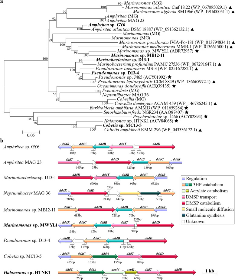
In contrast to dmdA, the RA of ddd genes increased to ~60% after the incubation with 13C-DMSP (13C_T samples), most of which (97%) were retrieved from the 13C-heavy fraction metagenomes (13C_H; Fig. 4a). Among the ddd genes, dddD was particularly enriched in the 13C-heavy fraction, with 95.8% of bacteria predicted to contain it (Fig. 4a and Table S1). Most of the dddD sequences were closely related to the Oceanispirillales genera Amphritea (84.5%) and Marinomonas (10.7%; Figs. 4b and 5a), supporting the 16S and MG data, which showed that these were the dominant genera in the 13C-heavy fraction (13C_H; Fig. 3, Table S2). dddD genes likely from Colwellia and Pseudovibrio were also retrieved from the 13C-heavy metagenomes, although with lower RA (<0.5%; Figs. 4b and 5a). Therefore, the DNA-SIP experiment with 13C-labelled DMSP clearly showed that Oceanospirillales were the key bacteria degrading DMSP for carbon via a DddD-mediated lysis pathway in these coastal seawater samples. This is not surprising since it has been previously reported that many Oceanospirillales bacteria with DddD, an enzyme with a high affinity for DMSP [57], can use DMSP as sole carbon source for growth [22, 31, 50].
Fig. 5.
Maximum likelihood phylogenetic tree of DddD proteins (a) and DMSP-catabolising gene clusters containing dddD from bacterial isolates and metagenome-assembled genomes (MAGs) retrieved from this study (b). a The tree shows DddD proteins from strains isolated in this study (in bold), previously ratified proteins (★), together with sequences retrieved from seawater metagenomes (MG), metagenome-assembled genomes (MAG), and reference strains (▲). Reference strains were selected as the most closely related strains to isolates from this study having publicly available genome sequences. Bootstrap values ≥50% (based on 100 replicates) are shown. Scale bar indicates 5% estimated phylogenetic divergence. b Gene function is indicated by a colour code detailed in legend. Bacteria in bold are those in which the gene clusters have been experimentally ratified
Four other DMSP lyase genes (dddP, dddL, dddW, dddX) were also detected in the metagenomes from the 13C-heavy fraction, but were much less abundant than dddD (RA of 0.2−0.6%; Fig. 4 and Table S1). These DMSP lyase genes were taxonomically affiliated to the Oceanospirillales genus Marinobacterium (dddX), and Rhodobacteraceae genera Sulfitobacter (dddL), Phaeobacter and Pseudophaeobacter (dddW; Fig. S6). Interestingly, the dddP sequences retrieved from the 13C-heavy fraction (13C_H) were closely related to Cobetia (Oceanospirillales) dddP, but no dddP genes from Rhodobacteraceae bacteria, including Amylibacter, whose dddP genes represented >98.8% of the dddP sequences in the T0 metagenomes, were detected (Fig. 4b and Fig. S5). These data imply that bacteria containing dddL, dddW, dddX and, notably, dddP, the most abundant DMSP lyase gene in marine environments [43], play a far less significant role in the assimilation of carbon from DMSP compared to the Oceanospirillales harbouring dddD, but may have important other roles in, e.g. generating DMS or acrylate as info-chemicals [12].
A complete suite of ancillary genes involved in the downstream catabolism of DMSP (prpE, acuI, dddB, dddC and dddT) [31–33] was enriched in the samples incubated with 13C-DMSP (13C_T) compared to the T0 samples (Fig. S10 and Table S1). These genes mostly originated from the 13C-heavy fractions (13C_H) and were affiliated to the Oceanospirillaceae genera Amphritea and Marinomonas (Fig. S10). Interestingly, dddA, whose product catalyses the same reaction as DddB in Halomonas [31], was present at a much lower RA in the 13C-DMSP incubations (13C_T) compared to the natural (T0) seawater samples, and it was much less abundant in the 13C-heavy fractions (13C_H) than dddB (4.6% vs 90.9%; Table S1). Thus, DddB likely represents the major route of malonate semialdehyde formation from DMSP in the seawater samples amended with 13C-DMSP (Fig. 1). Similarly, MG analysis revealed that Oceanospirillaceae bacteria assimilating carbon from 13C-DMSP in our incubations most likely metabolise acrylate via the PrpE-AcuI enzymes, rather than through AcuN-AcuK (Fig. 1). Although these four genes were mostly present in the 13C-heavy fractions (13C_H), the RA of acuN and acuK in the total microbial community (13C_T) from the 13C-DMSP incubations (1.8% and 21.9%, respectively) was lower than in the T0 samples (8.3 ± 1.7% and 34 ± 5.8%; Fig. S10 and Table S1). The opposite trend was observed with prpE and acuI, which had a 4- and 2-fold higher RA in the incubations with 13C-DMSP (13C_T) compared to the natural (T0) samples (Fig. S10 and Table S1).
Insights into DMSP cycling from metagenome-assembled genomes (MAGs)
To gain further insights into the microbial sulfur cycling pathways in this coastal environment, metagenome-assembled genomes (MAGs) were reconstructed and analysed. MAGs were screened for the presence of DMSP degradation genes and their RA in each sample calculated as described in Methods. dmdA, the dominant DMSP catabolic gene in T0 samples, was found in the most abundant MAG (5.1 ± 2.1% RA) from the T0 metagenomes (MAG 55, Alphaproteobacteria; Table S5) and also in several less abundant (RA <0.5% each) gammaproteobacterial (MAGs 11, 58 and 60) and Rhodobacterales MAGs (MAGs 3, 7, 18, 44, 68, 81; Fig. S4 and Table S5). MAG 44, taxonomically classified as Loktanella, also harboured a sequence encoding a DddW-like protein with 54% amino acid identity to R. pomeroyi DddW, which did not cluster with ratified DddW sequences (Fig. S6). Most of the dmdA-containing MAGs (except for MAGs 55, 58 and 68) also had dddP and were mainly classified as Rhodobacterales (Table S5). MAGs 55, 58 and 68 were only predicted to be 55.2 to 75.1% complete (Table S5), which might account for why no dddP homologues were detected. The Rhodobacterales MAGs whose taxonomy could be resolved at lower levels, belonged to the genera Loktanella, Lentibacter and Amylibacter (Table S5), which is consistent with these bacteria often containing both DMSP demethylation and lyase genes (Table S3). dddP was also found in a Planktomarina temperata MAG (MAG 73) that comprised 2.8 ± 1.9% of the T0 microbial community, and two gammaproteobacterial MAGs (<0.3% RA; Table S5 and Fig. S5). Most of the MAGs containing dmdA and/or dddP genes were not enriched after the 13C-DMSP incubations (13C_T) compared to the T0 samples except for the Rhodobacterales genera Lentibacter (MAG 81) and Pseudophaeobacter (MAG 68) and gammaproteobacterial Neptuniibacter MAGs (MAG 36, also containing dddD), which were mainly present in the 13C-heavy fraction (>17-fold higher RA than in 13C-light fraction; Table S5). These data further support the hypothesis that most Rhodobacterales and Gammaproteobacteria with DmdA and/or DddP are important DMSP degraders in coastal waters but not primarily for carbon assimilation.
Consistent with the 16S data, the RA of an Amphritea MAG harbouring dddD (MAG 23) dramatically increased in the samples incubated with 13C-DMSP (from 0.7 ± 0.5% in T0 to 35.3% in 13C_T samples) and was 12-fold more enriched in the 13C-heavy (13C_H) than 13C-light fractions (13C_L; Table S5). Prior to this study, Amphritea had not been shown to catabolise DMSP, perhaps because many Amphritea spp. lack dddD or other known DMSP-lyase genes (Table S3).
Another two Oceanospirillales MAGs (MAGs 30 and 85) enriched in the 13C-heavy fraction (13C_H) after incubation with 13C-DMSP (26-fold and 3-fold more abundant than in the 13C-light fraction 13C_L, respectively) were detected (Table S5). However, neither of these MAGs were assigned to Marinomonas, shown to be abundant in the 16S analysis described above, nor did they contain a known DMSP catabolic gene, again potentially due to their low completeness (61.4% for MAG 30 and 51.7% for MAG 85; Table S5).
As described above, Halobacteriovorax bacteria were enriched during incubation with13C-DMSP and one Halobacteriovorax MAG (MAG 43) was resolved from the metagenomic data. However, MAG 43 was more abundant in the 13C-light (13C_L) than in the 13C-heavy (13C_H) fractions after incubation and it lacked known DMSP catabolic genes (84.5% completeness; Table S5). Further studies are needed to evaluate if and how marine Halobacteriovorax catabolise DMSP.
Characterisation of bacterial strains isolated from seawater samples after incubation with DMSP
Sixty-six bacterial strains with distinct morphologies were isolated after incubations of T0 seawater with DMSP, 67% of which were able to catabolise DMSP yielding DMS or MeSH. All the DMSP-degrading isolates were either Alpha- (27%) or Gammaproteobacteria (73%; Table S6). Twenty-one representative strains of the different colony types were selected (Table 1), and publicly available genomes of closely related strains (herein described as reference strains) were analysed for the presence of dmd/ddd genes. In addition, we sequenced the genome of six isolates with high DMSP-dependent DMS production rates, but where no closely related reference strains were available, or where these did not contain dmd/ddd homologues (Table S6).
All DMSP-degrading alphaproteobacterial isolates were Rhodobacteraceae, well known for their ability to catabolise DMSP [23, 58]. The Sulfitobacter, Neptunicoccus and Pseudophaeobacter isolates, whose corresponding reference strains contained dddP, dddL and/or dddW DMSP lyase genes, released DMS from DMSP, with production rates ranging from 6 to 1411 nmol DMS·mg protein−1·h−1 (Table 1 and Table S6). Most Rhodobacterales, including all reference strains analysed here except for Sulfitobacter, contained dmdA and were predicted to demethylate DMSP [17]. However, only one isolate, Litoreibacter sp. GY12, with no DMSP lyase activity, showed MeSH production from DMSP. In addition, of the twelve Rhodobacterales isolates, only two strains MB13-6 and MC12-6 grew on DMSP as sole carbon source (Table S6). Thus, these data further support the hypothesis that most Rhodobacterales do not catabolise DMSP for carbon assimilation but likely use it as a sulfur source, in oxidative stress protection [8] or for signalling processes [12].
Of the DMSP-degrading gammaproteobacterial isolates, 43% were Oceanospirillales from the genera Cobetia, Amphritea, Marinobacterium and Marinomonas, all of which had relatively high DMSP lyase activity (Table S6). Indeed, Amphritea and Marinomonas were identified by the DNA-SIP experiments above, as being the two most dominant degraders of DMSP for their carbon demands (Fig. 3b and Table S2). Furthermore, 63% of the Oceanospirillales isolates grew on DMSP as sole carbon source (Table S6), with their reference strains mostly containing dddD genes (Table 1, Table S6 and Fig. 5). These data support the major finding from the DNA-SIP work that Oceanospirillales bacteria were the key DMSP degraders for carbon assimilation in these coastal waters.
Other gammaproteobacterial strains from the Alteromonadales (Alteromonas, Marinobacter and Pseudoalteromonas), Pseudomonadales (Pseudomonas) and Vibrionales (Vibrio) orders were also isolated from the seawater samples incubated with DMSP (Table S6). Alteromonas and Marinobacter strains had DMSP lyase activity and could use DMSP as sole carbon source, whereas Pseudoalteromonas sp. GY20 generated MeSH from DMSP but could not use DMSP as a carbon source. Although the genomes of the Marinobacter isolate and its reference strain harboured dddL, no DMSP catabolic genes were found in any genomes of the Alteromonas or Pseudoalteromonas reference strains (Table 1). All six Pseudomonas spp. isolates had DMSP lyase activity and grew on DMSP as sole carbon source (consistent with previous work [59]), except for isolate MC12-18 (Table S6). In addition, all the genomes of the Pseudomonas reference strains and isolates analysed contained dddD (Table 1). Finally, Vibrio sp. GY15 produced MeSH from DMSP and could use the latter as sole carbon source (Table 1). To our knowledge, no Vibrio spp. have been shown to contain dmdA. Thus, it will be interesting to further investigate the DMSP catabolic mechanisms in the Gammaproteobacteria that cleave and/or demethylate DMSP but lack the known ddd and dmdA genes in their genomes. Given the low RA of Alteromonadales, Pseudomonadales and Vibrionales bacteria (<1.3% in 16S and <1.1% in MG data; Fig. 3, Table S4) in the 13C-heavy fractions (13C_H) after incubation with 13C-DMSP, they are unlikely to be as important catabolisers of DMSP for carbon assimilation as the Oceanospirillales in the coastal samples studied here, but they may be in other environments.
The dddD genes in the genomes and MAGs from Oceanospirillaceae bacteria (Amphritea, Marinobacterium, Marinomonas and Neptuniibacter) and Pseudomonadales (Pseudomonas) were always linked to ancillary genes involved in downstream DMSP catabolism (Figs. 5b and 6), showing similar gene synteny (dddBCDRT) to dddD from Marinomonas sp. MWYL1, which assimilates carbon from DMSP for growth [22]. This was also the case for Halomonadaceae bacteria with DddD, including Halomonas sp. HTNK1 and Cobetia sp. MC13-5 but with dddA replacing dddB [31]. Such linkage of DMSP lyase genes to their ancillary metabolic and transport genes, and their coordinated gene expression, is likely important in allowing bacteria harbouring them to utilise DMSP as carbon source.
Fig. 6.
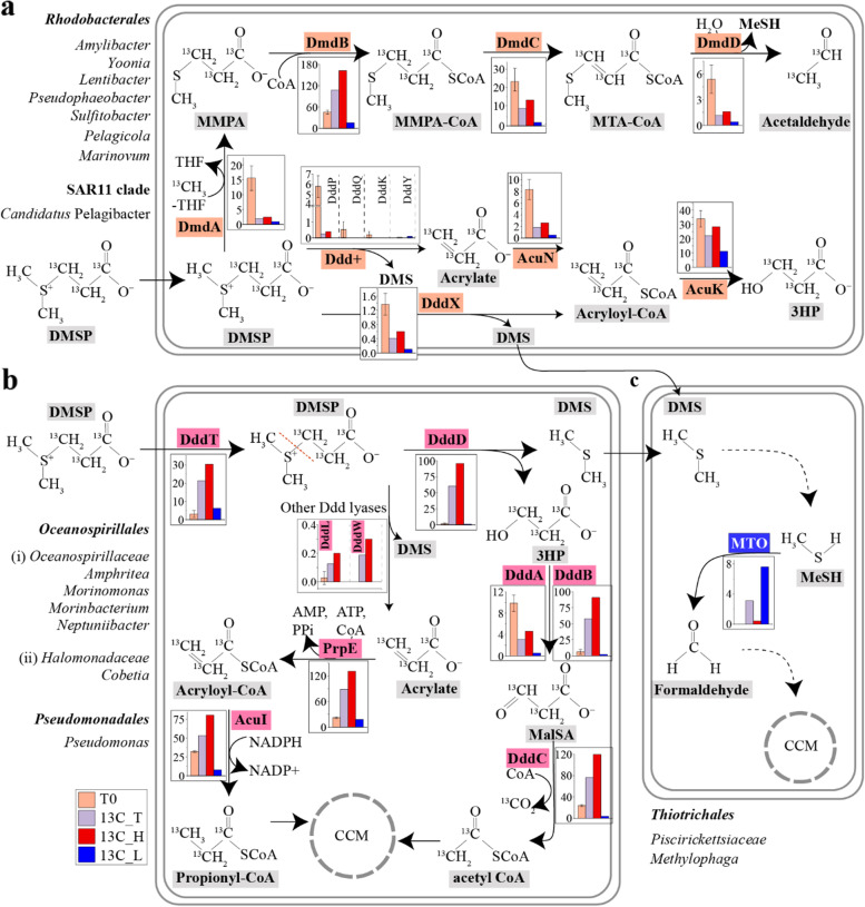
DMSP degradation pathways in microorganisms from coastal seawater samples. a Although Rhodobacterales (Roseobacter group) and SAR11 dominated the bacterial community of the natural (T0) seawater samples and their DMSP demethylation and cleavage genes (dmdA and dddP mainly) were relatively abundant in the T0 metagenomes (orange bars), the majority of the Roseobacter isolates were not able to grow on DMSP as sole carbon source. Thus, Rhodobacterales and SAR11 are predicted to use DMSP predominantly as a source of reduced sulfur and/or signalling in this coastal seawater. b Oceanospirillales were the major bacteria degrading DMSP for carbon requirements in the seawater incubations with 13C-DMSP. dddD from Oceanospirillales was the most abundant DMSP lyase gene in heavy fractions from samples incubated with 13C-DMSP (13C_H; red bars), although other DMSP lyase genes from Roseobacters, i.e. dddL and dddX were also present. Genes involved in the downstream catabolism of 3HP (dddBC in Oceanospirillaceae and dddAC in the Halomonadaceae) and acrylate (prpE and acuI) were also enriched in the 13C_H metagenomes compared to those from T0 samples. c DNA-SIP experiments showed that Methylophaga, a genus of the Piscirickettsiaceae family, and its gene encoding methanethiol oxidase (MTO) were highly abundant in the metagenomes from the 13C-light fraction (13C_L; blue bars), indicating that these bacteria were the major degraders of the DMS generated from the lysis of DMSP by Oceanospirillales. Bar charts represent the relative abundance of key genes involved in DMSP catabolism in metagenomes from natural (T0) seawater samples, 13C-DMSP incubations (13C_T) and heavy (labelled, 13C_H) and light (unlabelled, 13C_L) fractions from incubations with 13C- DMSP. T0 data show the average of three biological replicates, whereas replicates from 13C-heavy and 13C-light fractions were pooled prior to metagenomics analysis (see the “Methods” section). MMPA, methylmercaptopropionate; MTA-CoA, methylthioacryloyl-CoA; 3HP, 3-hydroxypropionate; MalSA, malonate semi-aldehyde; MeSH, methanethiol; CCM, central carbon metabolism
The role of other bacterial groups in DMS and MeSH cycling
The DNA-SIP strategy used in this study potentially allowed the identification of microorganisms assimilating the 12C-DMS component of 13C-DMSP, which should be preferentially represented in the 13C-light fractions (13C_L) from the incubations with 13C-labelled DMSP.
Sequencing data revealed that the RA of Thiotrichales from the 13C-DMSP incubations (13C_T) increased both in the 16S (from 0.5 ± 0.3% to 17.8 ± 8.6%) and MG (from 0.4 ± 0.1% to 15.6%) analyses compared to the T0 samples (Fig. 3a, Table S4). Furthermore, sequences from this order were >58-fold more abundant in the 13C-light (13C_L) than in the 13C-heavy (13C_H) fractions (Fig. 3a, Table S4). Such differences were not observed between light (12C_L) and heavy (12C_H) fractions of control samples incubated with 12C-DMSP (Fig. S8a). Analysis of 16S data showed that these Thiotrichales bacteria comprised Methylophaga and other unclassified genera from the Piscirickettsiaceae family which, based on the higher phylogenetic resolution of the MG analysis, may also be Methylophaga spp. (Fig. 3b). Similarly, the RA of gammaproteobacterial Vibrio and Glaciecola strains were respectively 5- and 9-fold more abundant after the incubations with 13C-DMSP (13C_T) than in the natural (T0) seawater samples and were mainly present in the 13C-light fraction (137- and 15-fold higher RA than in the 13C-heavy fraction; Fig. 3b, Table S2). These data imply that these Thiotrichales and Gammaproteobacteria can use the 12C-DMS generated from the lysis of the 13C-DMSP by Oceanospirillales bacteria with DddD for carbon assimilation. This is further supported by previous studies that have shown that several Methylophaga, Vibrio and Glaciecola strains catabolise DMS [60–63]. Indeed, the marine methylotroph M. thiooxydans, which was isolated from a DMS enrichment experiment and can use this compound as sole carbon and energy source for growth [64], is considered a model microorganism to study DMS degradation.
To investigate the underlying genetic mechanisms of DMS cycling, the MG data were interrogated with ratified genes involved in DMS metabolism (Fig. 1). Of these, tmm, whose product can generate dimethyl sulfoxide (DMSO) from DMS [65], was the most abundant DMS cycling gene in the T0 seawater samples (9.9 ± 1.7% RA) followed by DMSOR (5.3 ± 3.4% RA; Fig. S11a and Table S1), which encodes a DMSO reductase enzyme that catalyses the reverse reaction ([64], Fig. 1). Other genes involved in DMS metabolism such as mddA [66], ddhA [67] and dmoA [68] were present in <0.7% of the bacteria from the natural T0 samples (Fig. S11a and Table S1), suggesting that they have less important roles in this coastal seawater. mtoX, encoding MeSH oxidase [69], was the only known gene involved in DMS metabolism that was enriched in the incubations with 13C-DMSP (13C_T; 3.1% RA) compared to T0 samples (0.003 ± 0.006% RA), with the majority of sequences being from the 13C-light fractions (19-fold higher RA in 13C_L than 13C_H DNA; Table S1). As expected, all the mtoX sequences retrieved from the 13C-light fractions were closely related to DMS-catabolising Methylophaga spp (Fig. S11b). In addition, a Piscirickettsiaceae MAG (MAG 21) containing an mtoX sequence (Table S5) was more abundant in the 13C-DMSP samples (13C_T; RA 1.5%) compared to the natural (T0) seawater (RA 0.01 ± 0.004%) and was 34-fold more enriched in the 13C-light (13C_L) than in the 13C-heavy fraction (13C_H; Table S5), further supporting the notion that members of this family are key bacteria cycling DMS in this coastal seawater. These data are also consistent with previous work showing that mtoX, whose transcription and protein expression is upregulated by growth with DMS in Methylophaga thiooxydans, is the only known reporter gene for carbon assimilation from DMS in the Thiotrichales [70]. In M. thiooxydans and likely other microorganisms that use DMS as carbon source, DMS is proposed to be initially demethylated to MeSH [67]. However, the dmoA gene, whose product converts DMS into MeSH, was not enriched in the 13C-DMSP incubations (13C_T) compared to T0 samples nor was it present in the Piscirickettsiaceae MAG or many of the Methylophaga available genomes (Table S1 and Table S5). Therefore, it is possible that the initial generation of MeSH from DMS in Methylophaga might be catalysed by a novel enzyme and that it rapidly enters central metabolism via a reaction mediated by MTO (MeSH oxidase) [68].
Conclusions
This study represents the first attempt to identify, in tandem, distinct microorganisms degrading DMSP and/or DMS for carbon assimilation using DNA-SIP with 13C-labelled DMSP.
Given that Rhodobacterales and to a lesser extent SAR11 with dmdA and dddP genes were highly abundant in the coastal seawater studied here, and that representative strains of these bacteria can grow on DMSP as sole carbon source [28, 71], one would predict that they would play a major role in degrading DMSP for their carbon demands. However, DNA-SIP experiments showed that the less abundant Oceanospirillales bacteria in the natural (T0) samples were likely the key degraders of DMSP for carbon assimilation via DddD-mediated DMSP lysis. Oceanospirillales bacteria, e.g. the novel and dominant DMSP degraders in the DNA-SIP experiments, Amphritea and Marinomonas, were predicted to cleave DMSP into DMS and 3HP via their DddD enzyme, with the 3HP being assimilated and the DMS released for the benefit of a different suite of microorganisms (Fig. 6). Sequencing analysis also revealed that Rhodobacterales and SAR11 and their dmdA and dddP genes, though abundant in most surface waters, were scarcely present after incubation with 13C-DMSP, suggesting that these important bacteria likely utilise DMSP demethylation and DddP-mediated DMSP lysis either for their reduced sulfur requirements [56], to protect against oxidative stress [8] or to generate signalling molecules [12, 72]. These conclusions from SIP experiments with 13C-labelled DMSP were supported by culture-dependent work since (i) most Oceanospirillales isolates, e.g. Amphritea, yielded DMS from DMSP; (ii) these bacteria could use DMSP as a carbon source and in nearly all cases possessed DddD; and (iii) most Rhodobacterales isolates contained dmdA and could cleave DMSP but were not able to use it as a carbon source. However, it should be noted that our DNA-SIP experiments used artificially high (100 μM) levels of 13C-DMSP, which may have favoured the growth of, and carbon assimilation by, Oceanospirillales via DMSP cleavage rather than SAR11 and Rhodobacterales via DMSP lysis and/or demethylation. Indeed, marine bacteria are thought to favour DMSP cleavage over DMSP demethylation, upon exposure to high DMSP concentrations [21, 73]. However, note that a recent study showed that DMSP demethylation required μM DMSP levels, far higher than those for DMSP lysis (>35 nM), to induce transcription of these competing dmd and ddd catabolic genes [74].
This study also provides a new strategy to identify key microorganisms degrading DMS as well as DMSP for carbon assimilation in environmental samples. The use of 13C-DMSP where only the propionate was 13C-labelled and not the DMS moiety efficiently identified members of the Piscirickettsiaceae family, especially Methylophaga (well known for their ability to grow on DMS [70]), as important bacteria likely degrading the DMS generated from DMSP for carbon assimilation in these coastal seawater samples. Thus, it will be interesting to use the SIP strategy applied in this work on more varied marine environments to elucidate the variability in those distinct microorganisms using DMSP and/or DMS primarily for carbon requirements.
Although wide-ranging conclusions have been drawn from the experiments conducted here on one coastal site, it should be noted that there (i) may be considerable variation in the types of microorganisms using DMSP and/or DMS in distinct environments and with different levels of available DMSP and (ii) was no gene/protein expression work complementing the DNA-SIP experiments, and thus however plausible the conclusions made here are, further work is required to establish that, e.g. the Amphritea dddD genes are expressed in the presence of DMSP. Nevertheless, this study has deepened our understanding of microbial DMSP degradation, and it provides a note of caution that analysis of environmental ‘omics data for taxonomy and gene abundance alone can lead to significant misrepresentation of the importance and/or role of microbial groups in this process.
Methods
Sampling and quantification of DMSP and DMS in natural coastal seawater
North Sea surface seawater was collected from Great Yarmouth coast, UK, (52° 35′ 27.5928″ N; 1°44′ 17.9268″ E) on 27th January 2018. To study the composition of the natural (T0) microbial community, 3 L of coastal seawater was filtered through 0.22-μm polycarbonate membrane filters (Millipore Corporation) using a vacuum pump. Filters were then stored at −80 °C for DNA extraction.
DMS concentrations in natural (T0) coastal seawater samples were quantified using a purge-and-trap gas chromatography (GC) system [75] using a flame photometric detector (Agilent 7890A GC fitted with a 7693 autosampler) and a HP-INNOWAX 30 m × 0.320 mm capillary column (Agilent Technologies J&W Scientific). DMSP content was quantified indirectly via alkaline lysis as previously described [76]. An eight-point calibration curve with DMS standards was made as in [76]. The detection limit for DMS in the headspace was 0.8 pmol.
DNA-stable isotope probing experiments
Synthesis of 13C-DMSP
13C-DMSP was synthesised in house from acrylic acid-13C3 and DMS (Sigma-Aldrich) as described in [31].
DNA-SIP experiments with 13C-labelled DMSP
750 mL of coastal seawater were incubated in 2 L air-tight bottles containing 100 μM of either 12C- (control) or 13C-labelled DMSP (Fig. 2a). To ensure the recovery of enough 13C-labelled DNA for 16S and MG sequencing, six incubations with 12C- and six with 13C-DMSP were set up. All samples were incubated at 22 °C for 96 h (Fig. 2b) on a 12-h light (4000 lx) and 12-h dark cycle. After 96 h, when 77 μmol DMSP L−1 (231 μmol C L−1) were assimilated, the six biological replicates from 12C- and 13C-DMSP incubations were combined in pairs, respectively, and filtered through 0.22-μm polycarbonate membrane filters, resulting in triplicate samples used for subsequent DNA extraction.
Quantification of DMSP catabolism
DMSP, DMS and MeSH concentrations during SIP incubations with 12C- and 13C-DMSP were monitored by GC using the instrument and column cited above. To measure DMS and MeSH concentrations, 50 μl of headspace from 12C- and 13C-DMSP incubations were injected in the GC. To measure DMSP content, 1 mL seawater aliquots were taken from each replicate at selected timepoints and subsequently sparged with nitrogen to remove gaseous compounds. DMSP concentration was then measured via the addition of 10 M NaOH to 200 μL of the seawater samples in 2 mL gas-tight vials as described in [52]. Subsequent liberation of DMS was quantified by GC as above. An eight-point calibration curve of DMS and MeSH standards was used as in [52]. The detection limits for DMS and MeSH were 0.15 and 4 nmol, respectively.
Separation of labelled and unlabelled DNA
DNA from filters of natural (T0) seawater samples and incubations with 12C- and 13C-DMSP was extracted as in [77]. Four micrograms of DNA from 12C- and 13C-DMSP incubations was separated into heavy (13C-labelled) and light (12C-unlabelled) DNA by isopycnic ultracentrifugation as previously described [78]. DNA in each fraction was quantified using a Qubit dsDNA HS Assay kit (ThermoFisher Scientific) following the manufacturer’s instructions. The density of each fraction was determined by refractometry using a Reichert AR200 refractometer (Reichert Analytical Instruments). Heavy and light DNA fractions from each sample were identified by plotting DNA abundance vs refractive index (as a proxy for density; Fig. 2c) and used for subsequent downstream analysis.
16S rRNA gene amplicon sequencing
To investigate the microbial diversity in samples from DNA-SIP experiments, we used DNA from three biological replicates from unenriched (T0) samples and labelled (heavy; H) and unlabelled (light; L) fractions from 12C- and 13C-DMSP incubations. 16S rRNA genes were amplified with primers 515F and 806R [79, 80]. Triplicate PCR reactions for each sample were pooled before purification of PCR amplicons. Pyrosequencing was performed on Illumina MiSeq PE300 platform at Majorbio Bio-Pharm Technology Co. Ltd., (Shanghai, China), obtaining an average of 54936 quality-filtered reads per sample with an average length of 273 bp (Table S7). Sequences were analysed using Qiime [81], chimeras were excluded and OTUs assigned based on 97% similarity level. Taxonomic assignment was made using the SILVA database (Release 123 [82];) with 80% similarity threshold. Statistically significant differences in the RA of microbial groups from different samples were analysed using Student’s t-test. The total microbial community from samples incubated with 12C- (12C_T) and 13C-DMSP (13C_T) prior to fractionation was reconstructed in silico, by adding the contributions from both light and heavy DNA, in proportion to the relative amount of DNA in the fractions.
Denaturing gel gradient electrophoresis (DGGE)
Bacterial 16S rRNA genes from DNA fractions from SIP incubations with 12C- and 13C-DMSP were amplified using primers 314F-GC and 518R [83]. Denaturing gel gradient electrophoresis (DGGE) was performed to visualise 16S rRNA gene profiles of the bacterial communities from 12C- and 13C-DMSP incubations following the protocol described by Green et al. [84].
Metagenomic analysis of DNA-SIP samples
Three biological replicates from natural (T0) seawater samples were subjected to metagenomic sequencing. As biological replicates from 13C-heavy (13C_H) and 13C-light (13C_L) fractions from incubations with 13C-labelled DMSP showed highly similar 16S rRNA gene profiles in the DGGE analysis (Fig. S7), they were combined in equal proportions prior to metagenomic sequencing. Libraries from all samples were prepared by BGI (Shenzhen, China) for metagenomic sequencing without any amplification step. Shotgun sequencing was performed on an Illumina HiSeq X-Ten platform, with 2 × 150 bp paired-end reads. Metagenomic reads were quality-filtered and trimmed using SOAPnuke [85], obtaining a range of 13.2 to 14.1 Gb of high quality data per sample, with Q30 of each sample >90% (Table S8). Filtered reads were initially assembled by IDBA_UD [86] with different k-mer values for each sample. SOAPdenovo2 [87] was used to map reads from each sample to the assembled data, then combined with the N50 and mapping rates to choose the optimum k-mer and corresponding assembly results. Contigs shorter than 300 bp were excluded. Gene prediction was performed with MetaGeneMark [88]. Redundant sequences were removed using CD-Hit [89] at 95% identity and 90% coverage. Gene taxonomic annotation was determined by BLASTp (E ≤ 1e-5) against NCBI-nr databases using MEGAN software [90]. The RA of each gene was calculated as the percentage of its sequence coverage to the total sequence coverage (see Eq. 1). The sequence coverage was determined by the length of mapped reads relative to the reference sequence length.
| 1 |
where ai is the relative abundance of gene “i”; bi, the copy number of gene “i”; j, the total number of genes; Xi, the total reads number aligned to gene “i”; and Li, the length of gene “i”.
The sum of the RA of genes taxonomically assigned to a particular taxon was used to report the RA of that microbial group in metagenomic samples.
Profile Hidden Markov Model (HMM)-based searches for proteins of interest in metagenome datasets were performed using HMMER tools (v.3.1, http://hmmer.janelia.org/). Sequences of interest included DMSP synthesising enzymes (DSYB, TpMMT, DsyB and MmtN), DMSP lyases (DddD, DddL, DddP, DddQ, DddW, DddY, DddK, DddX and Alma1) and ancillary enzymes (DddA, DddB, DddC, DddT, AcuN, AcuK, PrpE and AcuI), DMSP-demethylase (DmdA, DmdB, DmdC and DmdD), DMS monooxgenase (DmoA), DMS dehydrogenase (DdhA), Trimethylamine monooxygenase (Tmm), Dimethyl sulfoxide reductase (DMSOR), MeSH S-methyltransferase (MddA) and methanethiol oxidase (MTO). Protein sequences ratified as functional in previous studies (Table S9) were used as training sequences to create the HMM profiles. HMM searches were performed against unique hits from seawater metagenomes using a cut-off value of E ≤ 1e−30 for DMSP cycling proteins and E ≤ 1e−5 for most DMS cycling proteins [3, 91] (Table S1). Each potential sequence of interest retrieved from the analysis of seawater metagenomes was manually curated by BLASTp against the database of reference sequences (Table S9) and discarded if they had <40% amino acid identity to the corresponding ratified proteins. Given the large size of known DddD polypeptides [22, 31], retrieved sequences <800 amino acids were excluded. Resultant sequences were used to construct a phylogenetic tree using MEGA v5.0 [92] with the Poisson model substitution model. Tree topology was checked using 100 bootstrap replicates. Only sequences whose top hits belonged to the same protein family and clustered with ratified sequences detailed in Table S9 were counted.
Finally, to determine the RA of bacteria and eukaryotes containing genes of interest, the number of unique hits of bacterial genes in seawater metagenomes was normalised to recA (a single copy marker gene possessed by the vast majority of bacteria), whereas unique hits of algal genes were normalised to ACTB (a phylogenetic marker gene for eukaryotes, encoding β-Actin protein) [3]. Hits of recA and ACTB were determined by HMM searches (E ≤ 1e−5) of sequences from a database obtained from RDP’s FunGene [93] for recA and from Uniprotkb/swiss-prot for ATCB [94]. The RA of bacteria containing genes of interest in the total microbial community from incubations with 13C-labelled DMSP prior fractionation (13C_T) was calculated as above.
Co-assembly of metagenomes from T0 samples and 13C-heavy and 13C-light fractions from incubations with 13C-DMSP was performed to reconstruct metagenome-assembled genomes (MAGs) using MetaBAT2 [95]. Completeness and contamination of MAGs was assessed using CheckM [96], and only MAGs with <5% contamination were considered for further analysis. The phylogeny of MAGs was determined using PhyloPhlAn [97], followed by average nucleotide identity (ANI) calculations to further confirm taxonomy. The RA of each MAG in seawater metagenomes was calculated by the formula “MAG coverage / genome equivalent”. The MAG coverage (copy number) of MAGs in each metagenome was evaluated using Anvi’o [98], by adding the coverage of each nucleotide in a MAG and dividing it by the MAG length. The genome equivalent of metagenomic samples (the total number of genomes per metagenome sample) was estimated by MicrobeCensus [99].
Isolation and characterisation of DMSP-degrading bacteria
Seawater from SIP incubations with 12C- and 13C-DMSP was serially diluted and plated onto marine broth agar, marine basal medium (MBM) supplemented with 10 mM mixed carbon source (glucose, succinate, sucrose, pyruvate and glycerol at 2 mM each) and MBM supplemented with 2 mM DMSP as sole carbon source [46]. After 48 h of incubation at 30 °C, sixty-six colonies with distinct morphologies were isolated and purified. To test the ability of the isolates to degrade DMSP, 300 μl of cultures grown on MBM with 10 mM mixed carbon source was transferred to 2-mL serum vials and supplemented with 0.5 mM DMSP. After 2-h incubation at 30 °C, DMS and MeSH liberated from DMSP was measured by GC. Media-only vials were set up as abiotic controls. Cellular protein content was estimated by Bradford assays (BioRad). Rates of DMS production are expressed as nmol·mg protein−1·h−1.
To determine if isolates could use DMSP as sole carbon source for growth, they were grown in MBM with mixed carbon source, pelleted and washed twice with MBM medium without any carbon source. Cultures were then inoculated 1% (v/v) in triplicate into fresh MBM medium containing no carbon source, 10 mM mixed carbon source or 2 mM DMSP. Cultures were incubated at 30 °C for 10 days and growth was estimated by measuring cell density at OD600 with a spectrophotometer. The statistical differences between the negative control (no carbon source) and cultures supplemented with DMSP or mixed carbon addition were determined by Student’s t-test (P < 0.05 signifying the ability to grow on DMSP).
For identification, genomic DNA from DMSP-degrading strains was extracted using a Wizard genomic DNA purification kit (Promega) and 16S rRNA genes were amplified using primers 27F/1492R [100]. Purified PCR products were sequenced by Eurofins Genomics (Munich, Germany) and isolates were taxonomically identified using the Ezbiocloud website (http://www.ezbiocloud.net/identify). Representative strains from each genera/species were selected for further bioinformatics analysis to predict their molecular mechanisms to cleave DMSP. Publicly available genomes of their most closely related reference strains were screened for the presence of dmd/ddd homologous genes. Homologue sequences to ratified proteins involved in DMSP cycling (Table S9) were identified using local BLASTp, with thresholds set as E ≤ 1e−30, ≥50% amino acid identity and ≥70% coverage.
Genomic sequencing of potential DddD-containing strains
The genomes of Pseudomonas sp. D13-4, Marinobacter sp. GY8 and four representative Oceanospirillales bacteria, Cobetia sp. MC13-5, Amphritea sp. GY6, Marinobacterium sp. D13-1 and Marinomonas sp. MB12-11, were sequenced using the PacBio RS II and Illumina HiSeq 4000 platforms at the Beijing Genomics Institute (GHI, Shenzhen, China). SMRT cells Zero-Mode Waveguide arrays of sequencing were used by the PacBio platform to generate the subreads set. Subreads <1 kb were removed. The Pbdagcon programme (https://github.com/PacificBiosciences/pbdagcon) was used for self-correction. Assembly of the combined short and long reads from Illumina and PacBio platform was done using Unicycler [101] to obtain full-length genomes.
Quality of assembled genomes was analysed with CheckM [96], resulting in ≥99.1% completeness and <1.3% contamination. Genes were predicted using Glimmer 3.02 [102]. Identification of genes encoding homologous proteins to ratified DMSP cycling enzymes was performed using BLASTp, as described above.
Supplementary Information
Additional file 1: Figure S1. Microbial taxonomic profiles of seawater samples at the domain, phylum and class levels. Figure S2. Potential eukaryotic sources of DMSP in the natural (T0) coastal seawater. Figure S3. Potential prokaryotic sources of DMSP in the natural (T0) coastal seawater. Figure S4. Maximum likelihood phylogenetic tree of DmdA proteins. Figure S5. Maximum likelihood phylogenetic tree of DddP proteins. Figure S6. Maximum likelihood phylogenetic trees of DddQ, DddL, DddK, DddY, DddW and DddX proteins. Figure S7. 16S rRNA gene profiles of seawater samples enriched with DMSP analysed by DGGE. Figure S8. Bacterial community profiles of coastal seawater samples analysed by 16S rRNA gene sequencing. Figure S9. Relative abundance and taxonomy of ancillary genes from the DMSP demethylation pathway in coastal seawater samples. Figure S10. Relative abundance and taxonomic affiliation of ancillary genes from the DMSP cleavage pathway in coastal seawater metagenomes. Figure S11. Relative abundance and taxonomic affiliation of DMS cycling genes in coastal seawater samples. Table S1. Relative abundance of genes encoding proteins involved in the cycling of DMSP, DMS and related compounds in metagenomes from seawater samples. Table S2. Relative abundance (RA) of main bacterial genera from seawater samples analysed by 16S rRNA gene amplicon (16S) and metagenomics (MG) sequencing. Table S3. Dominant bacterial genera in T0 seawater samples analysed by 16S rRNA gene amplicon sequencing. Table S4. Relative abundance (RA) of main bacterial orders from seawater samples analysed by 16S rRNA gene amplicon (16S) and metagenomics (MG) sequencing. Table S5. Metagenome-assembled genomes (MAGs) with homologous sequences to genes involved in DMSP cycling reconstructed from metagenomes from seawater samples. Table S6. Characteristics of bacterial strains with DMSP-degrading activity isolated from seawater incubations with DMSP. Table S7. 16S rRNA gene amplicon sequencing results for coastal seawater samples. Table S8. Statistics for metagenomic sequencing and assemblies. Table S9. Accession numbers of previously ratified enzymes involved in the cycling of DMSP and related compounds.
Acknowledgements
Not applicable
Authors’ contributions
JDT and X-HZ designed the experiments, analysed the data and wrote the paper. JLL and JDT sampled the seawater. JLL carried out all laboratory work, analysed the data and wrote the paper. C-XX contributed to the assembly of MAGs and bioinformatics analysis. JW contributed to the analysis of metagenomic data and bacterial genomes. ATC and OC contributed to the laboratory work and data analysis and wrote the paper. JCM designed experiments and with AWBJ edited the paper. JL and YZ helped with data analysis. The authors read and approved the final manuscript.
Funding
The work in X-HZ’s lab was funded by the National Natural Science Foundation of China (91751202 and 41730530), the National Key Research and Development Program of China (2016YFA0601303 and 2018YFE0124100), and the Fundamental Research Funds for the Central Universities (202172002). Work in JDT’s lab was funded by the Natural Environment Research Council (NE/P012671, NE/S001352) and the Leverhulme trust (RPG-2020-413) grants.
Availability of data and materials
16S rRNA gene amplicon sequencing and metagenomic data generated in this study were deposited to the sequence read archives (SRA) under Bioproject PRJNA685000. The accession numbers for genomes from bacterial strains isolated in this work and MAGs reconstructed from seawater metagenomes are listed in Table 1 and Table S5, respectively.
Declarations
Ethics approval and consent to participate
Not applicable
Consent for publication
Not applicable
Competing interests
The authors declare that they have no competing interests.
Footnotes
Publisher’s Note
Springer Nature remains neutral with regard to jurisdictional claims in published maps and institutional affiliations.
Contributor Information
Xiao-Hua Zhang, Email: xhzhang@ouc.edu.cn.
Jonathan D. Todd, Email: jonathan.todd@uea.ac.uk
References
- 1.Ksionzek KB, Lechtenfeld OJ, McCallister SL, Schmitt-Kopplin P, Geuer JK, Geibert W, et al. Dissolved organic sulfur in the ocean: Biogeochemistry of a petagram inventory. Science. 2016;354:456–459. doi: 10.1126/science.aaf7796. [DOI] [PubMed] [Google Scholar]
- 2.Sun H, Zhang Y, Tan S, Zheng Y, Zhou S, Ma QY, et al. DMSP-producing bacteria are more abundant in the surface microlayer than subsurface seawater of the East China Sea. Microb Ecol. 2020;80:350–365. doi: 10.1007/s00248-020-01507-8. [DOI] [PubMed] [Google Scholar]
- 3.Song D, Zhang Y, Liu J, Zhong H, Zheng Y, Zhou S, et al. Metagenomic insights into the cycling of dimethylsulfoniopropionate and related molecules in the eastern China marginal seas. Front Microbiol. 2020;11:157. doi: 10.3389/fmicb.2020.00157. [DOI] [PMC free article] [PubMed] [Google Scholar]
- 4.Zhang X-H, Liu J, Liu J, Yang G, Xue CX, Curson ARJ, et al. Biogenic production of DMSP and its degradation to DMS—their roles in the global sulfur cycle. Sci China Life Sci. 2019;62:1296–1319. doi: 10.1007/s11427-018-9524-y. [DOI] [PubMed] [Google Scholar]
- 5.Cosquer A, Pichereau V, Pocard JA, Minet J, Cormier M, Bernard T. Nanomolar levels of dimethylsulfoniopropionate, dimethylsulfonioacetate, and glycine betaine are sufficient to confer osmoprotection to Escherichia coli. Appl Environ Microbiol. 1999;65:3304–3311. doi: 10.1128/AEM.65.8.3304-3311.1999. [DOI] [PMC free article] [PubMed] [Google Scholar]
- 6.Wolfe GV, Steinke M, Kirst GO. Grazing-activated chemical defence in a unicellular marine alga. Nature. 1997;387:894–897. doi: 10.1038/43168. [DOI] [Google Scholar]
- 7.Strom S, Wolfe G, Slajer A, Lambert S, Clough J. Chemical defense in the microplankton II: Inhibition of protist feeding by β-dimethylsulfoniopropionate (DMSP) Limnol Oceanogr. 2003;48:230–237. doi: 10.4319/lo.2003.48.1.0230. [DOI] [Google Scholar]
- 8.Sunda W, Kieber DJ, Kiene RP, Huntsman S. An antioxidant function for DMSP and DMS in marine algae. Nature. 2002;418:317–320. doi: 10.1038/nature00851. [DOI] [PubMed] [Google Scholar]
- 9.Zheng Y, Wang J, Zhou S, Zhang Y, Liu J, Xue CX, et al. Bacteria are important dimethylsulfoniopropionate producers in marine aphotic and high-pressure environments. Nat Commun. 2020;11:1–12. doi: 10.1038/s41467-019-13993-7. [DOI] [PMC free article] [PubMed] [Google Scholar]
- 10.Sun L, Curson ARJ, Todd JD, Johnston AWB. Diversity of DMSP transport in marine bacteria, revealed by genetic analyses. Biogeochemistry. 2012;110:121–130. doi: 10.1007/s10533-011-9666-z. [DOI] [Google Scholar]
- 11.Simó R, Pedrós-Alló C. Role of vertical mixing in controlling the oceanic production of dimethyl sulphide. Nature. 1999;402:396–399. doi: 10.1038/46516. [DOI] [Google Scholar]
- 12.Seymour JR, Simó R, Ahmed T, Stocker R. Chemoattraction to dimethylsulfoniopropionate throughout the marine microbial food web. Science. 2010;329:342–345. doi: 10.1126/science.1188418. [DOI] [PubMed] [Google Scholar]
- 13.Kiene RP, Linn LJ. Distribution and turnover of dissolved DMSP and its relationship with bacterial production and dimethylsulfide in the Gulf of Mexico. Limnol Oceanogr. 2000;45:849–861. doi: 10.4319/lo.2000.45.4.0849. [DOI] [Google Scholar]
- 14.Simó R, Vila-Costa M, Alonso-Sáez L, Cardelús C, Guadayol Ó, Vázquez-Dominguez E, et al. Annual DMSP contribution to S and C fluxes through phytoplankton and bacterioplankton in a NW Mediterranean coastal site. Aquat Microb Ecol. 2009;57:43–55. doi: 10.3354/ame01325. [DOI] [Google Scholar]
- 15.Moran MA, Reisch CR, Kiene RP, Whitman WB. Genomic insights into bacterial DMSP transformations. Annu Rev Mar Sci. 2012;4:523–542. doi: 10.1146/annurev-marine-120710-100827. [DOI] [PubMed] [Google Scholar]
- 16.Andreae MO. Ocean-atmosphere interactions in the global biogeochemical sulfur cycle. Mar Chem. 1990;30:1–29. doi: 10.1016/0304-4203(90)90059-L. [DOI] [Google Scholar]
- 17.Howard EC, Henriksen JR, Buchan A, Reisch CR, Bürgmann H, Welsh R, et al. Bacterial taxa that limit sulfur flux from the ocean. Science. 2006;314:649–652. doi: 10.1126/science.1130657. [DOI] [PubMed] [Google Scholar]
- 18.Reisch CR, Moran MA, Whitman WB. Bacterial catabolism of dimethylsulfoniopropionate (DMSP) Front Microbiol. 2011;2:172. doi: 10.3389/fmicb.2011.00172. [DOI] [PMC free article] [PubMed] [Google Scholar]
- 19.Landa M, Burns AS, Durham BP, Esson K, Nowinski B, Sharma S, et al. Sulfur metabolites that facilitate oceanic phytoplankton–bacteria carbon flux. ISME J. 2019;13:2536–2550. doi: 10.1038/s41396-019-0455-3. [DOI] [PMC free article] [PubMed] [Google Scholar]
- 20.Kiene RP, Linn LJ. The fate of dissolved dimethylsulfoniopropionate (DMSP) in seawater: tracer studies using 35S-DMSP. Geochim Cosmochim Acta. 2000;64:2797–2810. doi: 10.1016/S0016-7037(00)00399-9. [DOI] [Google Scholar]
- 21.Kiene RP, Linn LJ, Bruton JA. New and important roles for DMSP in marine microbial communities. J Sea Res. 2000;43:209–224. doi: 10.1016/S1385-1101(00)00023-X. [DOI] [Google Scholar]
- 22.Todd JD, Rogers R, Li YG, Wexler M, Bond PL, Sun L, et al. Structural and regulatory genes required to make the gas dimethyl sulfide in bacteria. Science. 2007;315:666–669. doi: 10.1126/science.1135370. [DOI] [PubMed] [Google Scholar]
- 23.Curson ARJ, Rogers R, Todd JD, Brearley CA, Johnston AWB. Molecular genetic analysis of a dimethylsulfoniopropionate lyase that liberates the climate-changing gas dimethylsulfide in several marine α-proteobacteria and Rhodobacter sphaeroides. Environ Microbiol. 2008;10:757–767. doi: 10.1111/j.1462-2920.2007.01499.x. [DOI] [PubMed] [Google Scholar]
- 24.Todd JD, Curson ARJ, Kirkwood M, Sullivan MJ, Green RT, Johnston AWB. DddQ, a novel, cupin-containing, dimethylsulfoniopropionate lyase in marine roseobacters and in uncultured marine bacteria. Environ Microbiol. 2011;13:427–438. doi: 10.1111/j.1462-2920.2010.02348.x. [DOI] [PubMed] [Google Scholar]
- 25.Todd JD, Kirkwood M, Newton-Payne S, Johnston AWB. DddW, a third DMSP lyase in a model Roseobacter marine bacterium, Ruegeria pomeroyi DSS-3. ISME J. 2012;6:223–226. doi: 10.1038/ismej.2011.79. [DOI] [PMC free article] [PubMed] [Google Scholar]
- 26.Curson ARJ, Sullivan MJ, Todd JD, Johnston AWB. DddY, a periplasmic dimethylsulfoniopropionate lyase found in taxonomically diverse species of Proteobacteria. ISME J. 2011;5:1191–1200. doi: 10.1038/ismej.2010.203. [DOI] [PMC free article] [PubMed] [Google Scholar]
- 27.Sun J, Todd JD, Thrash JC, Qian Y, Qian MC, Temperton B, et al. The abundant marine bacterium Pelagibacter simultaneously catabolizes dimethylsulfoniopropionate to the gases dimethyl sulfide and methanethiol. Nat Microbiol. 2016;1:1–5. doi: 10.1038/nmicrobiol.2016.65. [DOI] [PubMed] [Google Scholar]
- 28.Todd JD, Curson ARJ, Dupont CL, Nicholson P, Johnston AWB. The dddP gene, encoding a novel enzyme that converts dimethylsulfoniopropionate into dimethyl sulfide, is widespread in ocean metagenomes and marine bacteria and also occurs in some Ascomycete fungi. Environ Microbiol. 2009;11:1376–1385. doi: 10.1111/j.1462-2920.2009.01864.x. [DOI] [PubMed] [Google Scholar]
- 29.Li CY, Wang XJ, Chen XL, Sheng Q, Zhang S, Wang P, et al. A novel ATP dependent dimethylsulfoniopropionate lyase in bacteria that releases dimethyl sulfide and acryloyl-CoA. Elife. 2021;10:e64045. doi: 10.7554/eLife.64045. [DOI] [PMC free article] [PubMed] [Google Scholar]
- 30.Alcolombri U, Ben-Dor S, Feldmesser E, Levin Y, Tawfik DS, Vardi A. Identification of the algal dimethyl sulfide-releasing enzyme: A missing link in the marine sulfur cycle. Science. 2015;348:1466–1469. doi: 10.1126/science.aab1586. [DOI] [PubMed] [Google Scholar]
- 31.Todd JD, Curson ARJ, Nikolaidou-Katsaraidou N, Brearley CA, Watmough NJ, Chan Y, et al. Molecular dissection of bacterial acrylate catabolism − unexpected links with dimethylsulfoniopropionate catabolism and dimethyl sulfide production. Environ Microbiol. 2010;12:327–343. doi: 10.1111/j.1462-2920.2009.02071.x. [DOI] [PubMed] [Google Scholar]
- 32.Wang P, Cao HY, Chen XL, Li CY, Li PY, Zhang XY, et al. Mechanistic insight into acrylate metabolism and detoxification in marine dimethylsulfoniopropionate-catabolizing bacteria. Mol Microbiol. 2017;105:674–688. doi: 10.1111/mmi.13727. [DOI] [PubMed] [Google Scholar]
- 33.Reisch CR, Crabb WM, Gifford SM, Teng Q, Stoudemayer MJ, Moran MA, et al. Metabolism of dimethylsulphoniopropionate by Ruegeria pomeroyi DSS-3. Mol Microbiol. 2013;89:774–791. doi: 10.1111/mmi.12314. [DOI] [PubMed] [Google Scholar]
- 34.González JM, Kiene RP, Moran MA. Transformation of sulfur compounds by an abundant lineage of marine bacteria in the α-subclass of the class Proteobacteria. Appl Environ Microbiol. 1999;65:3810–3819. doi: 10.1128/AEM.65.9.3810-3819.1999. [DOI] [PMC free article] [PubMed] [Google Scholar]
- 35.Yoch DC, Carraway RH, Friedman R, Kulkarni N. Dimethylsulfide (DMS) production from dimethylsulfoniopropionate by freshwater river sediments: phylogeny of Gram-positive DMS-producing isolates. FEMS Microbiol Ecol. 2001;37:31–37. doi: 10.1111/j.1574-6941.2001.tb00850.x. [DOI] [Google Scholar]
- 36.Howard EC, Sun S, Biers EJ, Moran MA. Abundant and diverse bacteria involved in DMSP degradation in marine surface waters. Environ Microbiol. 2008;10:2397–2410. doi: 10.1111/j.1462-2920.2008.01665.x. [DOI] [PubMed] [Google Scholar]
- 37.Curson ARJ, Todd JD, Sullivan MJ, Johnston AWB. Catabolism of dimethylsulphoniopropionate: microorganisms, enzymes and genes. Nat Rev Microbiol. 2011;9:849–859. doi: 10.1038/nrmicro2653. [DOI] [PubMed] [Google Scholar]
- 38.Stefels J. Physiological aspects of the production and conversion of DMSP in marine algae and higher plants. J Sea Res. 2000;43:183–197. doi: 10.1016/S1385-1101(00)00030-7. [DOI] [Google Scholar]
- 39.Radajewski S, Ineson P, Parekh NR, Murrell JC. Stable-isotope probing as a tool in microbial ecology. Nature. 2000;403:646–649. doi: 10.1038/35001054. [DOI] [PubMed] [Google Scholar]
- 40.Dumont MG, Murrell JC. Stable isotope probing — linking microbial identity to function. Nat Rev Microbiol. 2005;3:499–504. doi: 10.1038/nrmicro1162. [DOI] [PubMed] [Google Scholar]
- 41.Lee H, Park KT, Lee K, Jeong HJ, Yoo YD. Prey-dependent retention of dimethylsulfoniopropionate (DMSP) by mixotrophic dinoflagellates. Environ Microbiol. 2012;14:605–616. doi: 10.1111/j.1462-2920.2011.02600.x. [DOI] [PMC free article] [PubMed] [Google Scholar]
- 42.Keller MD, Bellows WK, Guillard RRL. Biogenic Sulfur in the Environment. In: Saltzman ES, Cooper WJ, editors. Vol. 761. Washington DC: American Chemical Society; 1989. p. 167–82.
- 43.Curson ARJ, Williams BT, Pinchbeck BJ, Sims LP, Martínez AB, Rivera PPL, et al. DSYB catalyses the key step of dimethylsulfoniopropionate biosynthesis in many phytoplankton. Nat Microbiol. 2018;3:430–439. doi: 10.1038/s41564-018-0119-5. [DOI] [PubMed] [Google Scholar]
- 44.Kageyama H, Tanaka Y, Shibata A, Waditee-Sirisattha R, Takabe T. Dimethylsulfoniopropionate biosynthesis in a diatom Thalassiosira pseudonana: Identification of a gene encoding MTHB-methyltransferase. Arch Biochem Biophys. 2018;645:100–106. doi: 10.1016/j.abb.2018.03.019. [DOI] [PubMed] [Google Scholar]
- 45.Curson ARJ, Liu J, Bermejo Martínez A, Green RT, Chan Y, Carrión O, et al. Dimethylsulfoniopropionate biosynthesis in marine bacteria and identification of the key gene in this process. Nat Microbiol. 2017;2:1–9. doi: 10.1038/nmicrobiol.2017.9. [DOI] [PubMed] [Google Scholar]
- 46.Williams BT, Cowles K, Bermejo Martínez A, Curson ARJ, Zheng Y, Liu J, et al. Bacteria are important dimethylsulfoniopropionate producers in coastal sediments. Nat Microbiol. 2019;4:1815–1825. doi: 10.1038/s41564-019-0527-1. [DOI] [PubMed] [Google Scholar]
- 47.Zeng Y. Phylogenetic diversity of dimethylsulfoniopropionatedependent demethylase gene dmdA in distantly related bacteria isolated from Arctic and Antarctic marine environments. Acta Oceanol Sin. 2019;38:64–71. doi: 10.1007/s13131-019-1393-7. [DOI] [Google Scholar]
- 48.González JM, Hernández L, Manzano I, Pedrós-Alió C. Functional annotation of orthologs in metagenomes: a case study of genes for the transformation of oceanic dimethylsulfoniopropionate. ISME J. 2019;13:1183–1197. doi: 10.1038/s41396-019-0347-6. [DOI] [PMC free article] [PubMed] [Google Scholar]
- 49.Morris RS, Rappé MS, Connon SA, Vergin KL, Siebold WA, Carlson CA, et al. SAR11 clade dominates ocean surface bacterioplankton communities. Nature. 2002;420:806–810. doi: 10.1038/nature01240. [DOI] [PubMed] [Google Scholar]
- 50.Nowinski B, Motard-Côté J, Landa M, Preston CM, Scholin CA, Birch JM, et al. Microdiversity and temporal dynamics of marine bacterial dimethylsulfoniopropionate genes. Environ Microbiol. 2019;21:1687–1701. doi: 10.1111/1462-2920.14560. [DOI] [PubMed] [Google Scholar]
- 51.Levine NM, Varaljay VA, Toole DA, Dacey JWH, Doney SC, Moran MA. Environmental, biochemical and genetic drivers of DMSP degradation and DMS production in the Sargasso Sea. Environ Microbiol. 2012;14:1210–1223. doi: 10.1111/j.1462-2920.2012.02700.x. [DOI] [PubMed] [Google Scholar]
- 52.Curson ARJ, Fowler EK, Dickens S, Johnston AWB, Todd JD. Multiple DMSP lyases in the γ-proteobacterium Oceanimonas doudoroffii. Biogeochemistry. 2012;110:109–119. doi: 10.1007/s10533-011-9663-2. [DOI] [Google Scholar]
- 53.Williams HN, Lymperopoulou DS, Athar R, Chauhan A, Dickerson TL, Chen H, et al. Halobacteriovorax, an underestimated predator on bacteria: Potential impact relative to viruses on bacterial mortality. ISME J. 2016;10:491–499. doi: 10.1038/ismej.2015.129. [DOI] [PMC free article] [PubMed] [Google Scholar]
- 54.Johnston AW, Green RT, Todd JD. Enzymatic breakage of dimethylsulfoniopropionate—a signature molecule for life at sea. Curr Opin Chem Biol. 2016;31:58–65. doi: 10.1016/j.cbpa.2016.01.011. [DOI] [PubMed] [Google Scholar]
- 55.Kudo T, Kobiyama A, Rashid J, Reza MS, Yamada Y, Ikeda Y, et al. Seasonal changes in the abundance of bacterial genes related to dimethylsulfoniopropionate catabolism in seawater from Ofunato Bay revealed by metagenomic analysis. Gene. 2018;665:174–184. doi: 10.1016/j.gene.2018.04.072. [DOI] [PubMed] [Google Scholar]
- 56.Tripp HJ, Kitner JB, Schwalbach MS, Dacey JWH, Wilhelm LJ, Giovannoni SJ. SAR11 marine bacteria require exogenous reduced sulphur for growth. Nature. 2008;452:741–744. doi: 10.1038/nature06776. [DOI] [PubMed] [Google Scholar]
- 57.Alcolombri U, Laurino P, Lara-Astiaso P, Vardi A, Tawfik DS. DddD is a CoA-transferase/lyase producing dimethyl sulfide in the marine environment. Biochemistry. 2014;53:5473–5475. doi: 10.1021/bi500853s. [DOI] [PubMed] [Google Scholar]
- 58.Wagner-Döbler I, Biebl H. Environmental biology of the marine Roseobacter lineage. Annu Rev Microbiol. 2006;60:255–280. doi: 10.1146/annurev.micro.60.080805.142115. [DOI] [PubMed] [Google Scholar]
- 59.Curson ARJ, Sullivan MJ, Todd JD, Johnston AWB. Identification of genes for dimethyl sulfide production in bacteria in the gut of Atlantic Herring (Clupea harengus) ISME J. 2010;4:144–146. doi: 10.1038/ismej.2009.93. [DOI] [PubMed] [Google Scholar]
- 60.Ansede JH, Friedman R, Yoch DC. Phylogenetic analysis of culturable dimethyl sulfide-producing bacteria from a Spartina-dominated salt marsh and estuarine water. Appl Environ Microbiol. 2001;67:1210–1217. doi: 10.1128/AEM.67.3.1210-1217.2001. [DOI] [PMC free article] [PubMed] [Google Scholar]
- 61.Neufeld JD, Schäfer H, Cox MJ, Boden R, McDonald IR, Murrell JC. Stable-isotope probing implicates Methylophaga spp and novel Gammaproteobacteria in marine methanol and methylamine metabolism. ISME J. 2007;1:480–491. doi: 10.1038/ismej.2007.65. [DOI] [PubMed] [Google Scholar]
- 62.Raina J-B, Dinsdale EA, Willis BL, Bourne DG. Do the organic sulfur compounds DMSP and DMS drive coral microbial associations? Trends Microbiol. 2010;18:101–108. doi: 10.1016/j.tim.2009.12.002. [DOI] [PubMed] [Google Scholar]
- 63.Vila-Costa M, Del Valle DA, González JM, Slezak D, Kiene RP, Sánchez O, et al. Phylogenetic identification and metabolism of marine dimethylsulfide- consuming bacteria. Environ Microbiol. 2006;8:2189–2200. doi: 10.1111/j.1462-2920.2006.01102.x. [DOI] [PubMed] [Google Scholar]
- 64.Schäfer H. Isolation of Methylophaga spp. from marine dimethylsulfide-degrading enrichment cultures and identification of polypeptides induced during growth on dimethylsulfide. Appl Environ Microbiol. 2007;73:2580–2591. doi: 10.1128/AEM.02074-06. [DOI] [PMC free article] [PubMed] [Google Scholar]
- 65.Lidbury I, Kröber E, Zhang Z, Zhu Y, Murrell JC, Chen Y, et al. A mechanism for bacterial transformation of dimethylsulfide to dimethylsulfoxide: a missing link in the marine organic sulfur cycle. Environ Microbiol. 2016;18:2754–2766. doi: 10.1111/1462-2920.13354. [DOI] [PubMed] [Google Scholar]
- 66.Carrión O, Curson ARJ, Kumaresan D, Fu Y, Lang AS, Mercadé E, et al. A novel pathway producing dimethylsulphide in bacteria is widespread in soil environments. Nat Commun. 2015;6:1–8. doi: 10.1038/ncomms7579. [DOI] [PubMed] [Google Scholar]
- 67.McDevitt CA, Hanson GR, Noble CJ, Cheesman MR, McEwan AG. Characterization of the redox centers in dimethyl sulfide dehydrogenase from Rhodovulum sulfidophilum. Biochemistry. 2002;41:15234–15244. doi: 10.1021/bi026221u. [DOI] [PubMed] [Google Scholar]
- 68.Boden R, Kelly DP, Murrell JC, Schäfer H. Oxidation of dimethylsulfide to tetrathionate by Methylophaga thiooxidans sp. nov.: a new link in the sulfur cycle. Environ Microbiol. 2010;12:2688–2699. doi: 10.1111/j.1462-2920.2010.02238.x. [DOI] [PubMed] [Google Scholar]
- 69.Suylen GMH, Large PJ, Van Dijken JP, Kuenen JG. Methyl mercaptan oxidase, a key enzyme in the metabolism of methylated sulphur compounds by Hyphomicrobium EG. Microbiology. 1987;133:2989–2997. doi: 10.1099/00221287-133-11-2989. [DOI] [Google Scholar]
- 70.Kröber E, Schäfer H. Identification of proteins and genes expressed by Methylophaga thiooxydans during growth on dimethylsulfide and their presence in other members of the genus. Front Microbiol. 2019;10:1132. doi: 10.3389/fmicb.2019.01132. [DOI] [PMC free article] [PubMed] [Google Scholar]
- 71.Reisch CR, Stoudemayer MJ, Varaljay VA, Amster IJ, Moran MA, Whitman WB. Novel pathway for assimilation of dimethylsulphoniopropionate widespread in marine bacteria. Nature. 2011;473:208–211. doi: 10.1038/nature10078. [DOI] [PubMed] [Google Scholar]
- 72.Johnson WM, Kido Soule MC, Kujawinski EB. Evidence for quorum sensing and differential metabolite production by a marine bacterium in response to DMSP. ISME J. 2016;10:2304–2316. doi: 10.1038/ismej.2016.6. [DOI] [PMC free article] [PubMed] [Google Scholar]
- 73.Simó R. Production of atmospheric sulfur by oceanic plankton: biogeochemical, ecological and evolutionary links. Trends Ecol Evol. 2001;16:287–294. doi: 10.1016/S0169-5347(01)02152-8. [DOI] [PubMed] [Google Scholar]
- 74.Gao C, Fernandez VI, Lee KS, Fenizia S, Pohnert G, Seymour JR, et al. Single-cell bacterial transcription measurements reveal the importance of dimethylsulfoniopropionate (DMSP) hotspots in ocean sulfur cycling. Nat Commun. 2020;11:1942. doi: 10.1038/s41467-020-15693-z. [DOI] [PMC free article] [PubMed] [Google Scholar]
- 75.Zhang S-H, Yang G-P, Zhang H-H, Yang J. Spatial variation of biogenic sulfur in the south Yellow Sea and the East China Sea during summer and its contribution to atmospheric sulfate aerosol. Sci Total Environ. 2014;488:157–167. doi: 10.1016/j.scitotenv.2014.04.074. [DOI] [PubMed] [Google Scholar]
- 76.Liu J, Liu J, Zhang S-H, Liang J, Lin H, Song D, et al. Novel insights into bacterial dimethylsulfoniopropionate catabolism in the East China Sea. Front Microbiol. 2018;9:3206. doi: 10.3389/fmicb.2018.03206. [DOI] [PMC free article] [PubMed] [Google Scholar]
- 77.Zhou J, Bruns MA, Tiedje JM. DNA recovery from soils of diverse composition. Appl Environ Microbiol. 1996;62:316–322. doi: 10.1128/aem.62.2.316-322.1996. [DOI] [PMC free article] [PubMed] [Google Scholar]
- 78.El Khawand M, Crombie AT, Johnston A, Vavlline DV, McAuliffe JC, Latone JA, et al. Isolation of isoprene degrading bacteria from soils, development of isoA gene probes and identification of the active isoprene-degrading soil community using DNA-stable isotope probing. Environ Microbiol. 2016;18:2743–2753. doi: 10.1111/1462-2920.13345. [DOI] [PubMed] [Google Scholar]
- 79.Apprill A, Mcnally S, Parsons R, Weber L. Minor revision to V4 region SSU rRNA 806R gene primer greatly increases detection of SAR11 bacterioplankton. Aquat Microb Ecol. 2015;75:129–137. doi: 10.3354/ame01753. [DOI] [Google Scholar]
- 80.Walters W, Hyde ER, Berg-lyons D, Ackermann G, Humphrey G, Parada A, et al. Transcribed spacer marker gene primers for microbial community surveys. mSystems. 2015;10:00009–00015. doi: 10.1128/mSystems.00009-15. [DOI] [PMC free article] [PubMed] [Google Scholar]
- 81.Caporaso JG, Kuczynski J, Stombaugh J, Bittinger K, Bushman FD, Costello EK, et al. QIIME allows analysis of high-throughput community sequencing data. Nat Methods. 2010;7:335–336. doi: 10.1038/nmeth.f.303. [DOI] [PMC free article] [PubMed] [Google Scholar]
- 82.Yilmaz P, Parfrey LW, Yarza P, Gerken J, Pruesse E, Quast C, et al. The SILVA and “all-species Living Tree Project (LTP)” taxonomic frameworks. Nucleic Acids Res. 2014;42:D643–D648. doi: 10.1093/nar/gkt1209. [DOI] [PMC free article] [PubMed] [Google Scholar]
- 83.Muyzer G, De Waal EC, Uitterlinden AG. Profiling of complex microbial populations by denaturing gradient gel electrophoresis analysis of polymerase chain reaction-amplified genes coding for 16S rRNA. Appl Environ Microbiol. 1993;59:695–700. doi: 10.1128/aem.59.3.695-700.1993. [DOI] [PMC free article] [PubMed] [Google Scholar]
- 84.Green SJ, Leigh MB, Neufeld JD. Denaturing gradient gel electrophoresis (DGGE) for microbial community analysis. In: McGenity T, Timmis K, Nogales B, editors. Hydrocarbon and Lipid Microbiology Protocols. Springer Protocols Handbooks. Berlin, Heidelberg: Springer; 2015. pp. 77–99. [Google Scholar]
- 85.Chen Y, Chen Y, Shi C, Huang Z, Zhang Y, Li S, et al. SOAPnuke: A MapReduce acceleration-supported software for integrated quality control and preprocessing of high-throughput sequencing data. Gigascience. 2018;7:1–6. doi: 10.1093/gigascience/gix120. [DOI] [PMC free article] [PubMed] [Google Scholar]
- 86.Peng Y, Leung HCM, Yiu SM, Chin FYL. IDBA-UD: a de novo assembler for single-cell and metagenomic sequencing data with highly uneven depth. Bioinformatics. 2012;28:1420–1428. doi: 10.1093/bioinformatics/bts174. [DOI] [PubMed] [Google Scholar]
- 87.Luo R, Liu B, Xie Y, Li Z, Huang W, Yuan J, et al. SOAPdenovo2: an empirically improved memory-efficient short-read de novo assembler. Gigascience. 2012;1:2047–217X. doi: 10.1186/2047-217X-1-18. [DOI] [PMC free article] [PubMed] [Google Scholar]
- 88.Zhu W, Lomsadze A, Borodovsky M. Ab initio gene identification in metagenomic sequences. Nucleic Acids Res. 2010;38:e132. doi: 10.1093/nar/gkq275. [DOI] [PMC free article] [PubMed] [Google Scholar]
- 89.Li W, Godzik A. Cd-hit: a fast program for clustering and comparing large sets of protein or nucleotide sequences. Bioinformatics. 2006;22:1658–1659. doi: 10.1093/bioinformatics/btl158. [DOI] [PubMed] [Google Scholar]
- 90.Huson DH, Auch AF, Qi J, Schuster SC. MEGAN analysis of metagenomic data. Genome Res. 2007;17:377–386. doi: 10.1101/gr.5969107. [DOI] [PMC free article] [PubMed] [Google Scholar]
- 91.Carrión O, Pratscher J, Richa K, Rostant WG, Haque MFU, Colin Murrell JC, et al. Methanethiol and dimethylsulfide cycling in Stiffkey saltmarsh. Front Microbiol. 2019;10:1040. doi: 10.3389/fmicb.2019.01040. [DOI] [PMC free article] [PubMed] [Google Scholar]
- 92.Tamura K, Peterson D, Peterson N, Stecher G, Nei M, Kumar S. MEGA5: molecular evolutionary genetics analysis using maximum likelihood, evolutionary distance, and maximum parsimony methods. Mol Biol Evol. 2011;28:2731–2739. doi: 10.1093/molbev/msr121. [DOI] [PMC free article] [PubMed] [Google Scholar]
- 93.Fish JA, Chai B, Wang Q, Sun Y, Brown CT, Tiedje JM, et al. FunGene: the functional gene pipeline and repository. Front Microbiol. 2013;4:291. doi: 10.3389/fmicb.2013.00291. [DOI] [PMC free article] [PubMed] [Google Scholar]
- 94.Boutet E, Lieberherr D, Tognolli M, Schneider M, Bairoch A. UniProtKB/Swiss-Prot: The manually annotated section of the UniProt KnowledgeBase. Methods Mol Biol. 2007;406:89–112. [Google Scholar]
- 95.Kang DD, Froula J, Egan R, Wang Z. MetaBAT, an efficient tool for accurately reconstructing single genomes from complex microbial communities. PeerJ. 2015;3:e1165. doi: 10.7717/peerj.1165. [DOI] [PMC free article] [PubMed] [Google Scholar]
- 96.Parks DH, Imelfort M, Skennerton CT, Hugenholtz P, Tyson GW. CheckM: assessing the quality of microbial genomes recovered from isolates, single cells, and metagenomes. Genome Res. 2015;25:1043–1055. doi: 10.1101/gr.186072.114. [DOI] [PMC free article] [PubMed] [Google Scholar]
- 97.Segata N, Börnigen D, Morgan XC, Huttenhower C. PhyloPhlAn is a new method for improved phylogenetic and taxonomic placement of microbes. Nat Commun. 2013;4:1–11. doi: 10.1038/ncomms3304. [DOI] [PMC free article] [PubMed] [Google Scholar]
- 98.Eren AM, Esen OC, Quince C, Vineis JH, Morrison HG, Sogin ML, et al. Anvi’o: An advanced analysis and visualization platformfor ’omics data. PeerJ. 2015;3:e1319. doi: 10.7717/peerj.1319. [DOI] [PMC free article] [PubMed] [Google Scholar]
- 99.Nayfach S, Pollard KS. Average genome size estimation improves comparative metagenomics and sheds light on the functional ecology of the human microbiome. Genome Biol. 2015;16:1–18. doi: 10.1186/s13059-015-0611-7. [DOI] [PMC free article] [PubMed] [Google Scholar]
- 100.Lane DJ. 16S/23S rRNA sequencing. In: Stackebrandt E, Goodfellow M, editors. Nucleic Acid Techniques in Bacterial Systematics;1991. p. 115–47.
- 101.Wick RR, Judd LM, Gorrie CL, Holt KE. Unicycler: resolving bacterial genome assemblies from short and long sequencing reads. PLoS Comput Biol. 2017;13:e1005595. doi: 10.1371/journal.pcbi.1005595. [DOI] [PMC free article] [PubMed] [Google Scholar]
- 102.Delcher AL, Bratke KA, Powers EC, Salzberg SL. Identifying bacterial genes and endosymbiont DNA with Glimmer. Bioinformatics. 2007;23:673–679. doi: 10.1093/bioinformatics/btm009. [DOI] [PMC free article] [PubMed] [Google Scholar]
Associated Data
This section collects any data citations, data availability statements, or supplementary materials included in this article.
Supplementary Materials
Additional file 1: Figure S1. Microbial taxonomic profiles of seawater samples at the domain, phylum and class levels. Figure S2. Potential eukaryotic sources of DMSP in the natural (T0) coastal seawater. Figure S3. Potential prokaryotic sources of DMSP in the natural (T0) coastal seawater. Figure S4. Maximum likelihood phylogenetic tree of DmdA proteins. Figure S5. Maximum likelihood phylogenetic tree of DddP proteins. Figure S6. Maximum likelihood phylogenetic trees of DddQ, DddL, DddK, DddY, DddW and DddX proteins. Figure S7. 16S rRNA gene profiles of seawater samples enriched with DMSP analysed by DGGE. Figure S8. Bacterial community profiles of coastal seawater samples analysed by 16S rRNA gene sequencing. Figure S9. Relative abundance and taxonomy of ancillary genes from the DMSP demethylation pathway in coastal seawater samples. Figure S10. Relative abundance and taxonomic affiliation of ancillary genes from the DMSP cleavage pathway in coastal seawater metagenomes. Figure S11. Relative abundance and taxonomic affiliation of DMS cycling genes in coastal seawater samples. Table S1. Relative abundance of genes encoding proteins involved in the cycling of DMSP, DMS and related compounds in metagenomes from seawater samples. Table S2. Relative abundance (RA) of main bacterial genera from seawater samples analysed by 16S rRNA gene amplicon (16S) and metagenomics (MG) sequencing. Table S3. Dominant bacterial genera in T0 seawater samples analysed by 16S rRNA gene amplicon sequencing. Table S4. Relative abundance (RA) of main bacterial orders from seawater samples analysed by 16S rRNA gene amplicon (16S) and metagenomics (MG) sequencing. Table S5. Metagenome-assembled genomes (MAGs) with homologous sequences to genes involved in DMSP cycling reconstructed from metagenomes from seawater samples. Table S6. Characteristics of bacterial strains with DMSP-degrading activity isolated from seawater incubations with DMSP. Table S7. 16S rRNA gene amplicon sequencing results for coastal seawater samples. Table S8. Statistics for metagenomic sequencing and assemblies. Table S9. Accession numbers of previously ratified enzymes involved in the cycling of DMSP and related compounds.
Data Availability Statement
16S rRNA gene amplicon sequencing and metagenomic data generated in this study were deposited to the sequence read archives (SRA) under Bioproject PRJNA685000. The accession numbers for genomes from bacterial strains isolated in this work and MAGs reconstructed from seawater metagenomes are listed in Table 1 and Table S5, respectively.


