ABSTRACT
African swine fever virus (ASFV) is responsible for an ongoing pandemic that is affecting central Europe, Asia, and recently the Dominican Republic, the first report of the disease in the Western Hemisphere in over 40 years. ASFV is a large, complex virus with a double-stranded DNA (dsDNA) genome that carries more than 150 genes, most of which have not been studied. Here, we assessed the role of the MGF110-5L-6L gene during virus replication in cell cultures and experimental infection in swine. A recombinant virus with MGF110-5L-6L deleted (ASFV-G-ΔMGF110-5L-6L) was developed using the highly virulent ASFV Georgia (ASFV-G) isolate as a template. ASFV-G-ΔMGF110-5L-6L replicates in swine macrophage cultures as efficiently as the parental virus ASFV-G, indicating that the MGF110-5L-6L gene is nonessential for virus replication. Similarly, domestic pigs inoculated with ASFV-G-ΔMGF110-5L-6L presented with a clinical disease undistinguishable from that caused by the parental ASFV-G, confirming that the MGF110-5L-6L gene is not involved in producing disease in swine. Sera from animals inoculated with an efficacious vaccine candidate, ASFV-G-ΔMGF, strongly recognized the protein encoded by the MGF110-5L-6L gene as a potential target for the development of an antigenic marker differentiation of infected from vaccinated animals (DIVA) vaccine. To test this hypothesis, the MGF110-5L-6L gene was deleted from the highly efficacious ASFV vaccine candidate ASFV-G-ΔI177L, generating the recombinant ASFV-G-ΔI177L/ΔMGF110-5L-6L. Animals inoculated with ASFV-G-ΔI177L/ΔMGF110-5L-6L developed an ASFV-specific antibody response detected by enzyme-linked immunosorbent assay (ELISA). The sera strongly recognized ASFV p30 expressed in eukaryotic cells but did not recognize ASFV MGF110-5L-6L protein, demonstrating that deletion of the MGF110-5L-6L gene can enable DIVA capabilities in preexisting vaccine candidates.
IMPORTANCE Currently, there are no African swine fever (ASF) commercial vaccines that can be used to prevent or control the spread of ASF. The only effective experimental vaccines against ASF are live-attenuated vaccines. However, these experimental vaccines, which rely on a deletion of a specific gene of the current circulating strain of ASF, make it hard to tell the difference between a vaccinated and an infected animal. In our search for a serological marker, we identified that the virus protein encoded by the MGF110-5L-6L gene induced an immune response, making a virus lacking this gene a vaccine candidate that allows the differentiation of infected from vaccinated animals (DIVA). Here, we show that deletion of MGF110-5L-6L does not affect virulence or virus replication. However, when the deletion of MGF110-5L-6L was added to vaccine candidate ASFV-G-ΔI177L, a reduction in the effectiveness of the vaccine occurred.
KEYWORDS: I177L, DIVA, vaccine, ASF, ASFV, African swine fever, African swine fever virus, MGF110-5L-6L
INTRODUCTION
African swine fever virus (ASFV) is the etiological agent of African swine fever (ASF), a disease of swine that is currently producing a pandemic affecting the swine production industry across Eurasia. Recently, the Dominican Republic and Haiti reported outbreaks of the disease after an absence of more than 40 years in the Western Hemisphere (1). Disease control is limited to culling all infected animals and strict measures to avoid the spread of the disease. No commercial vaccines are currently available.
ASFV is a complex virus with a large double-stranded DNA (dsDNA) genome carrying more than 150 genes, of which most have not been experimentally characterized (2, 3). Understanding the function of virus genes in the processes of replication and virulence is critical for the development of experimental vaccines and novel countermeasures (4–9). Deletion of specific genes from the genome of ASFV has been an efficacious tool to understand gene function. Recombinant viruses lacking specific genes permit the evaluation of gene function in important processes like virus replication and virulence in the natural host. This methodology had allowed for the rational development of ASFV experimental live-attenuated vaccine candidates by identifying specific virus genes involved in virulence. These attenuated viruses are effective in inducing protection against challenge with virulent strains (4–7, 9).
However, to date for all live-attenuated ASF vaccines, there is the inability to serologically differentiate between infected and vaccinated animals (DIVA concept). Introduction of a DIVA into an effective live-attenuated vaccine is critical for controlling ASF, particularly in areas that want to vaccinate for ASF but be declared free of ASF outbreaks. In the field of ASFV, there is very little information on viral proteins able to induce a strong immune response after administration of a live-attenuated vaccine. Those proteins would be candidates to be deleted to be used as serological DIVA markers. We have recently reported the identification of the first DIVA capable genomic deletion in ASF, deletion of a gene E184L (10). On its own, deletion of E184L partially attenuated ASF; however, when E184L was deleted in a candidate vaccine strain of ASF, the vaccine lost its efficacy (10). This result indicated that addition of a serological marker to current ASF vaccine candidates would have to be carefully selected and tested to not decrease vaccine efficacy. Based on the results from the inclusion of the E184L deletion, we hypothesized that including an additional deletion in preexisting ASF vaccine strains that by itself had a reduction in virulence when deleted could cause this decreased vaccine efficacy. Therefore, the better option for the inclusion of an additional deletion in current vaccine strains to be used as a DIVA candidate may be a deletion that does not adversely affect ASFV virulence.
Currently in the ASFV field, it is impossible to predict how deleting genes from the ASFV genome will affect virus virulence or, in the case of live-attenuated vaccines, the effect on vaccine efficacy. This is partially because there is a large gap in the knowledge on the molecular function of individual proteins in ASFV. As an example, ASFV protein CD2 has been successfully deleted in some vaccine platforms, increasing their safety and eliminating their residual virulence (9, 11), while in other vaccine platforms CD2 deletion completely abolished vaccine efficacy (12). To further complicate this scenario, deletion of CD2 in some ASFV isolates attenuates the virus (13, 14) and in other isolates has no effect on virus virulence (15). Due to the inability to predict the outcomes of gene deletions in ASFV, any new recombinant ASFV has to be tested for virulence, or in the case of incorporating additional gene deletions to vaccine strains, their protective efficacy needs to be assessed.
The ASFV genome contains several distinct multigene families (MGF), originally characterized as genes present in repetitive sequences in terminal genomic regions and named to reflect the average lengths of the predicted gene product (16–19). Several reports have demonstrated the involvement of members of the MGFs in disease production in domestic pigs; deletion of full or partial MGFs can lead to the development of attenuated strains of ASFV (6, 9, 20–22) or have no effect on virus virulence (23–25). This suggests that some of the single MGF family members could be a target for a potential DIVA marker and not adversely affect ASFV virulence.
This report assessed the role of a previously uncharacterized gene, MGF110-5L-6L, during virus replication in swine macrophage cultures (the natural cell target during infection in animals) and during experimental infection of domestic pigs. It is shown that a recombinant ASFV with a deletion of the MGF110-5L-6L gene (ASFV-G-ΔMGF110-5L-6L) has a replicative ability in primary swine macrophage cell cultures similar to that of parental virus ASFV-G and produces a disease similar to that of the parental virus during the infection of domestic pigs. It was also shown that the protein encoded by MGF110-5L-6L was highly immunogenic, supported by animals developing a strong antibody response during infection, making it a potential target for the development of an antigenic marker DIVA (differentiate infected from vaccinated animals) live vaccine. Deletion of the MGF110-5L-6L gene from the genome of the vaccine candidate ASFV-G-ΔI177L (7) generated a recombinant virus, ASFV-G-ΔI177L/ΔMGF110-5L-6L, that when inoculated into pigs induces a significant antibody response against major ASFV antigens but not to the MGF110-5L-6L protein. This demonstrates that deletion of the MGF110-5L-6L gene can potentially be used as a DIVA marker in preexisting vaccine candidates, providing them with DIVA capabilities.
RESULTS
Comparison of MGF110-5L-6L gene conservation across different ASFV isolates.
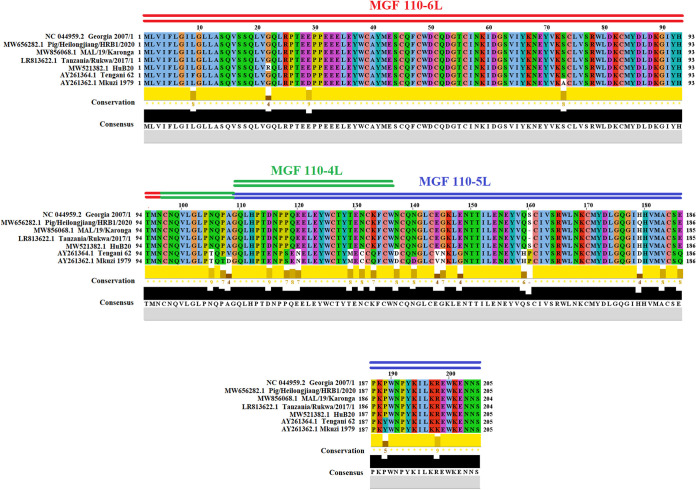
To assess the genetic identity between the gene MGF110-5L-6L present in the viral ASFV isolate Georgia 2007/1 and other ASFV isolates reported at the GenBank database, comparisons were carried out using the program BLAST. Interestingly, the results indicate that MGF110-5L-6L appears as a hallmark of all viral isolates associated with the pandemic Eurasia lineage (genotype II). Conversely, in the rest of the ASFV isolates, the genes MGF110-5L and MGF110-6L are present as individual genes. Overall, the nucleotide and amino acid identities among different isolates related with genotype II varied between 99.67% to 99.83% and 99.02% to 99.51%, respectively, indicating the high conservation of this gene during the evolution of this genotype. Single point mutations at amino acids G22R and H179Q were detected in the Chinese isolates HuB20 and Pig/Heilongjiang/HRB1/2020, respectively, while a deletion was found at residue 160 in the African isolates MAL/19/Karonga and Tanzania/Rukwa/2017/1 (Fig. 1).
FIG 1.
Representation of MGF110-5L-6L protein among field isolates. The alignment showing the 205 amino acid residues of MGF110-5L-6L protein was conducted in the software Jalview 2.11.1.6 and shows the diversity among isolates associated with genotype II (Georgia 2007/1, Pig/Heilongjiang/HRB1/2020, MAL/19/Karonga, Tanzania/Rukwa/2017/1, and HuB20). To set the limits of amino acids in MGF110-6L, MGF110-4L, and MGF110-5L present in MGF110-5L-6L protein, we included concatenated versions of these proteins present in Tengani 62 and Mkuzi 1979 isolates.
Furthermore, the BLAST analysis also revealed that 100% of the nucleotides comprising MGF110-5L-6L (618 nucleotides [nt]) are present exclusively at different sections in the genome of the ASFV isolates Tengani 62 and Mkuzi 1979. In this context, nucleotides 1 to 290 (associated with MGF110-6L), 291 to 411 (associated with MGF110-4L), and 319 to 618 (associated with MGF110-5L) were found at nucleotide positions 9097 to 9386, 8100 to 8221, 8447 to 8746 (Tengani 62), and 10624 to 10913, 8919 to 9040, and 9978 to 10272 (Mkuzi 1979), respectively. In this sense, overall amino acid identity of concatenated sections of the proteins MGF110-6L, MGF110-4L, and MGF110-5L of Tengani 62 and Mkuzi 1979 was calculated at 89.26% and 88.29%, respectively (Fig. 1).
Lastly, when we mapped different amino acid residues associated with MGF110-6L, MGF110-4L, and MGF110-5L genes, we found that truncated versions of these proteins were present in MGF110-5L-6L. In the case of MGF110-6L, we found that 22 amino acids at the N terminus of the native protein of other ASFV isolates are missing in MGF110-5L-6L, while in MGF110-5L we found 22 and 2 missing amino acids at the N and C terminus, respectively. Just a small section of MGF110-4L was found in MGF110-5L-6L protein.
In the light of these results, we may suggest that MGF110-5L-6L was the result of evolution of the ASFV genome originating from individual MGF110-4L, -5L, and -6L genes and not just the combining of the individual 5L and 6L genes found in other isolates.
Detection of MGF110-5L-6L transcription.
To determine the transcriptional kinetics of the MGF110-5L-6L gene during virus replication, a time course experiment was performed in primary swine macrophages infected with ASFV strain Georgia. Swine macrophage cultures were infected (MOI = 1) with ASFV-G, and samples were taken at 4, 6, 8, and 24 h postinfection (hpi). Presence of MGF110-5L-6L RNA was detected by two-step real-time PCR (RT-PCR) as described in Materials and Methods. Transcription of MGF110-5L-6L was detected at 4 hpi and remained stable until 24 hpi (Fig. 2). As a control of transcription, expression of the ASFV early protein p30 gene (CP204L) and the late protein p72 gene (B646L) was used as a reference for early and late transcription profiles, respectively. Expression of the MGF110-5L-6L gene is detected early throughout the tested time points, practically overlapping that of the CP204L gene that encodes the early ASFV p30 protein (Fig. 2).
FIG 2.
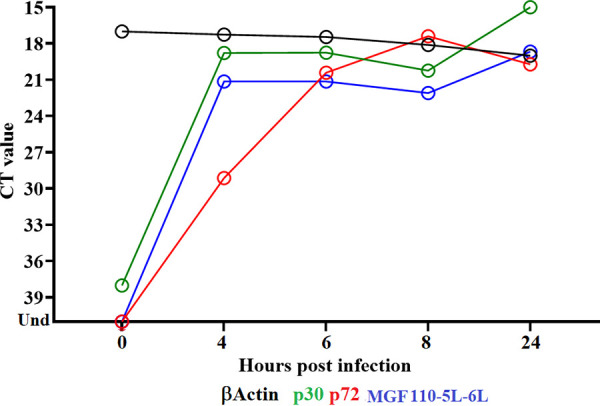
Expression profile of MGF110-5L-6L gene of ASFV during in vitro infection of porcine macrophages. Reverse transcription followed by qPCR was used to evaluate the expression profile of the MGF110-5L-6L gene during in vitro infection at different time points, up to 24 h. As a reference for this analysis, we use qPCR to specifically detect the expression of genes encoding ASFV proteins p30 (early expression) and p72 (late expression). Additionally, β-actin gene was used as a control to evaluate the quality and quantity of RNA throughout the infection. CT, threshold cycle.
Development of the ASFV-G-ΔMGF110-5L-6L deletion mutant.
To evaluate MGF110-5L-6L function during the process of ASFV replication in swine macrophage cell cultures and during the experimental infection in animals, a recombinant deletion mutant of the highly virulent ASFV Georgia 2007 strain (ASFV-G) lacking the MGF110-5L-6L gene was developed (ASFV-G-ΔMGF110-5L-6L). The MGF110-5L-6L gene was deleted by swapping 205 amino acid residues in the MGF110-5L-6L ORF with the p72mCherry cassette by homologous recombination (5). A region spanning 618 bp (between nucleotide positions 9490 and 10107) was deleted from the ASFV-G genome to delete the MGF110-5L-6L gene (see Materials and Methods) (Fig. 3). The recombinant ASFV-G-ΔMGF110-5L-6L stock was purified by limiting dilution using primary swine macrophage cell cultures.
FIG 3.
Schematic for the development of ASFV-G-ΔMGF110-5L-6L. The transfer vector contains the p72 promoter and a mCherry cassette; the gene positions are indicated. The homologous arms were designed to have flanking ends to both sides of the deletion/insertion cassette. The nucleotide positions of the area that was deleted in the ASFV-G genome are indicated by the dashed lines. The resulting ASFV-G-ΔMGF110-5L-6L virus with the cassette inserted is shown on the bottom.
The accuracy of the genetic modifications introduced into the ASFV-G-ΔMGF110-5L-6L genome and the integrity of the remaining virus genome were assessed by analyzing the full genome sequence using next-generation sequencing (NGS) on an Illumina NextSeq 500. The comparative analysis of ASFV-G-ΔMGF110-5L-6L and ASFV-G full-length genomes confirmed a deletion of 618 nucleotides, coinciding with the introduced genomic modifications. In addition, the ASFV-G-ΔMGF110-5L-6L genome possesses an insertion of 1,226 nucleotides corresponding with the insertion of the p72-mCherry cassette sequence. No additional genomic differences were detected between ASFV-G-ΔMGF110-5L-6L and ASFV-G, confirming that no undesired changes were introduced during the process of production and purification of ASFV-G-ΔMGF110-5L-6L. The NGS data also indicated the absence of parental ASFV-G genome as a potential contaminant in the ASFV-G-ΔMGF110-5L-6L stock.
Replication of ASFV-G-ΔMGF110-5L-6L in primary swine macrophages.
To assess the potential function of the MGF110-5L-6L gene during virus replication, the kinetics of ASFV-G-ΔMGF110-5L-6L replication was compared to that of the parental ASFV-G. A multistep growth curve using swine macrophage cultures was performed. Swine macrophages were infected at a multiplicity of infection (MOI) of 0.01 with either recombinant ASFV-G-ΔMGF110-5L-6L or parental ASFV-G. Virus yields were evaluated at 2, 24, 48, 72, and 96 hpi. Results demonstrated that ASFV-G-ΔMGF110-5L-6L has a growth kinetic that practically overlaps that of the parental ASFV-G. No significant virus yield differences were detected at any of the evaluated times postinfection (p.i.) (Fig. 4). Therefore, deletion of the MGF110-5L-6L gene from the ASFV-G genome does not affect the ability of ASFV-G-ΔMGF110-5L-6L to replicate in swine macrophages.
FIG 4.
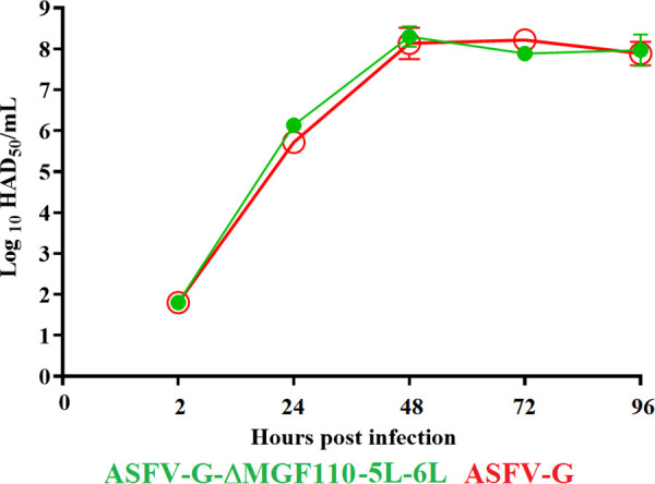
In vitro growth kinetics in primary swine macrophage cell cultures for ASFV-G-ΔMGF110-5L-6L and parental ASFV-G (MOI = 0.01). Samples were taken from three independent experiments at the indicated time points and titrated. Data represent means and standard deviations of three replicates. Sensitivity using this methodology for detecting virus is ≥log10 1.8 HAD50/mL. No significant differences in viral yields were observed at any time point tested determined using the Holm-Sidak method (α = 0.05), without assuming a consistent standard deviation. All calculations were conducted using the software GraphPad Prism version 8.
Assessment of ASFV-G-ΔMGF110-5L-6L virulence in swine.
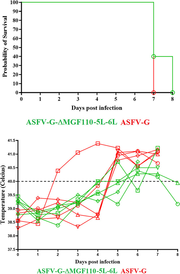
To evaluate the role of the MGF110-5L-6L gene in the virulence of ASFV-G, a group of five domestic pigs were inoculated intramuscularly (i.m.) with 102 HAD50. As a control, a similar group of animals was inoculated i.m. with 102 50% hemadsorption dose (HAD50) of ASFV-G. All animals inoculated with parental virulent ASFV-G presented with a rise in body temperature (>40°C) around day 6 p.i., quickly evolving to full clinical disease (depression, anorexia, staggering gait, diarrhea, and purple skin discoloration) (Table 1 and Fig. 5), with all animals being euthanized in extremis by day 7 p.i.
TABLE 1.
Swine survival and fever response following infection with ASFV-G-ΔMGF110-5L-6L and parental ASFV-G
| Virus (102 HAD50) | No. of survivors/total | Mean time to death, days (±SD)a | Fever characteristics |
||
|---|---|---|---|---|---|
| No. of days to onset (±SD)a | Duration no. of days (±SD)a | Maximum daily temp, °C (±SD)a | |||
| ASFV-G-ΔMGF110-5L-6L | 0/5 | 7.2 (1.34) | 5.4 (0.89) | 1.8 (1.67) | 40.54 (0.92) |
| ASFV-G | 0/5 | 7 (0) | 6.2 (2.28) | 2.6 (1.67) | 41 (0.47) |
SD, standard deviation.
FIG 5.
Evolution of mortality (top panel) and body temperature (bottom panel) in animals (5 animals/group) i.m. infected with 102 HAD50 of either ASFV-G-ΔMGF110-5L-6L or parental ASFV-G. No significant differences were found in the survival course between groups of pigs using the logrank test (Mantel-Cox test). No statistical differences were found in body temperatures between pigs in both groups when evaluated by the Holm-Sidak method (α = 0.05). All calculations were conducted using the software GraphPad Prism version 8.
All animals inoculated with ASFV-G-ΔMGF110-5L-6L developed a clinical disease practically indistinguishable from that of animals inoculated with parental ASFV-G. Appearance of clinical signs as well as their severity were very similar to those observed in the animals inoculated with ASFV-G. Therefore, deletion of the MGF110-5L-6L gene from the genome of ASFV-G does not alter the parental virus virulence during experimental infection in domestic swine.
The level of virus replication in the infected animals was assessed by quantifying titers of viremia after the experimental infection. Animals infected i.m. with parental ASFV-G presented titer values ranging between 103 to 108 HAD50/mL on day 4 p.i., increasing to titers of 107.5 to 108.5 HAD50/mL by day 7 p.i., when all animals were euthanized. All animals inoculated with 102 HAD50 of ASFV-G-ΔMGF110-5L-6L presented viremia values very similar to those of animals inoculated with the parental virus both at days 4 and 7 p.i. (Fig. 6). No statistical differences were found in the average of viremia titers at 4 days postinfection (dpi) and at 7 dpi between animals inoculated with either virus.
FIG 6.
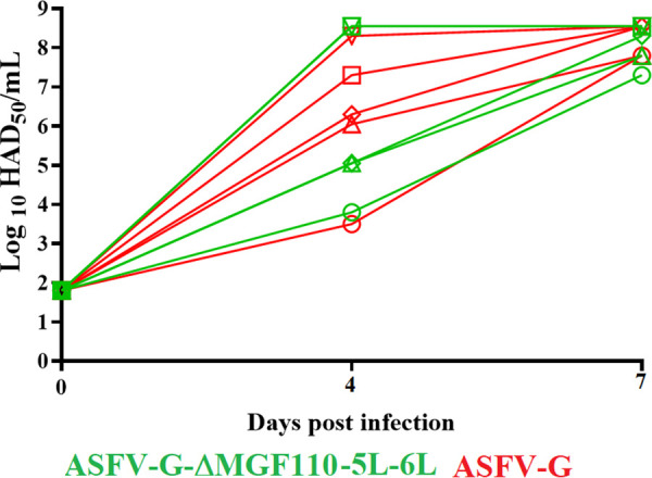
Viremia titers detected in pigs i.m. inoculated with 102 HAD50 of either ASFV-G-ΔMGF110-5L-6L or ASFV-G. Each symbol represents the average of animal titers in each of the groups. Sensitivity of virus detection: > log10 1.8 50% tissue culture infective dose (TCID50)/mL. No significant differences in viremia values between both groups of pigs were found during the course of the experiment using the Holm-Sidak method (α = 0.05). All calculations were conducted on the software GraphPad Prism version 8.
These results suggested that deletion of MGF110-5L-6L from the genome of ASFV-G does not significantly affect virus replication or virulence in domestic swine.
Development of an ASFV vaccine candidate with the deletion of MGF110-5L-6L gene as a potential DIVA marker.
To test the DIVA functionality of the MGF110-5L-6L gene, a novel recombinant vaccine strain was developed by deleting MGF110-5L-6L gene from the vaccine candidate ASFV-G-ΔI177L (7). The deletion of the MGF110-5L-6L gene was produced in the genome of ASFV-G-ΔI177L using a p30GFP reporter gene cassette with the same strategy and methodology used to develop ASFV-G-ΔMGF110-5L-6L. The resulting recombinant virus, ASFV-G-ΔI177L/ΔMGF110-5L-6L, has the MGF110-5L-6L gene replaced by the p30mGFP cassette, producing the same modifications in the ASFV-G-ΔI177L/ΔMGF110-5L-6L genome as those described in ASFV-G-ΔMGF110-5L-6L (Fig. 3).
The effect of the deletion of the MGF110-5L-6L gene on the ability of ASFV-G-ΔI177L/ΔMGF110-5L-6L to replicate was assessed by analyzing the growth kinetics in primary swine macrophage cultures and compared with that of ASFV-G in multistep growth curves (Fig. 7). ASFV-G-ΔI177L/ΔMGF110-5L-6L showed a decreased replication ability (approximately 100- to 10,000-fold, depending on the time point considered). These replication kinetics of ASFV-G-ΔI177L/ΔMGF110-5L-6L resemble those of ASFV-G-ΔI177L (7).
FIG 7.
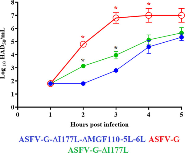
In vitro growth kinetics in primary swine macrophage cell cultures for ASFV-G-ΔI177L/ΔMGF110-5L-6L, ASFV-G-ΔI177L, and parental ASFV-G (MOI = 0.01). Samples were taken from three independent experiments at the indicated time points and titrated. Data represent means and standard deviations of three replicas. Sensitivity using this methodology for detecting virus is >log10 1.8 HAD50/mL. Significant differences in viral yields at specific time points between recombinant viruses are denoted by black asterisks, while red asterisks denote differences between recombinant viruses and ASFV-G. Analyses were conducted using the unpaired t test considering the two-stage step-up (Benjamini, Krieger, and Yekutieli) method. The significance of these discoveries was evaluated using the false-discovery rate method (FDR), with q values of <0.05 considered significant. All calculations were conducted using the software GraphPad Prism version 9.2.0.
The use of ASFV-G-ΔI177L/ΔMGF110-5L-6L as a potential DIVA marker experimental vaccine was assessed in domestic pigs. A group of five 80- to 90-pounds pigs were inoculated i.m. with 106 HAD50 of ASFV-G-ΔI177L/ΔMGF110-5L-6L. Inoculated animals were observed for 28 days without developing any clinical sign attributable to ASF. No viremia titers were detected in any of the ASFV-G-ΔI177L/ΔMGF110-5L-6L-inoculated animals during the first 28 days p.i. (Fig. 8).
FIG 8.
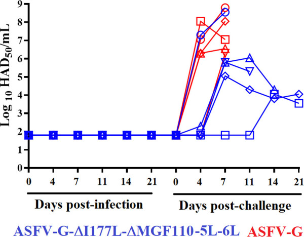
Viremia titers detected in pigs i.m. inoculated with 106 HAD50 of ASFV-G-ΔI177L/ΔMGF110-5L-6L and challenged 28 days later with 102 HAD50 of ASFV-G. A group of naive pigs was also challenged with 102 HAD50 of ASFV-G. Each symbol represents the average of animal titers in each of the groups. Sensitivity of virus detection: >log10 1.8 TCID50/mL.
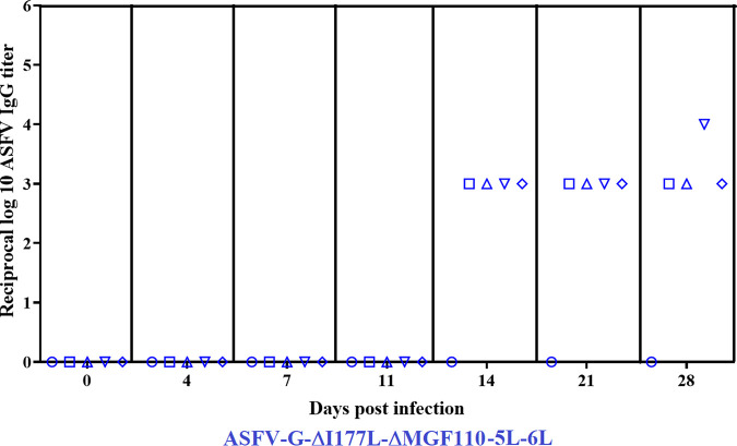
ASFV-specific antibody titers evaluated by ELISA were detected in ASFV-G-ΔI177L/ΔMGF110-5L-6L-infected animals started at day 14 p.i., remaining at similar titers until the day of challenge, at 28 day p.i. (Fig. 9). Interestingly, while one of the animals (no. 55) did not produce any detectable antibodies during the experimental period, the other four developed a detectable virus-specific antibody response.
FIG 9.
ASFV-specific antibody titers detected by ELISA, as described in Materials and Methods, in animals inoculated with 106 HAD50 of ASFV-G-ΔI177L/ΔMGF110-5L-6L. Antibody titers are presented as reciprocal of the highest dilution of serum that at least duplicates optical density (OD) reading of the preimmune serum. Each bar represents the titer of individual animals at different times p.i.
The specific reactivity of sera from these animals with the protein encoded by the MGF110-5L-6L gene was assessed by immunocytochemistry in Plum Island porcine epithelial (PIPEC) cells transfected with the MGF110-5L-6L gene. PIPEC cells were also transfected with the CP204L gene, encoding the well-characterized ASFV protein p30. While all four sera showed a clear antibody response against ASFV p30, none of the animals elicited antibodies recognizing the protein encoded by the MGF110-5L-6L gene. As control of expression and immunogenicity of the tested proteins, a pool of sera was used from animals immunized by vaccine candidates ASFV-G-ΔMGF (6), ASFV-G-Δ9GL/ΔUK (5), and ASFV-G-ΔI177L (7); all three of these vaccines contain the gene MGF110-5L-6L. All these pools of sera clearly recognized the expression of both proteins, p30 and MGF110-5L-6L, indicating that both proteins were efficiently expressed in the transfected cells and confirming the immunogenicity of both proteins (Fig. 10). These results showed that the MGF110-5L-6L gene can be a useful target gene to be deleted in a live-attenuated vaccine and can be used as the molecular basis for a DIVA antigenic marker. However, further studies will be required using large numbers of individual animal samples to determine the specificity and sensitivity of MGF110-5L-6L antibodies produced in individual animals.
FIG 10.
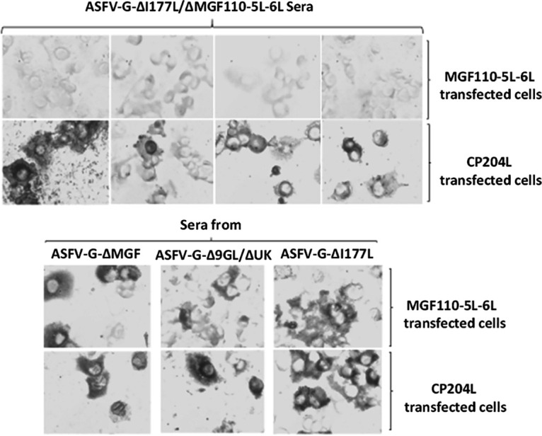
MGF110-5L-6L and CP204L antibody response in sera from animals inoculated with 106 HAD50 of ASFV-G-ΔI177L/ΔMGF110-5L-6L detected by immunocytochemistry. PIPEC cells transiently expressing either MGF110-5L-6L or p30 were used to test reactivity of individual serum from ASFV-G-ΔI177L/ΔMGF110-5L-6L animals. As a control, the reactivity of sera from animals infected with live-attenuated ASFV strains ASFV-G-ΔMGF, ASFV-G-Δ9GL/ΔUK, and ASFV-G-ΔI177L is shown.
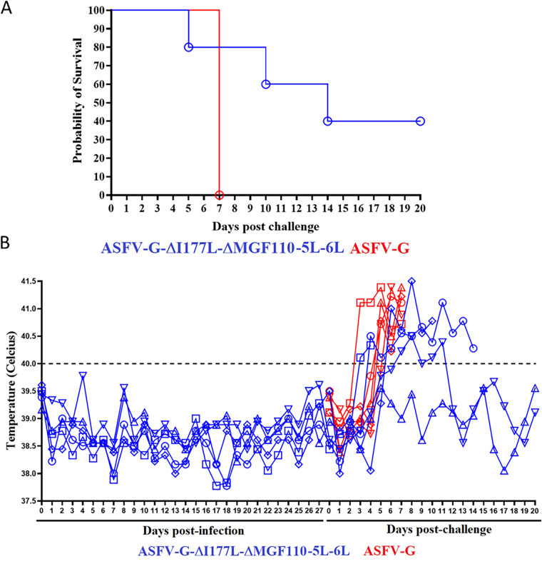
ASFV-G-ΔI177L/ΔMGF110-5L-6L animals were challenged at 28 dpi with virulent ASFV-G along with 5 naive pigs used as the control group. As expected, animals in the control group developed a typical clinical disease with a rise in body temperature by 4 days postchallenge (dpc) with a rapid progression in severity until all animals were euthanized by 7 dpc (Fig. 11). Conversely, after the challenge, animals infected with ASFV-G-ΔI177L/ΔMGF110-5L-6L presented a heterogeneous behavior. The animal that did not develop ASFV-specific antibodies quickly developed the clinical disease and was euthanized by day 5 postchallenge (p.c.). (Fig. 9 and 11). Two other animals developed a protracted form of the clinical disease and were euthanized on 10 and 14 dpc, respectively. The remaining two animals remained clinically normal during the 21-dpc observational period. Therefore, protective efficacy against challenge with virulent ASFV-G induced by ASFV-G-ΔI177L/ΔMGF110-5L-6L appears drastically reduced compared with that induced by its parental virus ASFV-G-ΔI177L. This result indicates that deletion of MGF110-5L-6L gene greatly affects ASFV-G-ΔI177L protective efficacy.
FIG 11.
Evolution of mortality (top panel) and body temperature (bottom panel) in animals (5 animals/group) i.m. infected with 106 HAD50 of ASFV-G-ΔI177L/ΔMGF110-5L-6L and challenged with 102 HAD50 of parental ASFV-G. A group of naive animals (ASFV-G) was included as the control group.
The profiles of viremia coincided with the clinical presentation of the disease (Fig. 8). As usual, animals in the control group developed high viremia titers (106 to 108 HAD50) by day 4 p.c., maintaining those values until they were euthanized at 7 dpc. All ASFV-G-ΔI177L/ΔMGF110-5L-6L-infected animals developed viremia as heterogenous as their clinical behavior. The animal euthanized at day 5 p.c. developed a viremia similar to that of the control animals. Animals euthanized at days 10 and 14 p.c. presented extended viremia lasting until they were euthanized. The two animals surviving the challenge also presented extended viremia with medium to low values until the end of the experimental period.
DISCUSSION
Currently, the development of an effective ASF vaccine has been based on the use of live-attenuated strains. These attenuated virus strains have used different approaches for their development. Some attempts have used natural attenuated field isolates (26). Others have adapted virulent field isolates to growth in cell cultures, resulting in virus attenuation (27, 28). The other methodology has been by genetic manipulation, deleting viral genes associated with virulence (4–9, 11, 20, 29–33). The latter appears to be the most effective and appears to offer a methodology safer than the use of naturally attenuated isolates. The referenced examples with deletions of single genes or a group of genes produced attenuated virus strains that induce protection against the virulent parental virus.
The inclusion of additional genetic mutations to these vaccine candidates, either to decrease residual attenuation or to increase the safety profile, has resulted in mixed success. Additions to the vaccine candidate ASFV-G-Δ9GL, which had residual virulence at high doses, resulted in a safe and effective vaccine when the deletion of UK was added (5). However, adding the MGF, EP153R or CD2 deletion caused the 9Gl vaccine to be ineffective, likely due to decreased replication in swine (12, 34). Interestingly, CD2 and the MGF deletion or the UK deletion could be combined using a derivative of ASFV-G isolated in China (9, 11). In another genotype vaccine candidate, ASFV-BA71ΔCD2 (8), additional deletions of either UK or EP153R decreased vaccine efficacy (35). The addition of gene deletions to existing vaccine platforms, while maintaining the original characteristics of the parental vaccine, appears to be partially unpredictable.
However, to date, for all live-attenuated ASF vaccines, there is the inability to serologically differentiate between infected and vaccinated animals (DIVA concept), as none of the genes deleted in the vaccine candidates have been shown to have any serological response that could be used as a DIVA marker. Introduction of a DIVA marker is critical for controlling ASF in determining which animals or farms are infected, and need to be culled, and which have the serological response because they were vaccinated. A DIVA-based vaccine is particularly of need in areas that want to vaccinate for ASF due to high risk of ASF introduction but want to be declared free of ASF outbreaks. We have previously reported the identification of the first DIVA-capable genomic deletion in ASF, deletion of a gene E184L (10). On its own, deletion of E184L partially attenuated ASF; however, when E184L was deleted in a vaccine strain of ASF, the vaccine lost its efficacy (10). This suggested that the addition of a virulence factor, as in the case of E184L, may cause problems in current vaccine platforms and perhaps the addition of a DIVA marker would have to be a deletion of a gene that was not a virulence factor.
In this study, we determined that the deletion of MGF110-5L-6L is a nonessential gene, since its deletion from the ASFV-G genome does not significantly alter virus replication in swine macrophage cultures or during in vivo infection and, importantly, is not critical for ASFV virulence in swine. We also showed that MGF110-5L-6L can be used as a target for the development of a DIVA marker vaccine if deleted in a live-attenuated vaccine candidate. It was expected that unlike E184L, which when deleted on its own affected virulence of ASFV-G, deletion of the MGF110-5L-6L gene had no observed effects on virus virulence, so it was assumed that it could have been deleted from vaccine candidate ASFV-G-ΔI177L without changing the vaccine efficacy. However, as reported here, deletion of MGF110-5L-6L affected the protective efficacy of the vaccine candidate ASFV-G-ΔI177L. A similar situation resulted by deleting another potential DIVA candidate gene, E184L, in the context of the vaccine candidate ASFV-G-ΔMGF (6). Our previous experience including additional deletions to vaccine candidates demonstrated that it is impossible to predict which combinations of gene deletions are compatible with maintaining vaccine efficacy. Here, we show that MGF110-5L-6L could perform as a DIVA marker for a live-attenuated ASFV vaccine; however, it was not compatible with the ASFV-G-ΔI177L vaccine platform. The discovery of MGF110-5L-6L as a DIVA marker could be useful in other live-attenuated ASFV vaccine platforms where adding this deletion could have an outcome different from that described here for ASFV-G-ΔI177L; however, to be a commercially viable DIVA marker, large trials of animals would be needed to determine the sensitivity and reliability of MGF110-5L-6L as a DIVA marker.
It appears that the deleterious effect on protective efficacy caused by additional deletions of potential DIVA target genes to current live-attenuated vaccine platforms could be a problem in the development of effective live-attenuated DIVA-compatible ASFV vaccine. The results presented here deleting MGF110-5L-6L along with our previous results in deletion of E184L suggest that with our current knowledge of ASFV, and particularly in the function of individual ASFV genes, it is hard to predict which serological markers could be deleted in current vaccine platforms, and the only way to determine the addition of a gene deletion to serve as a serological marker will be through testing of vaccine efficacy.
MATERIALS AND METHODS
Cell culture and viruses.
Primary cultures of blood-derived swine macrophages were performed using peripheral mononuclear adherent cells obtained by density gradients and adhesion to Primaria T25 6- or 96-well dishes at a density of 5 × 10⁶ cells per mL, as described previously (7). ASFV Georgia (ASFV-G) is a field isolate obtained in 2010 in the Republic of Georgia (27). Growth curves between ASFV-G-ΔMGF110-5L-6L or ASFV-G-ΔI177L/ΔMGF110-5L-6L and ASFV-G were performed in primary swine macrophage cell cultures at an MOI of 0.01 HAD50 as described previously (36).
Detection of MGF110-5L-6L transcription.
The transcriptional profile of the MGF110-5L-6L gene during the infection of ASFV-G in cultures of porcine macrophages was assessed as described previously, using real-time quantitative PCR (RT-qPCR) (37). As an internal control of transcription, the early CP204L (p30) and late B646L (p72) expressed genes of ASFV were used as references. Porcine macrophages were infected (MOI of 1) with ASFV-G followed by RNA extractions performed at different times postinfection (p.i.) using an RNeasy kit (Qiagen). Extracted material was treated with 2 units of DNase I (BioLabs) and then purified using the Monarch RNA cleanup kit (New England BioLabs, Inc.). One microgram of RNA was used to produce cDNA using qScript cDNA SuperMix (Quanta bio) that was used for the qPCR. Primers and probe for detection of the MGF110-5L-6L gene were designed using the ASFV Georgia 2007/1 strain (GenBank database NC_044959.2): forward, 5′-TCCGTTGGATGAAGTTGTCC-3′, reverse, 5′-TCATGTCTGGTTTCCCGTTG-3′, and probe, 5′-FAM/AGCCCTAAGACCTGGTTGCAATTCA/MGBNFQ-3′. Primers and probes for the detection of p72, p30, and β-actin genes were described previously (38). All qPCRs were conducted using the TaqMan universal PCR master mix (Applied Biosystems) using amplification conditions described elsewhere (38).
Construction of the ASFV MGF110-5L-6L deletion mutant.
ASFV lacking the MGF110.5L-6L gene (ASFV-G-ΔMGF110-5L-6L) was developed by homologous recombination between the genome of the parental ASFV-G and a recombination transfer vector as described elsewhere (5). The recombinant transfer vector (p72mCherryΔMGF110-5L-6L) harbors flanking genomic regions of the MGF110-5L-6L gene: the left and right arms are located between genomic positions 8489 to 9489 and 10108 to 11108, respectively. ASFV-G has the same sequence as GenBank accession number NC_044959.2. The recombinant transfer vector also harbors a reporter gene cassette containing the mCherry fluorescent protein (mCherry) gene under the control of the ASFV p72 late gene promoter (39). The recombinant transfer vector was obtained by DNA synthesis (Epoch Life Sciences, Sugar Land, TX, USA). As designed, this construction created a 618-nucleotide deletion between nucleotide positions 9490 to 10107, partially deleting the MGF110-5L-6L ORF sequence (Fig. 3). Recombinant mutant ASFV-G-ΔMGF110-5L-6L was purified to homogeneity by successive rounds of limiting dilution purification based on mCherry activity detection and was completely sequenced using next-generation sequencing (NGS).
Recombinant virus ASFV-G-ΔI177L/ΔMGF110-5L-6L was obtained by deleting MGF110-5L-6L gene from the vaccine candidate ASFV-G-ΔI177L (7) following the same procedure as that described above using green fluorescent protein (GFP) instead of mCherry as the selection marker.
Next-generation sequencing of ASFV.
ASFV DNA was extracted from the cytoplasm of infected macrophages as described previously (10). NGS of the extracted DNA was performed using the Nextera XT kit and the NextSeq platform (Illumina, San Diego, CA) following the manufacturer’s protocol. Sequence analysis was performed using CLC Genomics Workbench software (CLCBio, Aarthus, Denmark).
Animal experiments.
Virulence of ASFV-G-ΔMGF110-5L-6L was evaluated using 35 to 40 kg commercial breed swine. A group of five pigs was intramuscularly (i.m.) inoculated with 102 HAD50 of ASFV-G-ΔMGF110-5L-6L and compared with a group of five pigs inoculated with 102 HAD50 of ASFV-G. Clinical signs and body temperature were recorded daily throughout the experiment. Animal experiments were performed under biosafety level 3 conditions in the animal facilities at Plum Island Animal Disease Center, following a strict protocol approved by the Institutional Animal Care and Use Committee (225.01-16-R_090716). In protection experiments, animals inoculated with ASFV-G-ΔI177L/ΔMGF110-5L-6L were i.m. challenged 28 days later with 102 HAD50 of parental ASFV-G. Presence of clinical signs associated with the disease was recorded as described above (6).
Detection of ASFV-specific antibody response.
ASFV-specific antibody detection used an in-house ELISA performed as described previously in detail (40) using as the antigen a cell extract of ASFV-infected Vero cells. Each swine serum was tested at multiple dilutions against both infected and uninfected cell antigens. Serum titers were expressed as the log10 of the highest dilution where the optical density reading of the tested sera at least duplicates the reading of the mock-infected sera.
Presence of MGF110-5L-6-L or p30-specific antibodies was also detected by immunocytochemistry. Plum Island porcine epithelial (PIPEC) cells (29) transfected with overexpression plasmids corresponding to the desired ASFV gene under the control of the CMV promoter were used expressing the coding sequence from the ASFV-G genome for the MGF110-5L-6L or CP240L (p30) with the nucleotides optimized for expression in mammalian cells. Presence of MGF110-5L-6L or CP240L antibodies in pig sera was detected by a peroxidase-labeled anti-swine IgG reagent (Vector Laboratories, Burlingame, CA).
Data availability.
The next-generation sequencing of the recombinant ASFV viruses produced in this study was aligned to GenBank accession number NC_044959.2. The raw sequencing reads are available upon request.
ACKNOWLEDGMENTS
We thank the Plum Island Animal Disease Center Animal Care Unit staff for their excellent technical assistance. We wish to particularly thank Melanie V. Prarat for editing the manuscript. This research was supported in part by an appointment to the Plum Island Animal Disease Center (PIADC) Research Participation Program administered by the Oak Ridge Institute for Science and Education (ORISE) through an interagency agreement between the U.S. Department of Energy (DOE) and the U.S. Department of Agriculture (USDA). ORISE is managed by ORAU under DOE contract number DE-SC0014664. All opinions expressed in this paper are the authors’ and do not necessarily reflect the policies and views of USDA, ARS, APHIS, DOE, or ORAU/ORISE.
The authors Douglas Gladue and Manuel Borca have a patent for ASFV-G-ΔI177L as a live-attenuated vaccine for African swine fever.
This project was funded through an interagency agreement with the Science and Technology Directorate of the U.S. Department of Homeland Security under award number 70RSAT19KPM000056 and by the National Pork Board Project no. 21-137.
Contributor Information
Manuel V. Borca, Email: Manuel.Borca@usda.gov.
Douglas P. Gladue, Email: Douglas.Gladue@usda.gov.
Joanna L. Shisler, University of Illinois at Urbana Champaign
REFERENCES
- 1.Gonzales W, Moreno C, Duran U, Henao N, Bencosme M, Lora P, Reyes R, Nunez R, De Gracia A, Perez AM. 2021. African swine fever in the Dominican Republic. Transbound Emerg Dis 68:3018–3019. 10.1111/tbed.14341. [DOI] [PubMed] [Google Scholar]
- 2.Tulman ER, Delhon GA, Ku BK, Rock DL. 2009. African swine fever virus, p 43–87. In Etten JL, Lesser known large dsDNA viruses, vol 328. Springer-Verlag, Berlin Heidelberg. [DOI] [PubMed] [Google Scholar]
- 3.Costard S, Wieland B, de Glanville W, Jori F, Rowlands R, Vosloo W, Roger F, Pfeiffer DU, Dixon LK. 2009. African swine fever: how can global spread be prevented? Philos Trans R Soc Lond B Biol Sci 364:2683–2696. 10.1098/rstb.2009.0098. [DOI] [PMC free article] [PubMed] [Google Scholar]
- 4.O'Donnell V, Holinka LG, Krug PW, Gladue DP, Carlson J, Sanford B, Alfano M, Kramer E, Lu Z, Arzt J, Reese B, Carrillo C, Risatti GR, Borca MV. 2015. African swine fever virus Georgia 2007 with a deletion of virulence-associated gene 9GL (B119L), when administered at low doses, leads to virus attenuation in swine and induces an effective protection against homologous challenge. J Virol 89:8556–8566. 10.1128/JVI.00969-15. [DOI] [PMC free article] [PubMed] [Google Scholar]
- 5.O'Donnell V, Risatti GR, Holinka LG, Krug PW, Carlson J, Velazquez-Salinas L, Azzinaro PA, Gladue DP, Borca MV. 2017. Simultaneous deletion of the 9GL and UK genes from the African swine fever virus Georgia 2007 isolate offers increased safety and protection against homologous challenge. J Virol 91. 10.1128/JVI.01760-16. [DOI] [PMC free article] [PubMed] [Google Scholar]
- 6.O'Donnell V, Holinka LG, Gladue DP, Sanford B, Krug PW, Lu X, Arzt J, Reese B, Carrillo C, Risatti GR, Borca MV. 2015. African swine fever virus Georgia isolate harboring deletions of MGF360 and MGF505 genes is attenuated in swine and confers protection against challenge with virulent parental virus. J Virol 89:6048–6056. 10.1128/JVI.00554-15. [DOI] [PMC free article] [PubMed] [Google Scholar]
- 7.Borca MV, Ramirez-Medina E, Silva E, Vuono E, Rai A, Pruitt S, Holinka LG, Velazquez-Salinas L, Zhu J, Gladue DP. 2020. Development of a highly effective African swine fever virus vaccine by deletion of the I177L gene results in sterile immunity against the current epidemic Eurasia strain. J Virol 94. 10.1128/JVI.02017-19. [DOI] [PMC free article] [PubMed] [Google Scholar]
- 8.Monteagudo PL, Lacasta A, Lopez E, Bosch L, Collado J, Pina-Pedrero S, Correa-Fiz F, Accensi F, Navas MJ, Vidal E, Bustos MJ, Rodriguez JM, Gallei A, Nikolin V, Salas ML, Rodriguez F. 2017. BA71DeltaCD2: a new recombinant live attenuated African swine fever virus with cross-protective capabilities. J Virol 91. 10.1128/JVI.01058-17. [DOI] [PMC free article] [PubMed] [Google Scholar]
- 9.Chen W, Zhao D, He X, Liu R, Wang Z, Zhang X, Li F, Shan D, Chen H, Zhang J, Wang L, Wen Z, Wang X, Guan Y, Liu J, Bu Z. 2020. A seven-gene-deleted African swine fever virus is safe and effective as a live attenuated vaccine in pigs. Sci China Life Sci 63:623–634. 10.1007/s11427-020-1657-9. [DOI] [PMC free article] [PubMed] [Google Scholar]
- 10.Ramirez-Medina E, Vuono E, Rai A, Pruitt S, Espinoza N, Velazquez-Salinas L, Pina-Pedrero S, Zhu J, Rodriguez F, Borca MV, Gladue DP. 2022. Deletion of E184L, a putative DIVA target from the pandemic strain of African swine fever virus, produces a reduction in virulence and protection against virulent challenge. J Virol 96. 10.1128/JVI.01419-21. [DOI] [PMC free article] [PubMed] [Google Scholar]
- 11.Teklue T, Wang T, Luo Y, Hu R, Sun Y, Qiu HJ. 2020. Generation and evaluation of an African swine fever virus mutant with deletion of the CD2v and UK genes. Vaccines 8:763. 10.3390/vaccines8040763. [DOI] [PMC free article] [PubMed] [Google Scholar]
- 12.Gladue DP, O’Donnell V, Ramirez-Medina E, Rai A, Pruitt S, Vuono EA, Silva E, Velazquez-Salinas L, Borca MV. 2020. Deletion of CD2-like (CD2v) and C-type lectin-like (EP153R) genes from African swine fever virus Georgia-9GL abrogates its effectiveness as an experimental vaccine. Viruses 12:1185. 10.3390/v12101185. [DOI] [PMC free article] [PubMed] [Google Scholar]
- 13.Rodriguez JM, Yanez RJ, Almazan F, Vinuela E, Rodriguez JF. 1993. African swine fever virus encodes a CD2 homolog responsible for the adhesion of erythrocytes to infected cells. J Virol 67:5312–5320. 10.1128/JVI.67.9.5312-5320.1993. [DOI] [PMC free article] [PubMed] [Google Scholar]
- 14.Borca MV, Carrillo C, Zsak L, Laegreid WW, Kutish GF, Neilan JG, Burrage TG, Rock DL. 1998. Deletion of a CD2-like gene, 8-DR, from African swine fever virus affects viral infection in domestic swine. J Virol 72:2881–2889. 10.1128/JVI.72.4.2881-2889.1998. [DOI] [PMC free article] [PubMed] [Google Scholar]
- 15.Borca MV, O'Donnell V, Holinka LG, Risatti GR, Ramirez-Medina E, Vuono EA, Shi J, Pruitt S, Rai A, Silva E, Velazquez-Salinas L, Gladue DP. 2020. Deletion of CD2-like gene from the genome of African swine fever virus strain Georgia does not attenuate virulence in swine. Sci Rep 10:494. 10.1038/s41598-020-57455-3. [DOI] [PMC free article] [PubMed] [Google Scholar]
- 16.Imbery J, Upton C. 2017. Organization of the multigene families of African swine fever virus. Fine Focus 3:155–170. 10.33043/FF.3.2.155-170. [DOI] [Google Scholar]
- 17.Almendral JM, Almazan F, Blasco R, Vinuela E. 1990. Multigene families in African swine fever virus: family 110. J Virol 64:2064–2072. 10.1128/JVI.64.5.2064-2072.1990. [DOI] [PMC free article] [PubMed] [Google Scholar]
- 18.de la Vega I, Vinuela E, Blasco R. 1990. Genetic variation and multigene families in African swine fever virus. Virology 179:234–246. 10.1016/0042-6822(90)90293-z. [DOI] [PubMed] [Google Scholar]
- 19.Gonzalez A, Calvo V, Almazan F, Almendral JM, Ramirez JC, de la Vega I, Blasco R, Vinuela E. 1990. Multigene families in African swine fever virus: family 360. J Virol 64:2073–2081. 10.1128/JVI.64.5.2073-2081.1990. [DOI] [PMC free article] [PubMed] [Google Scholar]
- 20.Li D, Liu Y, Qi X, Wen Y, Li P, Ma Z, Liu Y, Zheng H, Liu Z. 2021. African swine fever virus MGF-110-9L-deficient mutant has attenuated virulence in pigs. Virol Sin 36:187–195. 10.1007/s12250-021-00350-6. [DOI] [PMC free article] [PubMed] [Google Scholar]
- 21.Sanchez-Cordon PJ, Jabbar T, Berrezaie M, Chapman D, Reis A, Sastre P, Rueda P, Goatley L, Dixon LK. 2018. Evaluation of protection induced by immunisation of domestic pigs with deletion mutant African swine fever virus BeninDeltaMGF by different doses and routes. Vaccine 36:707–715. 10.1016/j.vaccine.2017.12.030. [DOI] [PMC free article] [PubMed] [Google Scholar]
- 22.Zsak L, Lu Z, Burrage TG, Neilan JG, Kutish GF, Moore DM, Rock DL. 2001. African swine fever virus multigene family 360 and 530 genes are novel macrophage host range determinants. J Virol 75:3066–3076. 10.1128/JVI.75.7.3066-3076.2001. [DOI] [PMC free article] [PubMed] [Google Scholar]
- 23.Ramirez-Medina E, Vuono E, Pruitt S, Rai A, Silva E, Espinoza N, Zhu J, Velazquez-Salinas L, Borca MV, Gladue DP. 2021. Development and in vivo evaluation of a MGF110-1L deletion mutant in African swine fever strain Georgia. Viruses 13:286. 10.3390/v13020286. [DOI] [PMC free article] [PubMed] [Google Scholar]
- 24.Ramirez-Medina E, Vuono EA, Rai A, Pruitt S, Silva E, Velazquez-Salinas L, Zhu J, Gladue DP, Borca MV. 2020. Evaluation in swine of a recombinant African swine fever virus lacking the MGF-360-1L gene. Viruses 12:1193. 10.3390/v12101193. [DOI] [PMC free article] [PubMed] [Google Scholar]
- 25.Ramirez-Medina E, Vuono EA, Velazquez-Salinas L, Silva E, Rai A, Pruitt S, Berggren KA, Zhu J, Borca MV, Gladue DP. 2020. The MGF360-16R ORF of African swine fever virus strain Georgia encodes for a nonessential gene that interacts with host proteins SERTAD3 and SDCBP. Viruses 12:60. 10.3390/v12010060. [DOI] [PMC free article] [PubMed] [Google Scholar]
- 26.Barasona JA, Gallardo C, Cadenas-Fernandez E, Jurado C, Rivera B, Rodriguez-Bertos A, Arias M, Sanchez-Vizcaino JM. 2019. First oral vaccination of Eurasian wild boar against African swine fever virus genotype II. Front Vet Sci 6:137. 10.3389/fvets.2019.00137. [DOI] [PMC free article] [PubMed] [Google Scholar]
- 27.Krug PW, Holinka LG, O'Donnell V, Reese B, Sanford B, Fernandez-Sainz I, Gladue DP, Arzt J, Rodriguez L, Risatti GR, Borca MV. 2015. The progressive adaptation of a Georgian isolate of African swine fever virus to Vero cells leads to a gradual attenuation of virulence in swine corresponding to major modifications of the viral genome. J Virol 89:2324–2332. 10.1128/JVI.03250-14. [DOI] [PMC free article] [PubMed] [Google Scholar]
- 28.Sereda AD, Balyshev VM, Kazakova AS, Imatdinov AR, Kolbasov DV. 2020. Protective properties of attenuated strains of African swine fever virus belonging to seroimmunotypes I-VIII. Pathogens 9:274. 10.3390/pathogens9040274. [DOI] [PMC free article] [PubMed] [Google Scholar]
- 29.Borca MV, Rai A, Ramirez-Medina E, Silva E, Velazquez-Salinas L, Vuono E, Pruitt S, Espinoza N, Gladue DP. 2021. A cell culture-adapted vaccine virus against the current African swine fever virus pandemic strain. J Virol 95:e0012321. 10.1128/JVI.00123-21. [DOI] [PMC free article] [PubMed] [Google Scholar]
- 30.Lewis T, Zsak L, Burrage TG, Lu Z, Kutish GF, Neilan JG, Rock DL. 2000. An African swine fever virus ERV1-ALR homologue, 9GL, affects virion maturation and viral growth in macrophages and viral virulence in swine. J Virol 74:1275–1285. 10.1128/jvi.74.3.1275-1285.2000. [DOI] [PMC free article] [PubMed] [Google Scholar]
- 31.Zsak L, Caler E, Lu Z, Kutish GF, Neilan JG, Rock DL. 1998. A nonessential African swine fever virus gene UK is a significant virulence determinant in domestic swine. J Virol 72:1028–1035. 10.1128/JVI.72.2.1028-1035.1998. [DOI] [PMC free article] [PubMed] [Google Scholar]
- 32.Zhang J, Zhang Y, Chen T, Yang J, Yue H, Wang L, Zhou X, Qi Y, Han X, Ke J, Wang S, Yang J, Miao F, Zhang S, Zhang F, Wang Y, Li M, Hu R. 2021. Deletion of the L7L-L11L genes attenuates ASFV and induces protection against homologous challenge. Viruses 13:255. 10.3390/v13020255. [DOI] [PMC free article] [PubMed] [Google Scholar]
- 33.Borca MV, Ramirez-Medina E, Silva E, Vuono E, Rai A, Pruitt S, Espinoza N, Velazquez-Salinas L, Gay CG, Gladue DP. 2021. ASFV-G-I177L as an effective oral nasal vaccine against the Eurasia strain of Africa swine fever. Viruses 13:765. 10.3390/v13050765. [DOI] [PMC free article] [PubMed] [Google Scholar]
- 34.O’Donnell V, Holinka LG, Sanford B, Krug PW, Carlson J, Pacheco JM, Reese B, Risatti GR, Gladue DP, Borca MV. 2016. African swine fever virus Georgia isolate harboring deletions of 9GL and MGF360/505 genes is highly attenuated in swine but does not confer protection against parental virus challenge. Virus Res 221:8–14. 10.1016/j.virusres.2016.05.014. [DOI] [PubMed] [Google Scholar]
- 35.Lopez E, Bosch-Camos L, Ramirez-Medina E, Vuono E, Navas MJ, Munoz M, Accensi F, Zhang J, Alonso U, Argilaguet J, Salas ML, Anachkov N, Gladue DP, Borca MV, Pina-Pedrero S, Rodriguez F. 2021. Deletion mutants of the attenuated recombinant ASF virus, BA71DeltaCD2, show decreased vaccine efficacy. Viruses 13:1678. 10.3390/v13091678. [DOI] [PMC free article] [PubMed] [Google Scholar]
- 36.Reed LJM, Muench H. 1938. A simple method of estimating fifty percent endpoints. The American J Hygiene 27:493–497. 10.1093/oxfordjournals.aje.a118408. [DOI] [Google Scholar]
- 37.Gladue DP, Ramirez-Medina E, Vuono E, Silva E, Rai A, Pruitt S, Espinoza N, Velazquez-Salinas L, Borca MV. 2021. Deletion of A137R gene from the pandemic strain of African swine fever virus is attenuated and offers protection against virulent pandemic virus. J Virol 95. 10.1128/JVI.01139-21. [DOI] [PMC free article] [PubMed] [Google Scholar]
- 38.Ramirez-Medina E, Vuono EA, Pruitt S, Rai A, Espinoza N, Velazquez-Salinas L, Gladue DP, Borca MV. 2021. Evaluation of an ASFV RNA helicase gene A859L for virus replication and swine virulence. Viruses 14:10. 10.3390/v14010010. [DOI] [PMC free article] [PubMed] [Google Scholar]
- 39.Borca MV, O'Donnell V, Holinka LG, Sanford B, Azzinaro PA, Risatti GR, Gladue DP. 2017. Development of a fluorescent ASFV strain that retains the ability to cause disease in swine. Sci Rep 7:46747. 10.1038/srep46747. [DOI] [PMC free article] [PubMed] [Google Scholar]
- 40.Carlson J, O’Donnell V, Alfano M, Velazquez Salinas L, Holinka L, Krug P, Gladue D, Higgs S, Borca M. 2016. Association of the host immune response with protection using a live attenuated African swine fever virus model. Viruses 8:291. 10.3390/v8100291. [DOI] [PMC free article] [PubMed] [Google Scholar]
Associated Data
This section collects any data citations, data availability statements, or supplementary materials included in this article.
Data Availability Statement
The next-generation sequencing of the recombinant ASFV viruses produced in this study was aligned to GenBank accession number NC_044959.2. The raw sequencing reads are available upon request.


