ABSTRACT
OBJECTIVE
To analyze the blood oxygen concentrations (StO2) of different stages of pressure injury (PI) tissue using hyperspectral images to serve as a guideline for the treatment and care of PIs.
METHODS
This study used a prospective design. A total of 30 patients with sacral PIs were recruited from the rehabilitation ward of a teaching hospital. The authors used a hyperspectral detector to collect wound images and the Beer-Lambert law to estimate changes in tissue StO2 in different stages of PI.
RESULTS
The tissue StO2 of healthy skin and that of stage 1 PI skin were similar, whereas the tissue StO2 of the wound in stage 2 PIs was significantly higher than that of healthy skin and scabbed tissue (medians, 82.5%, 74.4%, and 68.3%; P < .05). In stage 3 PIs, StO2 was highest in subcutaneous tissue and adipose tissue (82.5%) and lowest in peripheral scabs (68.35%). The tissue StO2 was highest in subcutaneous tissue in stage 4 PIs, and this tissue was red in the hyperspectral spectrum. The scab-covered area of unstageable PIs had the lowest StO2 of all PI tissue types (median, 44.3%).
CONCLUSIONS
Hyperspectral imaging provides physiologic information on wound microcirculation, which can enable better evaluation of healing status. Assessing tissue StO2 data can provide a clinical index of wound healing.
KEYWORDS: blood oxygen concentration, hyperspectral imaging, pressure injury, sacral pressure injury, StO2, wound healing
INTRODUCTION
Pressure injury (PI) development is a common wound care problem in patients with chronic diseases, older adults, and immobilized patients in acute wards, nursing homes, long-term care, or home care.1 Risk factors for PI are diverse and include poor nutrition status, decreased sensory perception, multiple invasive medical devices, and poor tissue perfusion, with immobility being the greatest risk factor.2,3 The prevalence of PI in acute care settings ranges from 4.94% to 54%.4 Because PIs cause patient discomfort, reduced quality of life, prolonged hospital stays, and increased financial burdens, medical personnel focus on PI prevention.4
Pressure injuries are staged based on the extent of tissue damage and may be classified as stage 1, 2, 3, 4, unstageable, or deep-tissue PI.5 In clinical practice, healthcare or wound care staff visually evaluate the PI, noting its size and depth, secretions, color, and the wound bed tissue, as well as signs of infection, swelling, and so on.6 Wound monitoring typically involves visual inspection of the wound site for changes and physical measurements of the wound size and depth. However, noninvasive imaging techniques can provide additional wound information beyond what can be determined from visual assessment alone. Healthcare providers can use optical imaging techniques to assess underlying processes within the tissue and identify whether the wound is healing.7 For example, both blood oxygenation and wound microcirculation may vary depending on the degree of wound healing. Different optical measurements provide clinical medical staff with an objective, quantitative assessment of wounds.
Hyperspectral imaging (HSI) is a new, noninvasive, noncontact, automated optical measurement technology.8 It combines spatial and spectral wavelength information (spectroscopy)9 to obtain both a two-dimensional image and three-dimensional hyperspectral information. Each pixel contains hyperspectral information with light wavelengths of 480 to 640 nm; the blood oxygen concentration (StO2) value of each pixel can be calculated.8,10 As a result, HSI captures hundreds of spectral bands and can detect information that is commonly unavailable in visible light images. Therefore, the characteristics of the target can be detected by means of the correlations or differences between these various spectral bands. For example, the features of oxyhemoglobin may show differences in HSI information with some spectral bands. The advantage lies in collecting information on continuous spectral bands that cannot be seen by the human eye.8 In recent years, research has been conducted on HSI related to evaluating burn healing,11 skin melanoma,12 tissue blood oxygen, and the risk of diabetic foot ulcer formation.10
In the present study, the authors used HSI with wavelengths of 480 to 640 nm to evaluate the StO2 of PI tissues in different stages, obtain accurate data on the progress of wound healing, describe wound changes, and inform PI treatment guidelines.
METHODS
This prospective study was conducted in the rehabilitation ward of a regional teaching hospital in central Taiwan. Patients were recruited by intentional sampling; they were included in the study if they were 18 years or older, were admitted to the rehabilitation ward with chronic disease, and had a sacral PI. Patients with scabies, fractures, and shock were excluded.
Research Tool, Experimental Steps, and Parameters
The authors captured HSI data at wavelengths of 480 to 640 nm using an HSI device with a snapshot mosaic hyperspectral image sensor (IMEC Inc, Ghent, Belgium) to calculate the concentrations of oxygenated and nonoxygenated hemoglobin based on the Beer-Lambert law. Because of the obvious characteristic changes of oxygenated and nonoxygenated hemoglobin in the wavelength range of visible light, investigators could calculate the StO2 of tissue blood to evaluate changes during wound healing.
To capture HSI data, researchers: (1) explained the HSI test to the patient; (2) focused the HSI device and performed background image correction; (3) captured HSI images of the soles of the patient’s feet as baseline values of healthy skin; (4) captured the HSI images of the PI; and (5) calculated the concentrations of oxyhemoglobin (CHbO2), deoxyhemoglobin (CHb), melanin (Cmelanin), and tissue StO2 from the HSI images.
Calculating CHbO2, CHb, and Cmelanin
After capturing HSI images of the soles of the patient’s feet, these data were used with the Beer-Lambert law equation (1) to obtain the concentrations of CHbO2, CHb, and Cmelanin:
| (1) |
where ε is the molar extinction coefficient, L is the length of skin, I and Io are intensities of HSI data, and C is concentration of the absorbing species.
Calculating Tissue StO2
Thirteen bands of the wavelengths of hyperspectral data were used to calculate tissue StO2 and the concentration parameters. The final tissue StO2 was calculated as follows:
| (2) |
Data Collection
Data were collected from December 5, 2019 to April 25, 2020. Three surgeons independently staged the sacral PIs of 30 patients before they were admitted to the rehabilitation department of the hospital. The doctors referred to the National Pressure Ulcer Advisory Panel 2016 pressure injury staging guideline as the standard for training, discussion, and patient assessment. All three doctors provided the same assessments for 28 of the 30 patients (93.3%). For the remaining two patients, the doctors discussed the assessments to reach consensus. The surgeons were trained in hyperspectroscopy. Nursing staff caring for the patients took pictures of the sacral PIs.
Data Analysis
The authors used SPSS Statistics version 22.0 (IBM, Armonk, New York) to analyze the basic characteristics. Sigma Plot 12.5 (Alfasoft, Gothenburg, Sweden) data analysis software was used for statistical analysis, and box plots were used to perform a one-way analysis of variance to calculate P values. Because the data were considered to have a nonnormal distribution, both the Kruskal-Wallis test and the Student-Newman-Keuls method were used for the mode of variance analysis and postvalidation.
Ethical Considerations
The authors obtained approval from the regional hospital review committee (no. 108-75) before the study commenced. Eligible patients or their guardians were informed of the purpose and method of the study; those who agreed to participate signed an informed consent form. To protect patient privacy, the collected data were archived with numeric identification. If patients or their guardians wished to terminate their study participation, they could withdraw at any time, and the data would immediately be deleted.
RESULTS
Hyperspectral image data of 30 patients were included in the analysis. The color map showed that more than 80% of the distribution range was red, indicating that the hemoglobin in the tissue was oxygenated (StO2 = 80%). Tissue containing 70% to 80% oxygenated hemoglobin was indicated by yellow, 60% to 69% by green, 50 to 59% by blue, and so on. Greater redness represents higher tissue StO2.
The average age of the participants was 71.7 ± 5.97 years, and 20 (66.7%) of them were men. Most of the patients (93.3%) were married, and the majority (83.4%) had less than a junior high school education. All the PIs analyzed were in the sacral region; physicians identified 2 cases (6.67%) of stage 1 PIs, 13 cases (43.33%) of stage 2 PIs, 10 cases (33.33%) of stage 3 PIs, and 1 case (3.33%) of a stage 4 PI. Four cases (13.33%) of PIs were unstageable (Table).
Table.
DEMOGRAPHIC CHARACTERISTICS (N = 30)
| Variable | n | % |
|---|---|---|
| Sex | ||
| Male | 20 | 66.7 |
| Female | 10 | 33.3 |
| Age (mean ± SD), y | 71.7 ± 15.97 | |
| Marital status | ||
| Married | 28 | 93.3 |
| Single | 2 | 6.7 |
| Education level | ||
| Below middle school | 25 | 83.4 |
| Senior high school (vocational) | 4 | 13.3 |
| University | 1 | 3.3 |
| Pressure injury stage | ||
| Stage 1 | 2 | 6.6 |
| Stage 2 | 13 | 43.3 |
| Stage 3 | 10 | 33.3 |
| Stage 4 | 1 | 3.3 |
| Unstageable | 4 | 13.3 |
StO2 Distributions
Stage 1 PI Tissue
Under visual evaluation, stage 1 PI skin was completely red (Figure 1A). The authors calculated that the StO2 of the stage 1 PI tissue ranged from 79.4% to 81.2%; in the surrounding healthy skin, the StO2 was 83.8% (Figure 1A). No significant differences were observed in the StO2 distributions of the PI versus healthy tissues under HSI, and the color was light red (Figure 1B). According to Yafi et al,13 most of the tissue StO2 distribution in healthy groups is uniform, and the StO2 of a stage 1 PI is difficult to distinguish from healthy skin.
Figure 1.
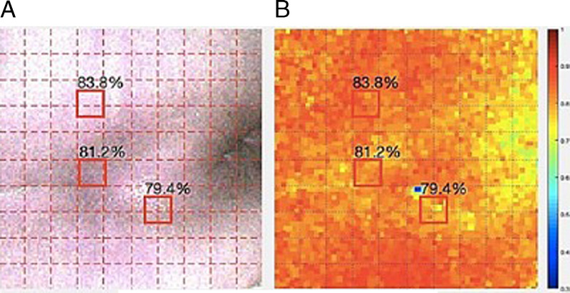
BLOOD OXYGEN CONCENTRATION DISTRIBUTION IN STAGE 1 PRESSURE INJURY TISSUE
Stage 2PI Tissue
Stage 2 PIs are characterized by partial cortical defect and visible dermis, as shown in Figure 2A. The tissue StO2 of the core area of the PI was 81.0% and 83.1%, whereas the StO2 of the healthy skin tissue on the upper left half of the PI site was 70.6%. In the scabbed tissue on the lower left side of the PI site, the StO2 was 65.9% and 66.5% (Figure 2A). The estimated tissue StO2 data indicated that the tissue StO2 was higher in the wound area than in other areas. The core part of the PI was uniformly red under the HSI, healthy skin was light red, and the scabbed area was light blue (Figure 2B).
Figure 2.
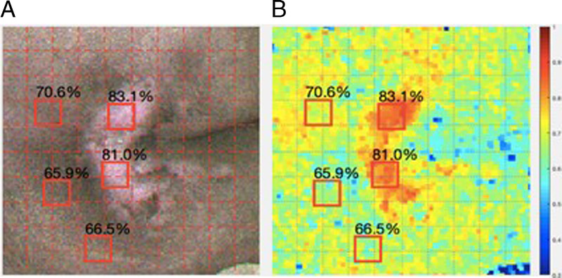
BLOOD OXYGEN CONCENTRATION DISTRIBUTION IN STAGE 2 PRESSURE INJURY TISSUE
Stage 3 PI Tissue
Stage 3 PIs are characterized by subcutaneous tissue and fat being visible to the naked eye, as shown in Figure 3A. The StO2 of the subcutaneous and fat tissue of the stage 3 sacral PI was 84.7%. The StO2 of the surrounding healthy skin was 74.4% and 76.1%, and the StO2 of the scab around the wound was 67.9% and 71.0% (Figure 3A). The PI subcutaneous tissue showed a wide range of red under HSI. Healthy skin was light red, and the scab, which had the lowest oxygen content, was light blue (Figure 3B). These results indicate that StO2 was higher in the wound area than in other areas.
Figure 3.
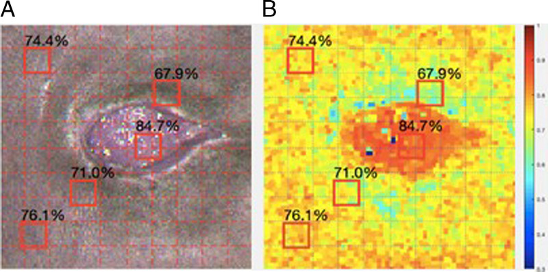
BLOOD OXYGEN CONCENTRATION DISTRIBUTION IN STAGE 3 PRESSURE INJURY TISSUE
Stage 4 PI Tissue
The stage 4 PI wound presents full-thickness skin and tissue defects as well as exposed fascia and muscle, as shown in Figure 4A. The tissue StO2 of the stage 4 PI subcutaneous tissue was the highest among all regions, 83.7% and 84.9%, followed by the tissue StO2 of the fascia at 79.9% and healthy skin at 75.5%. The StO2 of the scab around the wound was 64.1% (Figure 4A). The tissue StO2 in the wound area had a clear distribution and was higher than in other areas. With HSI, the fascia was red, the subcutaneous tissue was orange, the healthy skin was light red, and the scab, with the lowest oxygen content, was light blue (Figure 4B).
Figure 4.
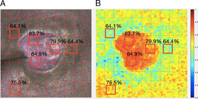
BLOOD OXYGEN CONCENTRATION DISTRIBUTION IN STAGE 4 PRESSURE INJURY TISSUE
Unstageable PI Tissue
As shown in Figure 5A, the overall area covered by scabbing had the lowest StO2 (44.3%) of all the PI tissues. The StO2 of the healthy skin around the wound was higher (73.6% and 74.7%), and the tissue with the highest StO2 (78.6%) was the new tender skin. This was a significant difference (P < .05) as compared with the StO2 of the wound tissue in the crusted area. The scab-covered area, which had the lowest oxygen content, was dark blue in the HSI, and the new skin was bright red (Figure 5B).
Figure 5.
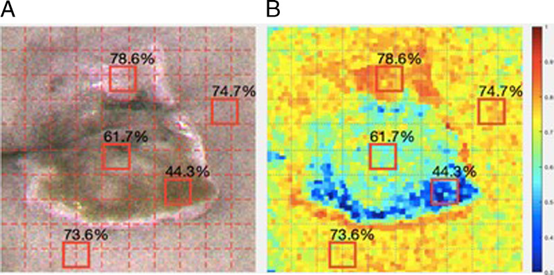
BLOOD OXYGEN CONCENTRATION DISTRIBUTION IN UNSTAGEABLE PRESSURE INJURY TISSUE
Tissue StO2 Analysis
Figure 6 shows box-and-whisker diagrams analyzing the StO2 values (which indicate the fraction of oxygen-saturated hemoglobin relative to total hemoglobin [unsaturated + saturated] in the blood as a percentage) of PI tissues by stage for all 30 cases. The stage 1 PI box-and-whisker diagram (n = 3) shows a median StO2 value of approximately 79.30%, indicating 79.30% oxygenated hemoglobin in the tissue. The tissue StO2 values of healthy skin and erythema skin were similar in stage 1 PIs. The stage 2 PI box-and-whisker chart (n = 10) shows statistically significant differences among median StO2 values in the wound tissue (82.50%), healthy skin (74.40%), and scab tissue (68.30%; P < .05).
Figure 6.
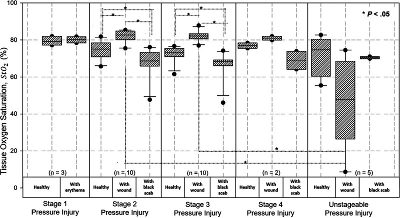
TISSUE BLOOD OXYGEN CONCENTRATION BOX-AND-WHISKER DIAGRAM OF PRESSURE INJURY STAGING
Similarly, the stage 3 PI StO2 box chart (n = 10) revealed that the subcutaneous tissue and adipose tissue in the core area of the wound had the highest median StO2 (82.50%), followed by healthy skin (73.75%) and skin with scabs (68.35%). The difference in StO2 between tissue types was statistically significant (P < .05).
In stage 4 PIs (n = 2), the median StO2s of healthy skin, wounded skin, and scabbed skin were 76.95%, 80.95%, and 69.00%, respectively (P = .067). In cases of unstageable PI (n = 5), the median StO2 values of healthy skin, wounded skin, and scabbed skin were 74.7%, 47.7%, and 69.9% respectively (P = .093). There were no significant differences in the distribution of the stage 4 and unstageable PIs because of the small number of cases.
Assessing StO2 Values According to Skin Condition
As shown in Figure 7, the authors divided the PI skin condition data of all 30 cases into wounded skin, scabbed skin, and healthy skin. Wounded skin had a higher median StO2 (81.9%) compared with healthy skin (75.3%) and scabbed skin (68.65%). The differences were statistically significant (P < .05).
Figure 7.
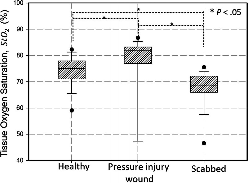
BOX-AND-WHISKER DIAGRAM OF BLOOD OXYGEN CONCENTRATIONS OF PRESSURE INJURY TISSUE BY SKIN CONDITION
DISCUSSION
The purpose of this study was to use noninvasive HSI to evaluate PI healing. The authors captured at least three HSI images in series to calculate StO2, rather than using only one data point. In accordance with the findings of Khaodhiar et al,14 the StO2 value of wound tissue was higher than that of the healthy surrounding skin or scabbed tissue. The results of the study provide objective information on the StO2 of PI tissues in different stages of wound healing. They also supplement the clinical subjective visual assessment of PI healing with empirical evidence and can inform guidelines for proper wound care.
The researchers used HSI to obtain StO2 data of PI tissues in different stages. The StO2 of the stage 1 PI tissue (79.4%–81.2%) was evenly distributed and close to that of healthy skin tissue (83.8%). In the stage 2 PI, the StO2 of the wound tissue was highest (82.5%), indicating that the injured tissue needed more oxygen during the healing process, and StO2 was lowest (68.3%) in the scab tissue of the unhealed wound. In the stage 3 PI, the StO2 of the subcutaneous and adipose tissues in the wound area was higher (82.5%) than that of the surrounding scab (68.35%). This finding is somewhat consistent with results of previous studies. Khaodhiar et al14 reported that the mean StO2 of wound tissue in a nonhealing diabetic foot ulcer was 38% ± 2%, and the StO2 of the wound tissue of the healing ulcer was 50% ± 3%.9 In addition, Yafi et al,13 who used the spatial frequency domain imaging method, reported that the StO2 of stage 1 PIs was evenly distributed as compared with healthy skin, whereas the StO2 of stage 2 PI tissue was higher compared with other areas and unstageable wounds. The StO2 of wound tissue was lower than that of healthy tissue.13
Khaodhiar et al14 also reported that ulceration could be observed by calculating tissue StO2 from hyperspectral images. Jayachandran et al7 reported that noninvasive optical imaging technology could provide larger quantities of accurate, more objective wound information about wound healing and treatment processes than visual inspection. In addition, a study on HSI in wound care by Saiko et al15 found that measuring tissue oxygenation may help predict the risk of wound formation or delayed healing. According to Yafi et al,13 the concentrations of oxygenated hemoglobin, nonoxygenated hemoglobin, and melanin should be calculated at the same time as tissue StO2. In the calculation of hyperspectral data, the three parameters are independent and do not affect one another, so skin color does not affect the calculation of tissue StO2.
Limitations
Because HSI is an emerging technology, it not yet popular in clinical practice. Considering that most patients with PIs are mobility challenged, and the impact of the COVID 19 pandemic, few patients were available for recruitment in this study. It took half a year to examine just 30 cases of patients with PIs. In addition, because of the limited data sources, the present study could not provide StO2 data for deep-tissue PIs.
CONCLUSIONS
The results of the study show that noninvasive HSI technology can be used for wound examination and healing assessment. These findings indicate that StO2 values are higher in the subcutaneous and fat tissue of stage 2 PI wound sites and in the core area of stage 3 PIs compared with healthy skin, which is helpful for the assessment of wound healing. This study also confirmed that HSI technology can provide a larger quantity of more accurate wound information during the PI healing process than is available from visual assessment to inform timely protective measures and treatment plans for PI decompression. Future areas for research are as follows: (1) adjusting PI preventive measures (eg, turning and repositioning) based on his data; (2) evaluating if HSI enables nurses to assess wound healing more quickly and objectively, saving time and resources; (3) assessing if early screening and prevention with HSI can reduce medical costs; and (4) investigating if HSI data can help prevent PIs in long-term care.
References
- 1.Berlowitz B. Epidemiology, pathogenesis and risk assessment of pressure ulcers. UpToDate. 2020. www.uptodate.com/contents/epidemiology-pathogenesis-and-risk-assessment-of-pressure-induced-skin-and-soft-tissue-injury. Last accessed April 18, 2022. [Google Scholar]
- 2.Liu Y Wu X Ma Y, et al. The prevalence, incidence, and associated factors of pressure injuries among immobile inpatients: a multicentre, cross-sectional, exploratory descriptive study in China. Int Wound J 2019;16:459–66. [DOI] [PMC free article] [PubMed] [Google Scholar]
- 3.National Pressure Ulcer Advisory Panel, European Pressure Ulcer Advisory Panel, and Pan Pacific Pressure Injury Alliance. Prevention and Treatment of Pressure Ulcers: Clinical Practice Guideline . Haesler E, ed. Cambridge Media: Osborne Park, Western Australia; 2014. [Google Scholar]
- 4.Choi JS, Hyun SY, Chang SJ. Comparing pressure injury incidence based on repositioning intervals and support surfaces in acute care settings: a quasi-experimental pragmatic study. Adv Skin Wound Care 2021;34(8):1–6. [DOI] [PubMed] [Google Scholar]
- 5.European Pressure Ulcer Advisory Panel . 2019 International EPUAP NPIAP PPPIA Pressure injury guideline. 2019. www.epuap.org/pu-guidelines. Last accessed April 18, 2022.
- 6.Daeschlein G Langner I Wild T, et al. Hyperspectral imaging as a novel diagnostic tool in microcirculation of wounds. Clin Hemorheol Microcirc 2017;67:467–74. [DOI] [PubMed] [Google Scholar]
- 7.Jayachandran M, Rodriguez S, Solis E, Lei J, Godavarty A. Critical review of noninvasive optical technologies for wound imaging. Adv Wound Care 2016;5:349–59. [DOI] [PMC free article] [PubMed] [Google Scholar]
- 8.Yudovsky D, Nouvong A, Pilon L. Hyperspectral imaging in diabetic foot wound care. J Diabetes Sci Technol 2010;4:1099–113. [DOI] [PMC free article] [PubMed] [Google Scholar]
- 9.Chen T, Yuen P, Richardson M, She Z, Liu G. Wavelength and model selection for hyperspectral imaging of tissue oxygen saturation. Imaging Sci J 2015;63:290–5. [Google Scholar]
- 10.Nouvong A, Hoogwerf B, Mohler E, Davis B, Tajaddini A, Medenilla E. Evaluation of diabetic foot ulcer healing with hyperspectral imaging of oxyhemoglobin and deoxyhemoglobin. Diabetes Care 2009;32:2056–61. [DOI] [PMC free article] [PubMed] [Google Scholar]
- 11.Paul DW Ghassemi P Ramella-Roman JC, et al. Noninvasive imaging technologies for cutaneous wound assessment: a review. Wound Repair Regen 2015;23:149–62. [DOI] [PubMed] [Google Scholar]
- 12.Dicker DT Lerner J Van Belle P, et al. Differentiation of normal skin and melanoma using high resolution hyperspectral imaging. Cancer Biol Ther 2006;5:1033–8. [DOI] [PubMed] [Google Scholar]
- 13.Yafi A Muakkassa FK Pasupneti T, et al. Quantitative skin assessment using spatial frequency domain imaging (SFDI) in patients with or at high risk for pressure ulcers. Lasers Surg Med 2017;49:827–34. [DOI] [PubMed] [Google Scholar]
- 14.Khaodhiar L Dinh T Schomacker KT, et al. The use of medical hyperspectral technology to evaluate microcirculatory changes in diabetic foot ulcers and to predict clinical outcomes. Diabetes Care 2007;30:903–10. [DOI] [PubMed] [Google Scholar]
- 15.Saiko G, Lombardi P, Au Y, Queen D, Armstrong D, Harding K. Hyperspectral imaging in wound care: a systematic review. Int Wound J 2020;17(6):1840–56. [DOI] [PMC free article] [PubMed] [Google Scholar]


