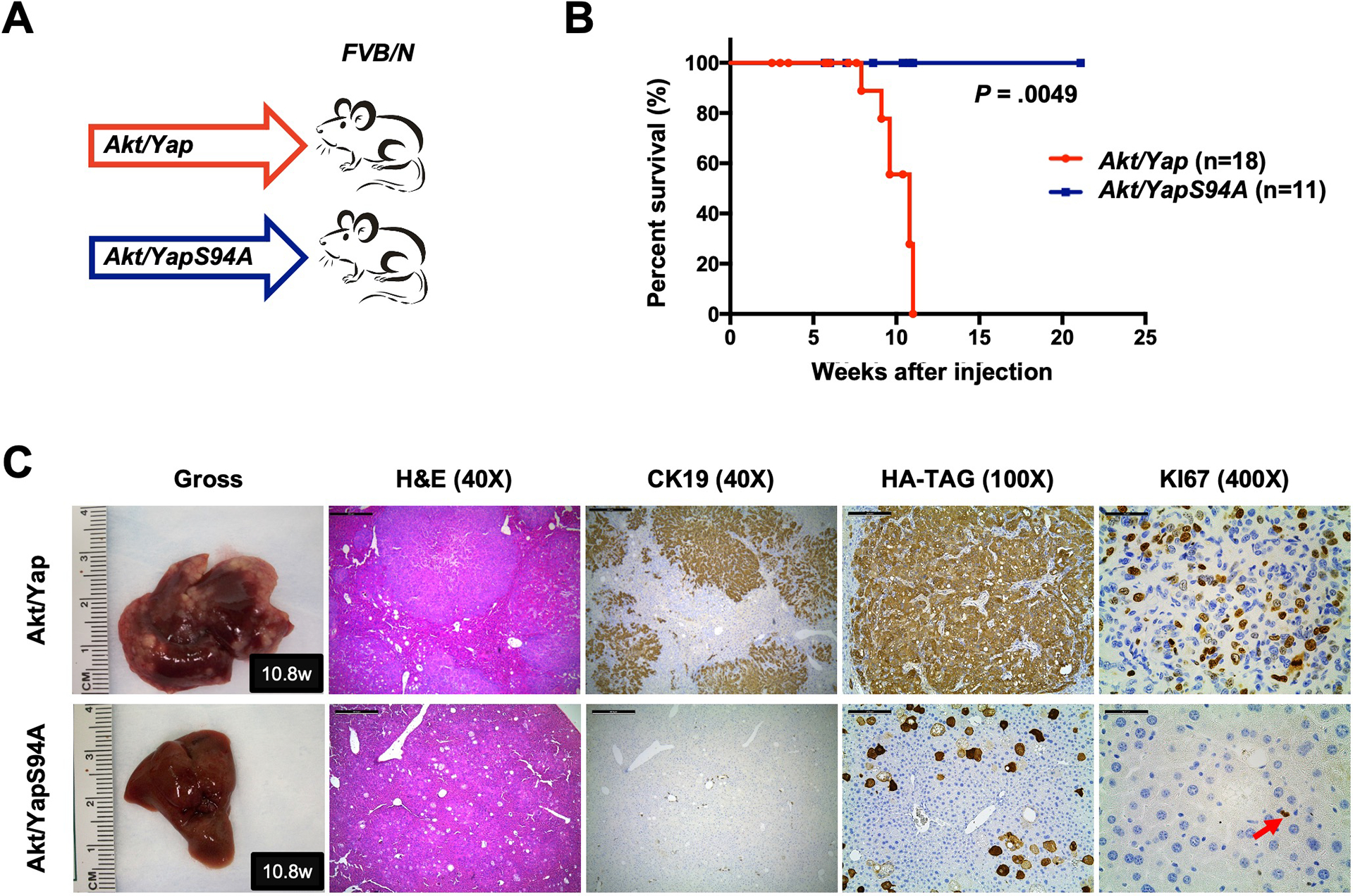
Figure 1. TEADs is required for Yap-driven iCCA formation in mice.

(A) Study design. FVB/N mice were subjected to HTVi of either Akt/Yap (n=18) or Akt/YapS94A (n = 11) plasmids. (B) Mouse survival curves. (C) Representative gross images, H&E, and immunohistochemistry for CK19, HA-TAG and KI67 of liver sections from Akt/Yap (M10.8w.p.i) and Akt/YapS94A (F10.8w.p.i) mice. The red arrow indicates KI67 positive stained nuclei. Scale bar: 500 μm (40x), 100 μm (100x), and 50 μm (400x). Abbreviations: H&E, hematoxylin and eosin staining; w.p.i, weeks post-injection.
