



Abstract
Purpose:
We sought to analyze biologic, clinical, and prognostic differences according to pattern of failure at the time of first relapse in neuroblastoma.
Patients and Methods:
Children <21 years diagnosed with neuroblastoma between 1989–2017 with known site of first relapse (isolated local vs. distant only vs. combined local and distant sites) were identified from the INRG database. Data were compared between sites of relapse according to clinical features, biologic features, initial treatment, time to first relapse, and overall survival (OS) from time of first relapse.
Results:
Pattern of first relapse among 1,833 children was: 19% isolated local; 65% distant only; and 16% combined sites. All evaluated clinical and biologic variables with exception of tumor diagnosis differed statistically by relapse pattern, with patients with isolated local failure having more favorable prognostic features. Patients with stage 3 disease were more likely to have isolated local failure compared to all other stages (49% vs. 16%; p<0.001). OS significantly differed by relapse pattern (5-year OS ± SE): isolated local, 64% ± 3%; distant only, 23 ± 2%; and combined, 26 ± 4% (p<0.001). After controlling for age, stage, and MYCN status, patients with isolated local failure (adjusted HR=0.46; 95% CI, 0.33–0.62; p<0.001) and distant only failure (adjusted HR=0.57; 95% CI, 0.45–0.71; p<0.001) remained at decreased risk for death as compared to patients with combined failure.
Conclusion:
Patients with distant only and combined failures have a higher proportion of unfavorable clinical and biological features, and a lower survival than those with isolated local relapse.
Keywords: neuroblastoma, relapse, pattern of failure, survival
INTRODUCTION
The combination of myeloablative therapy, anti-GD2 immunotherapy, and differentiation therapy with 13-cis-retinoic acid has led to an improvement in survival for high-risk neuroblastoma.1 However, >50% of these children will still relapse. As relapse remains the major obstacle to cure for patients with high-risk neuroblastoma, it is imperative to assess the patterns of relapse and the clinical and biologic predictors of specific patterns of failure. Recurrence exclusively in sites of previous disease might suggest the need for more intensive local treatment with irradiation or more aggressive surgery. Alternatively, recurrence at distant sites suggests the possibility of resistance to systemic therapies and biologic tumor or host factors favoring metastases.
Previous smaller studies have investigated the patterns of local and distant sites of relapse in neuroblastoma patients who present with either low-risk or high-risk disease.2–8 Recurrences of low stage neuroblastoma are infrequent events and the pattern of progression has rarely been reported, while other larger studies have concentrated on the clinical and biological factors at diagnosis.3,4
These studies suggest that there may be clinical and biologic characteristics that are associated with patterns of failure. Moreover, a clear understanding of the impact of the sites of relapse has not been possible from prior studies. We therefore performed a comprehensive analysis using the largest available cohort of patients with neuroblastoma, the International Neuroblastoma Risk Group (INRG) database, to assess whether clinical features, tumor biologic features, and survival differ according to pattern of failure.
PATIENTS AND METHODS
Patients
Patients younger than 21 years of age with pathologically confirmed neuroblastoma or ganglioneuroblastoma who were diagnosed/enrolled starting in 1989 comprise the INRG database.9 Patients provided consent and were enrolled into one or more neuroblastoma clinical trials or biologic studies in Japan, Germany, Italy, Spain, or the United Kingdom or into a North American Children’s Oncology Group (COG) study or an International Society of Paediatric Oncology Europe Neuroblastoma Group (SIOPEN) study. Each country, cooperative group, and treating institution obtained institutional review board approval and informed patient consent for their respective studies. In addition to survival outcomes, information on 35 potential risk factors is included in the INRG database.9
Only those patients diagnosed from 1989–2017 with known site of first relapse/progression (referred to simply as “relapse” for the remainder of this report) were included in the analytic cohort (N=1,833). Sites of first relapse were categorized as isolated local failure for relapse at original primary site including local/regional lymph nodes, distant only failure for relapse only at distant sites even if previously involved at original diagnosis, and combined local and distant failure. Patients with refractory disease without prior progression were excluded.
Statistical Analysis
Site of relapse was the predictor variable of interest in this analysis. Clinical and biological dependent variables described in the INRG database at initial diagnosis and analyzed in this study are listed in Table 1. For ferritin and LDH, median values from this analytic cohort (172 units/L and 1,012 ng/mL, respectively) were used to dichotomize patients as elevated or not elevated following the convention used for previous INRG analyses.9 The INRG database includes data on LOH at 1p, gain of 17q, and 11q aberration. We created a pooled variable reflecting the presence of segmental chromosomal aberration if at least one of these aberrations was present.10 Clinical and biologic features were compared between groups defined by sites of relapse using chi-squared tests (categorical variables); Student’s t-test or Mann-Whitney U test (two-group comparisons of continuous variables); and analysis of variance between groups (ANOVA) or Kruskal-Wallis test (three-group comparisons of continuous variables).
Table 1:
Clinical and biologic characteristics of the INRG analytic cohort by site of first relapse (isolated local vs. distant only vs. combined; N = 1833)
| Factora | All (N = 1833)b | Site of First Relapse |
p-valuec | ||
|---|---|---|---|---|---|
| Isolated Local (N = 347) | Distant Only (N = 1185) | Combined (N = 301) | |||
|
| |||||
| Mean age at diagnosis, months | 43.7 | 31.6 | 46.8 | 45.1 | <0.001 |
|
| |||||
| Age at diagnosis | |||||
| ≥ 18 months | 1363/1833 (74.4%) | 182/347 (52.5%) | 957/1185 (80.8%) | 224/301 (74.4%) | <0.001 |
|
| |||||
| Tumor diagnosisd | |||||
| Neuroblastoma or ganglio-neuroblastoma, nodular | 1286/1302 (98.8%) | 288/292 (98.7%) | 779/787 (99%) | 219/223 (98.2%) | 0.63 |
|
| |||||
| INSS Stage | |||||
| 4 | 1320/1808 (73%) | 113/346 (32.6%) | 999/1162 (86%) | 208/300 (69.3%) | <0.001 |
|
| |||||
| INSS Stage 3 (vs. all others) | 181/1808 (10%) | 88/346 (25.4%) | 53/1162 (4.6%) | 40/300 (13.3%) | <0.001 |
|
| |||||
| Serum ferritin (ng/mL) | |||||
| ≥ 172 | 154/307 (50.2%) | 17/59 (28.8%) | 108/198 (54.6%) | 29/50 (58%) | 0.001 |
|
| |||||
| LDH (U/L) | |||||
| ≥ 1012 | 194/388 (50%) | 23/70 (32.9%) | 136/256 (53.1%) | 35/62 (56.5%) | 0.006 |
|
| |||||
| MYCN status | |||||
| Amplified | 538/1634 (32.9%) | 66/329 (20%) | 392/1037 (37.8%) | 80/268 (29.9%) | <0.001 |
|
| |||||
| Ploidy | |||||
| ≤ 1 (diploid, hypodiploid) | 728/1498 (48.6%) | 106/286 (37.1%) | 490/970 (50.5%) | 132/242 (54.6%) | <0.001 |
|
| |||||
| Pooled segmental chromosomal aberration | |||||
| LOH at 1p, gain of 17q, and/or 11q aberration | 347/630 (55.1%) | 53/155 (34.2%) | 226/361 (62.6%) | 68/114 (59.7%) | <0.001 |
|
| |||||
| INPC pathology classification | |||||
| Unfavorable | 1114/1435 (77.6%) | 163/314 (51.9%) | 761/879 (86.6%) | 190/242 (78.5%) | <0.001 |
|
| |||||
| MKI | |||||
| High | 364/1207 (30.2%) | 61/282 (21.6%) | 247/720 (34.3%) | 56/205 (27.3%) | <0.001 |
|
| |||||
| Grade of differentiation | |||||
| Undifferentiated/Poorly differentiated | 1286/1357 (94.8%) | 264/292 (90.4%) | 801/835 (95.9%) | 221/230 (96.1%) | 0.001 |
|
| |||||
| Initial Treatment | |||||
| Intensive multi-modal therapye | 888/1089 (81.5%) | 71/151 (47%) | 676/765 (88.4%) | 141/173 (81.5%) | <0.001 |
|
| |||||
| Median time to first relapse, months | 12.8 (N=1020) | 10.1 | 14.6 | 10.8 | <0.001 |
– For each variable, only the percent with the adverse risk factor is shown.
– Adverse risk factor sample size over the total sample size with data available for the variable of interest.
– p-value refers to a one-way ANOVA test (for continuous age variable), Kruskal-Wallis test (for continuous time to first relapse variable), or X2 test for all other variables (age, tumor diagnosis, INSS Stage, serum ferritin, LDH, MYCN status, ploidy, pooled segmental chromosomal classification, INPC pathology classification, MKI, grade of differentiation and initial treatment categories).
– International Neuroblastoma Pathology Classification diagnostic category. Neuroblastoma or ganglioneuroblastoma, nodular vs. ganglioneuroblastoma, intermixed, maturing subtype; or ganglioneuroblastoma, well differentiated.
– Intensive multi-modal therapy vs. all other types of therapy (no therapy, surgery only, or chemotherapy and surgery)
Abbreviations: INSS, International Neuroblastoma Staging System; LDH, lactate dehydrogenase; MYCN, N-myc protooncogene; LOH, loss of heterozygosity; INPC, International Neuroblastoma Pathology Classification; MKI, Mitosis Karyorrhexis Index.
We fit logistic regression models to describe the clinical and biological predictors of isolated local failure after controlling for key potential confounders. Determined a priori, clinical and biologic variables were included in the logistic regression model if p<0.05 and had <30% missing data. Given the striking differences in the clinical and biological predictors between sites of relapse in Table 1, we used a more stringent p-value of <0.05 for inclusion in our logistic regression model. We also excluded variables that were dependent on other variables included in this model. For example, the International Neuroblastoma Pathology Classification (INPC) histology classification system incorporates age as well as grade, MKI, and tumor diagnosis. To avoid confounding of INPC histology and age in multivariate models, only the underlying components (i.e., age, grade) were included if they met the above a priori parameters.
Overall survival (OS) was defined as time from first relapse until death, with living patients censored at time of last contact. OS was estimated using Kaplan-Meier methods with survival distributions compared according to pattern of relapse using a two-sided log-rank test.11 Cox proportional hazards regression models were used to calculate the hazard ratio (HR) for increased risk of death while controlling for key potential confounders. Time-dependent covariates were used to test the proportional hazards assumption. Any variables that did not satisfy the proportional hazards assumption were removed as covariates from the model and instead used as stratification variables, including age, stage, and MYCN status as these three variables have been shown to be independently predictive of survival after relapse in neuroblastoma.12 All statistical analyses were performed using STATA, version 13.
RESULTS
Patient Characteristics
The clinical and biological characteristics at diagnosis of the 1,833 patients in the INRG analytic cohort with a known site of first relapse are listed in Table 1. As the cohort included only patients with relapse, features are enriched for unfavorable prognostic factors, although, some of the patients in the analytic cohort may have been classified as low- and intermediate-risk at the time of diagnosis. There were 19% isolated local failure, 65% distant only failure, and 16% combined local and distant failure. Histograms showing the distribution of the time to first relapse by site of failure are shown in Figure 1 (N=1020 with available data). The median time from diagnosis to first relapse was 12.8 months (Table 1). Timing of first relapse differed according to pattern of relapse with patients who had distant only relapse occurring later than those with isolated local or combined relapses (p<0.001).
Figure 1:
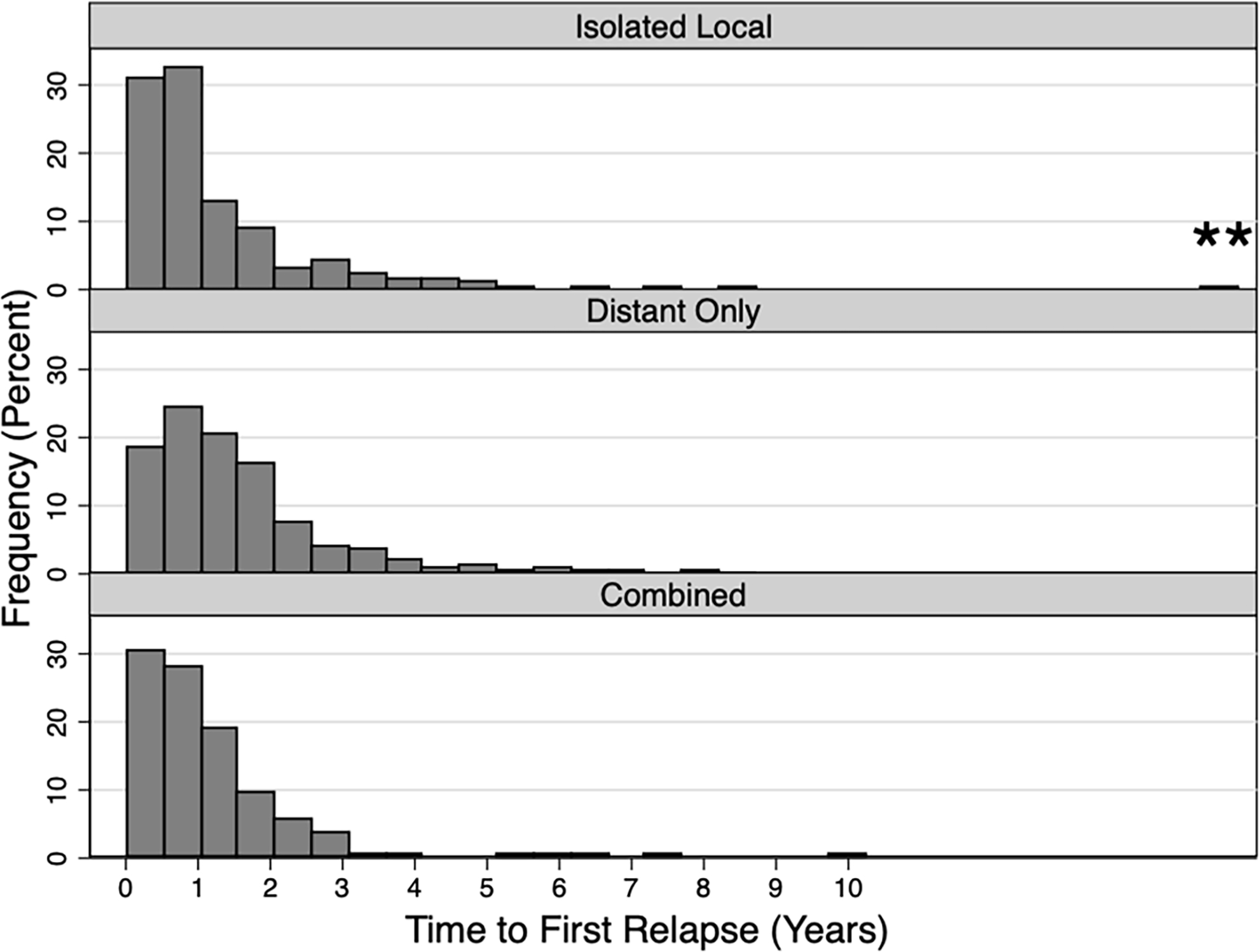
Distribution of the time to first relapse of the INRG analytic cohort according to site of failure (isolated local vs. distant only vs. combined; N = 1020). The median time from diagnosis to first relapse was 12.8 months
**Patient outlier who relapsed after 10 years
Clinical and Biological Features Differ by Site of Relapse
Each of the evaluated clinical and biologic features evaluated in Table 1 showed statistically significant differences when compared across all three sites of relapse categories (p<0.05), with the exception of the INPC diagnostic category (Table 1). We observed that patients with isolated local failure had more favorable prognostic features. We also analyzed clinical and biologic features between patients with any local failure (i.e., isolated local or combined failure) and those with no local failure (i.e., distant only failure; Supplemental Table 1). Each of the evaluated clinical and biologic features showed statistically significant differences when compared across these two categories (p<0.05), with exception of the INPC diagnostic category and LDH (Supplemental Table 1). We again observed that patients with any local failure had more favorable prognostic features.
INSS stage was known for 1,808 of patients in our analytic cohort: 5% with stage 1; 9% with stage 2; 10% with stage 3; 73% with stage 4; and 3% with stage 4s. In 255 patients with INSS stages 1 and 2 in our analytic cohort, 133 (52%) had isolated local failure, 84 (33%) had distant only failure, and 38 (15%) had combined local and distant failure. In patients with stage 3 disease, there was a higher proportion of patients who had isolated local failure (88/181, 49%) compared to patients with all other stages (258/1,627, 16%; p<0.001).
Given the striking differences in the incidence of clinical and biologic features according to pattern of relapse, we used multivariate logistic regression analysis to identify predictors of isolated local failure while controlling for key confounders (Table 2). After adjusting for age at diagnosis, stage, MYCN status, ploidy, and grade of differentiation, we found that patients with age ≥ 18 months (adjusted odds ratio=0.53; 95% CI, 0.37–0.74; <0.001) and stage 4 disease (adjusted odds ratio=0.14; 95% CI, 0.10–0.20; <0.001) had statistically lower odds of having isolated local failure (Table 2).
Table 2:
Logistic regression analysis of clinical and biologic predictors of isolated local failure for age at diagnosis, INSS stage, MYCN status, ploidy, and grade of differentiation. The reference group for each of the clinical and biologic predictors is the favorable risk factor (not shown)
| Isolated Local Failure | Adjusted Odds Ratio (95% CI) | p-value |
|---|---|---|
| Age at diagnosis ≥ 18 months | 0.53 (0.37–0.74) | <0.001 |
| INSS Stage 4 | 0.14 (0.10–0.20) | <0.001 |
| MYCN amplification | 0.90 (0.60–1.34) | 0.60 |
| Ploidy ≤ 1 (diploid, hypodiploid) | 0.98 (0.69–1.39) | 0.89 |
| Undifferentiated/Poorly differentiated grade | 0.58 (0.32–1.04) | 0.069 |
We also evaluated the clinical and biologic characteristics of patients with any distant relapse by specific sites of first relapse, including bone/bone marrow, liver, lung, and CNS (N=1,379; Supplemental Table 2). In general, patients who relapsed in the bone/bone marrow had a statistically higher proportion of adverse prognostic factors, including age ≥ 18 months, stage 4 disease, unfavorable INPC pathology classification, and intensive multi-modal initial treatment compared to those who did not relapse in the bone/bone marrow (p<0.001). Interestingly, MYCN amplification was seen less frequently in patients who relapsed in the bone/bone marrow compared to those who did not relapse in the bone/bone marrow (32% vs. 43%, p<0.001). In patients who relapsed in the CNS, there was a statistically lower proportion of older patients (≥ 18 months at diagnosis) compared to those who did not relapse in the CNS (50% vs. 80%, p=0.005).
Overall Survival Differs According to Pattern of Relapse
We next evaluated potential differences in OS according to pattern of first relapse. Patients with isolated local failure had significantly improved OS from time of first relapse (p<0.001; Figure 2A). Five-year OS rates according to the three relapse categories were as follows (± standard error, SE): isolated local failure, 64 ± 3%; distant only failure, 23 ± 2%; and combined local and distant failure, 26 ± 4%. A different pattern was observed in patients age ≥ 18 months with stage 4 disease in whom patients with combined local and distant failure had inferior outcomes and isolated local failure was no longer associated with more favorable outcomes (p<0.001; Figure 2B). Five-year OS rates were as follows (± SE): isolated local failure, 14 ± 5%; distant only failure, 12 ± 2%; and combined local and distant failure, 5 ± 3%. Comparatively, in patients with stage 1 and 2 disease, their 5-year OS rates were as follows (± SE): isolated local failure, 92 ± 3%; distant only failure, 65 ± 7%; and combined local and distant failure, 61 ± 10% (Figure 3).
Figure 2A:

Kaplan-Meier estimated overall survival from time of first relapse according to site of relapse
Figure 2B:
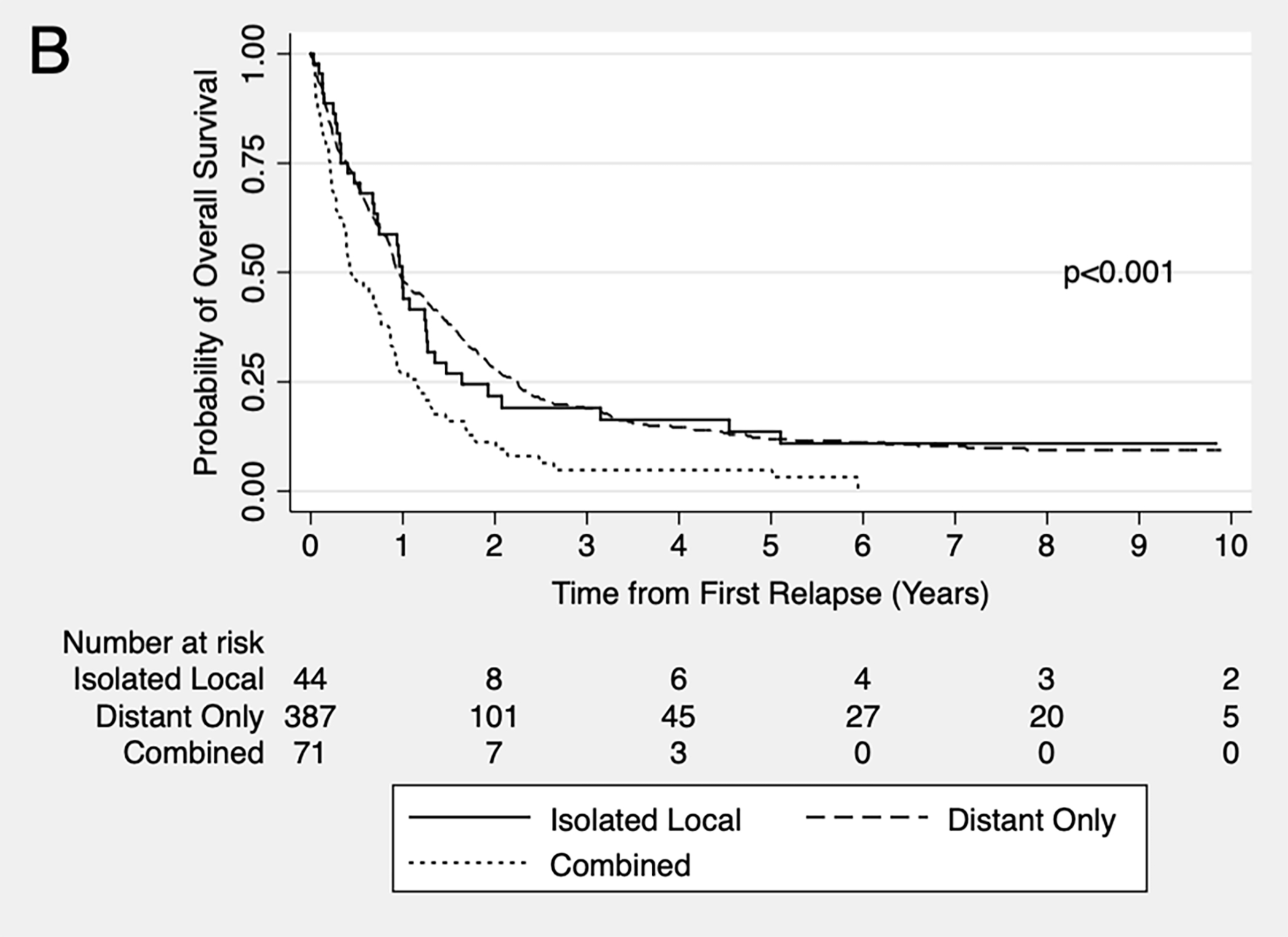
Kaplan-Meier estimated overall survival from time of first relapse according to site of relapse in patients age ≥ 18 months and INSS stage 4 disease
Figure 3:

Kaplan-Meier estimated overall survival from time of first relapse according to site of relapse in patients with INSS stage 1 and 2 disease
Our finding that the three main prognostic factors in neuroblastoma (age, stage, and MYCN status) also differed significantly according to pattern of relapse raised the possibility that these differences confounded our univariate observation of differential OS according to pattern of relapse. We therefore constructed Cox proportional hazards models to control for these differences in age, stage, and MYCN (Table 3). Using this model, we evaluated OS from time of first relapse and found that patients with isolated local failure (adjusted HR=0.46; 95% CI, 0.33–0.62; p<0.001) and distant only failure (adjusted HR=0.57; 95% CI, 0.45–0.71; p<0.001) remained at a decreased risk for death as compared to the reference group of patients with combined local and distant failure.
Table 3:
Cox proportional hazards regression analysis of the association between site of first relapse and overall survival from first relapse without (unadjusted hazard ratio) and with adjustment (adjusted hazard ratio) for age at diagnosis, INSS stage and MYCN status
| Site of Relapse | Unadjusted Hazard Ratio (95% CI) | p-value | Adjusted Hazard Ratio (95% CI) | p-value |
|---|---|---|---|---|
| Combined local and distant failure | 1 (Reference) | Reference | 1 (Reference) | Reference |
| Distant only failure | 0.91 (0.73–1.11) | 0.34 | 0.57 (0.45–0.71) | <0.001 |
| Isolated local failure | 0.31 (0.23–0.41) | <0.001 | 0.46 (0.33–0.62) | <0.001 |
DISCUSSION
In this large comprehensive analysis of patterns of relapse in neuroblastoma, we observed that the majority of relapses include failure at distant sites, emphasizing the need for improved systemic therapies in this disease. Still, 35% of patients experienced any local failure at first relapse, highlighting the need for efforts to improve locoregional control as well. We found statistically significant differences in clinical and biological characteristics according to pattern of relapse, with more favorable features enriched in patients with isolated local failure. Importantly, we also observed that patients with combined local and metastatic relapse had inferior OS independent of age at diagnosis, stage, and MYCN status in our analytic cohort. The time to relapse in this analysis was similarly seen in other studies.12,13 In a sub-analysis of patients with INSS stage 1 and 2 disease, patients with any distant failure had inferior outcomes to those with isolated local failure, though all groups had five-year OS rates > 50%, suggesting that these patients are salvageable with additional therapy.
Our findings that clinical and biologic features differ according to site of relapse confirm and extend previous observations. Clinical and biological predictors of survival after first relapse were further explored in a recent retrospective study that proposed a new risk score system in a patients with stage 4 disease and age ≥ 18 months.8 Comparable to our study, isolated local failure was seen in 15% (72/469) of patients. However, a higher proportion of patients with combined failure were seen in that study (28%, 131/469). The prognostic index included the following adverse variables which predicted secondary EFS: number of recurrence organs (> 1 vs. 1), time to recurrence, presence of liver metastasis at diagnosis, first recurrence at site of the primary tumor, and age at diagnosis (< 42 months vs. ≥ 42 months).
Prior studies have examined the role of radiation in treating distant metastatic sites in high-risk neuroblastoma in an attempt to reduce distant failure.6,7 These observations support irradiating metastases that persist after induction chemotherapy in high-risk patients. However, we were unable to compare disease recurrence in new versus prior discrete metastatic sites in our study.
Recent trials have evaluated the extent of surgical resection and radiation dose to reduce local failure rates and improve survival in high-risk neuroblastoma patients. Greater extent of surgical resection appears to decrease cumulative incidence of local progression (CILP) and improve EFS.14,15 However, there were no significant differences in CILP or survival based on the extent of lymph node irradiation regardless of the degree of surgical resection.16 Recently, Liu et al. evaluated the impact of increasing the radiation dose to residual primary tumor on CILP in patients enrolled on COG ANBL0532.17,18 Strategies to boost radiotherapy to gross residual tumor did not significantly improve CILP or survival.18 These results highlight the need for new strategies to decrease the risk of locoregional failure.
There are several limitations to analyzing data from a tumor registry. We were limited to the available variables in the database. There were patients who relapsed who did not have a site of relapse available in the INRG database and were not included in the analytic cohort. We were also unable to report on important treatment variables, such as extent of local control and immunotherapy, which might impact sites of relapse. Finally, some variables were missing, as noted in Table 1, and we could only report on what was available in the database. Determined a priori, clinical and biologic variables with <30% missing data were included in the logistic regression model as described in our statistical analysis plan. Within each variable, we also performed an assessment to determine whether the primary variable of interest was missing at random as previously described.19 Variables with 0–10% missing data were considered to have trivial effect on the analysis. Of variables with 10–30% missing data included in the analysis, INPC diagnostic classification, MYCN, and grade variables all had data that were missing not at random. We did not specifically exclude these variables in our models.
A key advantage of our study is our ability to control for potential confounding variables that might be associated both with site of relapse and prognosis. As the largest multivariable analysis addressing the prognostic impact of site of relapse, our study demonstrated that the inferior outcomes for patients with any distant failure compared to isolated local failure are independent of differences in age, stage, and MYCN status.
We conclude that there are significant differences in clinical features, biologic characteristics, and outcomes according to patterns of relapse in neuroblastoma. Significantly, survival in patients with combined local and metastatic failure is worse than patients with isolated local or distant only failure, independent of age at diagnosis, stage, and MYCN status.
Supplementary Material
ACKNOWLEDGEMENTS
The INRG database is supported in part by the William Guy Forbeck Research Foundation, the Little Heroes Cancer Research Fund, Children’s Neuroblastoma Cancer Foundation, Neuroblastoma Children’s Cancer Foundation, and the Super Jake Foundation. Data included in the INRG database were provided by Children’s Oncology Group (COG), Pediatric Oncology Group (POG), Children’s Cancer Study Group (CCSG), German Gesellschaft für Pädiatrische Onkologie und Hämatologie (GPOH), European Neuroblastoma Study Group (ENSG), International Society of Paediatric Oncology Europe Neuroblastoma Group (SIOPEN), Japanese Advanced Neuroblastoma Study Group (JANB), Japanese Infantile Neuroblastoma Co-operative Study Group (JINCS), Spanish Neuroblastoma Group and the Italian Neuroblastoma Group. Supported also in part by the Alex’s Lemonade Stand Foundation (KTV, SGD, KKM), Frank A. Campini Foundation (KTV), Posey Family Foundation (KTV), Edward Conner Fund (KKM), and the Mildred V. Strouss Chair (KKM). This publication was also supported by the National Center for Advancing Translational Sciences, National Institutes of Health (NIH), through UCSF-CTSI Grant KL2 TR001870 (KTV). The contents are solely the responsibility of the authors and do not necessarily represent the official views of the NIH.
Abbreviations:
- ANOVA
analysis of variance between groups
- CI
confidence interval
- CILP
cumulative incidence of local progression
- COG
Children’s Oncology Group
- CNS
central nervous system
- EFS
event-free survival
- HR
hazard ratio
- INPC
International Neuroblastoma Pathology Classification
- INRG
International Neuroblastoma Risk Group
- INSS
International Neuroblastoma Staging System
- LOH
loss of heterozygosity
- LDH
lactate dehydrogenase
- MKI
mitotic-karyorrhectic index
- OS
overall survival
- SE
standard error
- SIOPEN
International Society of Paediatric Oncology Europe Neuroblastoma Group
Footnotes
CONFLICTS OF INTEREST
None
This work was previously presented in abstract form at the Advances in Neuroblastoma Research (ANR) virtual meeting in January 2021.
DATA SHARING AND DATA ACCESSIBILITY
The data for this study are publicly available in the INRG Data Commons; see http://inrgdb.org/publication-policy/apply/ for access.
REFERENCES
- 1.Maris JM, Hogarty MD, Bagatell R, Cohn SL. Neuroblastoma. Lancet 2007;369:2106–20. [DOI] [PubMed] [Google Scholar]
- 2.Matthay KK, Atkinson JB, Stram DO, Selch M, Reynolds CP, Seeger RC. Patterns of relapse after autologous purged bone marrow transplantation for neuroblastoma: a Childrens Cancer Group pilot study. J Clin Oncol 1993;11:2226–33. [DOI] [PubMed] [Google Scholar]
- 3.Berthold F, Hero B, Breu H, et al. The recurrence patterns of stages I, II and III neuroblastoma: experience with 77 relapsing patients. Ann Oncol 1996;7:183–7. [DOI] [PubMed] [Google Scholar]
- 4.Garaventa A, Parodi S, De Bernardi B, et al. Outcome of children with neuroblastoma after progression or relapse. A retrospective study of the Italian neuroblastoma registry. Eur J Cancer 2009;45:2835–42. [DOI] [PubMed] [Google Scholar]
- 5.Simon T, Berthold F, Borkhardt A, Kremens B, De Carolis B, Hero B. Treatment and outcomes of patients with relapsed, high-risk neuroblastoma: results of German trials. Pediatr Blood Cancer 2011;56:578–83. [DOI] [PubMed] [Google Scholar]
- 6.Polishchuk AL, Li R, Hill-Kayser C, et al. Likelihood of bone recurrence in prior sites of metastasis in patients with high-risk neuroblastoma. Int J Radiat Oncol Biol Phys 2014;89:839–45. [DOI] [PubMed] [Google Scholar]
- 7.Li R, Polishchuk A, DuBois S, et al. Patterns of Relapse in High-Risk Neuroblastoma Patients Treated With and Without Total Body Irradiation. Int J Radiat Oncol Biol Phys 2017;97:270–7. [DOI] [PubMed] [Google Scholar]
- 8.Kreitz K, Ernst A, Schmidt R, et al. A new risk score for patients after first recurrence of stage 4 neuroblastoma aged ≥18 months at first diagnosis. Cancer medicine 2019;8:7236–43. [DOI] [PMC free article] [PubMed] [Google Scholar]
- 9.Cohn S, Pearson A, London W, al e. The International Neuroblastoma Risk Group (INRG) classification system: An INRG Task Force report. J Clin Oncol 2009;27:289–97. [DOI] [PMC free article] [PubMed] [Google Scholar]
- 10.Schleiermacher G, Mosseri V, London WB, al e. Segmental chromosomal alterations have prognostic impact in neuroblastoma: a report from the INRG project. Br J Cancer 2012;107:1418–22. [DOI] [PMC free article] [PubMed] [Google Scholar]
- 11.Kaplan EL, Meier P. Nonparametric estimation from incomplete observations. J Am Stat Assoc 1958;53:457–81. [Google Scholar]
- 12.London WB, Castel V, Monclair T, et al. Clinical and biologic features predictive of survival after relapse of neuroblastoma: a report from the International Neuroblastoma Risk Group project. Journal of clinical oncology : official journal of the American Society of Clinical Oncology 2011;29:3286–92. [DOI] [PMC free article] [PubMed] [Google Scholar]
- 13.Kreissman SG, Seeger RC, Matthay KK, et al. Purged versus non-purged peripheral blood stem-cell transplantation for high-risk neuroblastoma (COG A3973): a randomised phase 3 trial. The Lancet Oncology 2013;14:999–1008. [DOI] [PMC free article] [PubMed] [Google Scholar]
- 14.Holmes K, Potschger U, Pearson ADJ, et al. Influence of Surgical Excision on the Survival of Patients With Stage 4 High-Risk Neuroblastoma: A Report From the HR-NBL1/SIOPEN Study. J Clin Oncol 2020:JCO1903117. [DOI] [PubMed] [Google Scholar]
- 15.von Allmen D, Davidoff AM, London WB, et al. Impact of Extent of Resection on Local Control and Survival in Patients From the COG A3973 Study With High-Risk Neuroblastoma. J Clin Oncol 2017;35:208–16. [DOI] [PMC free article] [PubMed] [Google Scholar]
- 16.Braunstein SE, London WB, Kreissman SG, et al. Role of the extent of prophylactic regional lymph node radiotherapy on survival in high-risk neuroblastoma: A report from the COG A3973 study. Pediatr Blood Cancer 2019;66:e27736. [DOI] [PMC free article] [PubMed] [Google Scholar]
- 17.Park JR, Kreissman SG, London WB, et al. Effect of Tandem Autologous Stem Cell Transplant vs Single Transplant on Event-Free Survival in Patients With High-Risk Neuroblastoma: A Randomized Clinical Trial. Jama 2019;322:746–55. [DOI] [PMC free article] [PubMed] [Google Scholar]
- 18.Liu KX, Naranjo A, Zhang FF, et al. Prospective Evaluation of Radiation Dose Escalation in Patients With High-Risk Neuroblastoma and Gross Residual Disease After Surgery: A Report From the Children’s Oncology Group ANBL0532 Study. J Clin Oncol 2020:JCO1903316. [DOI] [PMC free article] [PubMed] [Google Scholar]
- 19.Thompson D, Vo KT, London WB, et al. Identification of patient subgroups with markedly disparate rates of MYCN amplification in neuroblastoma: A report from the International Neuroblastoma Risk Group project. Cancer 2016;122:935–45. [DOI] [PMC free article] [PubMed] [Google Scholar]
Associated Data
This section collects any data citations, data availability statements, or supplementary materials included in this article.
Supplementary Materials
Data Availability Statement
The data for this study are publicly available in the INRG Data Commons; see http://inrgdb.org/publication-policy/apply/ for access.