Abstract
The essential oil (EO) of Calycolpus goetheanus (Myrtaceae) specimens (A, B, and C) were obtained through hydrodistillation. The analysis of the chemical composition of the EOs was by gas chromatography coupled with mass spectrometry CG-MS, and gas chromatography coupled with a flame ionization detector CG-FID. The phytotoxic activity of those EOs was evaluated against two weed species from common pasture areas in the Amazon region: Mimosa pudica L. and Senna obtusifolia (L.) The antioxidant capacity of the EOs was determined by (DPPH•) and (ABTS•+). Using molecular docking, we evaluated the interaction mode of the major EO compounds with the molecular binding protein 4-hydroxyphenylpyruvate dioxygenase (HPPD). The EO of specimen A was characterized by β-eudesmol (22.83%), (E)-caryophyllene (14.61%), and γ-eudesmol (13.87%), while compounds 1,8-cineole (8.64%), (E)-caryophyllene (5.86%), δ-cadinene (5.78%), and palustrol (4.97%) characterize the chemical profile of specimen B’s EOs, and specimen C had α-cadinol (9.03%), δ-cadinene (8.01%), and (E)-caryophyllene (6.74%) as the majority. The phytotoxic potential of the EOs was observed in the receptor species M. pudica with percentages of inhibition of 30%, and 33.33% for specimens B and C, respectively. The EOs’ antioxidant in DPPH• was 0.79 ± 0.08 and 0.83 ± 0.02 mM for specimens A and B, respectively. In the TEAC, was 0.07 ± 0.02 mM for specimen A and 0.12 ± 0.06 mM for specimen B. In the results of the in silico study, we observed that the van der Waals and hydrophobic interactions of the alkyl and pi-alkyl types were the main interactions responsible for the formation of the receptor–ligand complex.
Keywords: natural products, volatile compounds, terpenes, allelopathy, antioxidant capacity
1. Introduction
In the last decades, EOs have been applied in several industry sectors, among which are the cosmetics, pharmaceutical, and food industries, being used primarily as food flavoring, medication, and in the preparation of fragrances [1,2,3,4,5]. Aromatic and medicinal plants produce EOs that are recognized for their aroma and flavor characteristics, and their antioxidant and biological properties such as: antimicrobial, anticancer, and cytotoxic [2,6].
The biological properties of EOs are strongly influenced by their chemical composition [7], containing complex mixtures of volatile and low-molecular-weight organic compounds. Within the composition of EOs, there are several chemical structures that encompass two groups with distinct biosynthetic origins: terpenes (monoterpenes and sesquiterpenes) and another group of aliphatic and aromatic compounds (for example, aldehydes, phenols, among others) [8]. Within this context, we can highlight that the Amazon region is a source of species rich in EOs, among which are species of Myrtaceae that are widely distributed in the tropical and subtropical regions of the planet [9,10].
The Myrtaceae family is comprised of approximately 132 genera and over 6000 tree and shrub species [10,11,12], and in Brazil, we can find 29 genera and 1192 species [13]. Recent studies show that EOs from the Myrtaceae family have a great potential to solve problems in several industries, such as health, food, and even agricultural production [14], as they have important properties, including antioxidant, insecticide, parasiticide, antimicrobial [14,15].
Studies highlight Myrtaceae as a source of compounds of biological interest against pests or pathogens due to its essential oil content [16,17]. With regard to the herbicidal properties of EOs, the search for alternative sources of natural origin to replace synthetic herbicides is increasing nowadays, as the excessive use of synthetic herbicides causes serious damage to human health and the environment, due to their high toxicity and low biodegradability [18]. Moreover, according to Zhou et al. [19], the essential oil obtained from Eucalyptus grandis of the Myrtaceae family has an excellent phytotoxic activity. Phytotoxicity is a biological phenomenon that affects the growth and development of plants, using secondary metabolites produced in nature. Among natural sources, EOs are strong candidates as they are sources of highly phytotoxic allelochemicals [20,21]. Allelochemicals are molecules that may have a natural origin and are considered important substances for the control of invasive plant species. In the Amazon, for example, two species of invasive plants can be found in management areas, Mimosa pudica and Senna obtusifolia, these species are described in the literature as species that can change the dynamics of the areas, as they exert interactions and competition with plants; in addition, they are plants that can cause damage to the oral mucosa of small and large ruminants and poisoning in these animals [22,23].
In addition, another property of pharmacological interest of the Myrtaceae family is its antioxidant capacity. The antioxidant properties of EOs from the Myrtaceae family are reported for some species [24]. Due to the high potential for scavenging free radicals, the search for new natural antioxidants has grown strongly, especially in view of the great pharmacological benefits arising from both the control of oxidative stress and its promising use in food preservation [25]. However, within the Myrtaceae family, there are species whose reports of their antioxidant and phytotoxic potential are unknown in the literature, such as Calycolpus goetheanus (Mart. ex DC.) O. Berg. The Calycolpus genus (Myrtaceae) has 15 species that are distributed in Central America until Minas Gerais (Brazil) and concentrated in northern South America [26], of those species, 10 occur in Brazil [27]. Calycolpus goetheanus (ameixa-da-praia, or “beach plum”), a shrub or tree species that bears edible fruits, native to Brazil, not endemic, occurring in the Amazon and the Brazilian Cerrado [28,29].
There are few reports on the chemical composition of C. goetheanus EOs. Studies carried out by [30] and [31], show the predominance of mono- and sesquiterpenes. Regarding the biological activities of C. goetheanus, there are no reports in the literature. In order to contribute to the scientific and economic knowledge of native plant species in the Amazon region, this study aimed to evaluate the chemical composition and phytotoxic and antioxidant potential of EOs from C. goetheanus specimens.
2. Results and Discussion
2.1. Yield
The highest yield of C. goetheanus essential oil obtained through hydrodistillation was from specimen B (1.10%), followed by specimen A (0.69%) and specimen C (0.20%). The yield obtained in specimen B’s essential oil was higher than that found in the essential oil of a sample collected in Maracana, State of Pará, Brazil, with a content equal to (1.0%) [30]. However, in the circadian study conducted by [31], with a specimen collected in Salvaterra on Marajó Island, State of Pará, Brazil, the contents were higher than those found in our study (1.2–2.3%).
2.2. Chemical Composition of the EOs
The chemical constituents identified in the EOs of the dried leaves of C. goetheanus specimens are listed in Table 1. In total, 103 compounds were identified and quantified through gas chromatography coupled to mass spectrometry GC-MS.
Table 1.
Chemical composition of EOs isolated from Calycolpus goetheanus leaves through hydrodistillation. The concentration values of the compounds are relative to the percentage (%).
| RIL | RIC | Constituents | Specimen | ||
|---|---|---|---|---|---|
| A (%) | B (%) | C (%) | |||
| 932 | 933 | α-Pinene | 0.33 | ||
| 988 | 990 | Myrcene | 0.17 | ||
| 1014 | 1016 | α-Terpinene | 0.08 | ||
| 1020 | 1024 | ρ-Cymene | 0.03 | ||
| 1026 | 1033 | 1,8-Cineole | 8.64 | ||
| 1044 | 1046 | (E)-β-Ocimene | 0.03 | ||
| 1054 | 1057 | γ-Terpinene | 0.28 | ||
| 1086 | 1088 | Terpinolene | 0.09 | ||
| 1095 | 1099 | Linalool | 0.77 | 0.36 | |
| 1162 | 1166 | δ-Terpineol | 0.04 | ||
| 1174 | 1177 | Terpinen-4-ol | 0.24 | ||
| 1186 | 1192 | α-Terpineol | 2.5 | ||
| 1335 | 1340 | δ-Elemene | 2.91 | ||
| 1345 | 1352 | α-Cubebene | 0.16 | 0.91 | |
| 1369 | 1369 | Cyclosativene | 0.16 | 0.07 | |
| 1373 | 1374 | α-Ylangene | 0.31 | 0.06 | 0.04 |
| 1374 | 1379 | α-Copaene | 0.97 | 2.53 | 2.92 |
| 1390 | 1393 | Sativene | 0.11 | ||
| 1389 | 1394 | β-Elemene | 2.71 | ||
| 1400 | 1398 | β-Longipinene | 0.04 | ||
| 1409 | 1414 | α-Gurjunene | 0.17 | 2.24 | 0.24 |
| 1417 | 1426 | (E)-Caryophyllene | 14.61 | 5.86 | 6.74 |
| 1421 | 1428 | β-Duprezianene | 0.01 | ||
| 1430 | 1432 | β-Copaene | 0.19 | 0.27 | 0.74 |
| 1434 | 1442 | γ-Elemene | 0.14 | 2.91 | |
| 1439 | 1442 | Aromadendrene | 0.52 | 0.24 | |
| 1442 | 1446 | 6,9-Guaiadiene | 0.47 | ||
| 1448 | 1449 | cis-Muurola-3,5-diene | 0.77 | 0.02 | |
| 1451 | 1454 | trans-Muurola-3,5-diene | 0.61 | 0.87 | 0.53 |
| 1452 | 1458 | α-Humulene | 1.85 | 2.35 | 4.73 |
| 1458 | 1459 | allo-Aromadendrene | 0.66 | 0.24 | |
| 1464 | 1465 | 9-epi-(E)-Caryophyllene | 1.17 | 0.12 | |
| 1471 | 1473 | 4,5-di-epi-aristolochene | 0.11 | ||
| 1475 | 1478 | trans-Cadina-1(6),4-diene | 3.83 | ||
| 1478 | 1480 | γ-Muurolene | 1.33 | ||
| 1471 | 1483 | Dauca-5,8-diene | 1.82 | 0.58 | |
| 1483 | 1484 | α-Amorphene | 0.4 | 0.22 | |
| 1484 | 1486 | Germacrene D | 0.64 | 6.34 | |
| 1492 | 1488 | cis-β-Guaiene | 0.6 | 0.23 | |
| 1489 | 1491 | β-Selinene | 3.21 | 2.48 | |
| 1492 | 1494 | δ-Selinene | 1.99 | ||
| 1493 | 1496 | trans-Muurola-4(14),5-diene | 0.62 | ||
| 1498 | 1500 | α-Selinene | 2.89 | ||
| 1500 | 1504 | α-Muurolene | 0.89 | 2.91 | 2.11 |
| 1503 | 1505 | β-Dihydro agarofuran | 0.93 | ||
| 1496 | 1506 | Viridiflorene | 1.7 | 6.7 | |
| 1501 | 1509 | Epizonarene | 0.75 | ||
| 1513 | 1518 | γ-Cadinene | 1.07 | 1.01 | 0.65 |
| 1511 | 1518 | δ- Amorphene | 2.31 | 1.13 | |
| 1514 | 1521 | β-Curcumene | 0.12 | ||
| 1520 | 1522 | 7-epi-α-Selinene | 0.15 | 0.49 | |
| 1521 | 1528 | trans-Calamenene | 0.63 | ||
| 1522 | 1531 | δ-Cadinene | 5.69 | 5.78 | 8.01 |
| 1528 | 1533 | Zonarene | 2.73 | 1.35 | |
| 1533 | 1538 | trans-Cadina-1,4-diene | 0.81 | 1.77 | 0.51 |
| 1532 | 1539 | γ-Cuprene | 0.17 | ||
| 1537 | 1542 | α-Cadinene | 0.47 | 0.53 | |
| 1540 | 1546 | Selina-4(15),7(11)-diene | 2.01 | ||
| 1544 | 1547 | α-Calacorene | 0.49 | 1.05 | 0.1 |
| 1548 | 1551 | α-Agarofuran | 0.04 | ||
| 1448 | 1552 | Elemol | 0.38 | ||
| 1545 | 1552 | Selina-3,7(11)-diene | 1 | 0.24 | |
| 1447 | 1556 | Italicene epoxide | 0.02 | ||
| 1562 | 1559 | epi-Longipinanol | 0.07 | ||
| 1559 | 1563 | Germacrene B | 0.11 | 1.26 | |
| 1561 | 1568 | (E)-Nerolidol | 1.93 | 1.23 | |
| 1567 | 1575 | Palustrol | 4.97 | 1.09 | |
| 1577 | 1581 | Spathulenol | 1.34 | ||
| 1570 | 1581 | Dendrolasin | 0.13 | ||
| 1582 | 1585 | Caryophyllene oxide | 0.08 | ||
| 1586 | 1589 | Gleenol | 2.46 | ||
| 1586 | 1595 | Thujopsan-2-α–ol | 0.52 | ||
| 1590 | 1598 | Globulol | 0.43 | 4.02 | |
| 1592 | 1598 | Viridiflorol | 0.36 | 2.58 | 3.68 |
| 1600 | 1606 | Rosifoliol | 0.91 | ||
| 1602 | 1611 | Ledol | 0.58 | 3.6 | |
| 1608 | 1613 | Humulene epoxide II | 0.2 | ||
| 1607 | 1620 | 5-epi-7-epi-α-Eudesmol | 0.16 | ||
| 1618 | 1623 | Junenol | 0.94 | ||
| 1618 | 1626 | 1,10-di-epi-Cubenol | 0.41 | ||
| 1622 | 1628 | 10-epi-γ-Eudesmol | 4.81 | ||
| 1629 | 1630 | Eremoligenol | 0.41 | 2.57 | |
| 1630 | 1633 | γ-Eudesmol | 13.87 | 3.33 | 1.56 |
| 1630 | 1633 | Muurola-4,10(14)-dien-1-β-ol | 5.31 | ||
| 1627 | 1637 | 1-epi-Cubenol | 3.3 | ||
| 1640 | 1639 | Hinesol | 0.94 | 2.16 | |
| 1640 | 1647 | epi-α-Muurolol | 5.69 | ||
| 1645 | 1650 | Cubenol | 1.81 | 4.03 | |
| 1644 | 1655 | α-Muurolol | 1.63 | 1.62 | |
| 1652 | 1659 | α-Eudesmol | 2.79 | ||
| 1652 | 1660 | α-Cadinol | 9.03 | ||
| 1656 | 1663 | Valerianol | 3 | 3.98 | |
| 1658 | 1664 | Selin-11-en-4-α-ol | 0.19 | 0.54 | |
| 1662 | 1667 | 7-epi-α-Eudesmol | 0.64 | ||
| 1658 | 1667 | neo-Intermedeol | 0.12 | ||
| 1649 | 1667 | β-Eudesmol | 22.83 | ||
| 1670 | 1669 | Bulnesol | 8.09 | ||
| 1665 | 1670 | Intermedeol | 0.16 | ||
| 1679 | 1673 | Khusinol | 0.28 | ||
| 1675 | 1679 | Cadalene | 0.12 | ||
| 1685 | 1687 | α-Bisabolol | 0.52 | ||
| 1700 | 1708 | Eudesm-7(11)-en-4-ol | 0.73 | 0.6 | |
| 1702 | 1715 | 10-nor-Calamenen-10-one | 0.03 | ||
| Hydrocarbon monoterpenes | 1.01 | ||||
| Oxygenated Monoterpenes | 0.77 | 11.78 | |||
| Hydrocarbons sesquiterpenes | 43.16 | 43.38 | 60.17 | ||
| Oxygenated Sesquiterpenes | 56.07 | 43.83 | 39.83 | ||
| Total | 100 | 100 | 100 | ||
The terpene class characterized the essential oil of C. goetheanus specimens. Specimen A was characterized by the presence of oxygenated sesquiterpenes (56.07%) and hydrocarbons (43.19%). Specimen B also showed high concentrations of oxygenated sesquiterpenes (43.83%) and hydrocarbons (43.38%), in addition to oxygenated monoterpenes (11.78%). The presence of oxygenated monoterpenes was also found in specimen A, but in low concentrations (0.77%). Specimen C was represented by hydrocarbon (60.17%) and oxygenated (39.83%) sesquiterpenes.
Specimen A had the oxygenated sesquiterpene β-eudesmol (22.83%) as its main compound, followed by (E)-caryophyllene (14.61), γ-eudesmol (13.87%), and bulnesol (8.09%). The oxygenated monoterpene 1,8-cineole (8.64%) and the hydrocarbon sesquiterpene (E)-Caryophyllene (5.86%) and δ-cadinene (5.78%) were the primary constituents of specimen B. Specimen C’s essential oil was characterized by the high concentration of the oxygenated sesquiterpene α-cadinol (9.03%) and the hydrocarbon sesquiterpenes δ-cadinene (8.01%), (E)-caryophyllene (6.74%), viridiflorene (6.7%), and germacrene D (6.34%).
Dos Santos et al. [31] evaluated the seasonal and circadian rhythms of the essential oil of a C. goetheanus specimen collected on Marajó Island (Pará) and obtained, as main constituents, 1,8-cineole (14.5–33.0%), followed by limonene (5.4–11.7%), δ-cadinene (0.0–9.9%), α-terpineol (3.5–7.9%), α-copaene (3.5–7.3%), and (E)-Caryophyllene (0.0–4.9%) in samples collected in the rainy (January) and dry (July) seasons, every 3 h (starting at 6 a.m. and ending at 9 p.m.). The limonene compound was not detected in any of the samples studied in this work, α-copaene was observed in the three studied specimens, but in low concentrations (0.97–2.92%). Furthermore, the α-terpineol compound was only identified in specimen B, with a low content (0.04%).
Pereira et al. [30] evaluated the chemical composition of a C. goetheanus sample collected in the municipality of Maracanã (Pará) and obtained, as main constituents, 1,8-cineole (44.75%), limonene (6.78%), α-terpineol (6.59%), and (E)-caryophyllene (6.26%). β-eudesmol and γ-eudesmol were the main constituents found in the essential oil of C. goetheanus specimen A, absent in the oil studied by [30]; however, they were observed, in low concentrations, in the samples studied by dos Santos, [31]. Germacrene D, one of the main constituents of the specimen C essential oil, and bulnesol from specimen A were not found in any of these works presented.
In addition, these identified compounds have potential for other applications, for example, 1,8-cineole is added to many cosmetic products due to its pleasant aroma and taste. This compound is reported in the literature as having several properties, such as: antioxidant, anti-inflammatory [34], insecticide [35], and antiproliferative [36]. (E)-Caryophyllene has a characteristic woody odor and is used in cosmetics and as food additives [37]. Its biological activities are widely reported in the literature and include antimicrobial [14], antiproliferative, and antiprotozoal [38]. There are also reports of its anticonvulsant [39], analgesic, and anti-inflammatory properties [40].
Germacrene D has antimicrobial [2,41] and cytotoxic [42] activities described in the literature. δ-Cadinene has acaricidal activity against Psoroptes cuniculi [43] and antimicrobial properties against the Streptococcus pneumoniae bacterium, the main etiological agent of respiratory infections [44]. Furthermore, this sesquiterpene has antiproliferative activity against human ovarian cancer cells (OVCAR-3) [45]. Oxygenated sesquiterpene β-eudesmol is reported to have cytotoxic activities against cells that cause cholangiocarcinoma or bile duct cancer [46,47].
2.3. Antioxidant Activity
The results of the ABTS•+ and DPPH• radical scavenging assays were presented as Trolox Equivalent Antioxidant Capacity (TEAC) using Trolox as a reference standard. Additionally, to demonstrate the directly dependent values, the antioxidant activity was calculated from the equations of the straight line obtained from the standard (ABTS•+ y = 0.455x + 0.0002 R2 = 0.998; DPPH• y = 0.2261x − 0.0094 R2 = 0.9831). Due to insufficient essential oil for specimen C, it was not possible to determine the antioxidant potential of this sample.
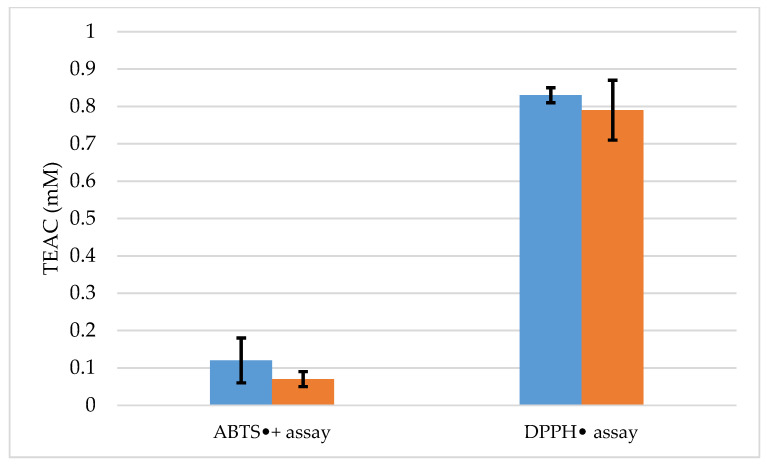
The DPPH• assay values were 0.79 ± 0.08 and 0.83 ± 0.02 mM, respectively, for specimens A and B (Figure 1). The DPPH• assay data confirm that both EOs from the specimens are active in the presence of the DPPH• radical and have a good antioxidant capacity. On the other hand, ABTS•+ values were 0.07 ± 0.02 mM for specimen A and 0.12 ± 0.06 mM for specimen B. These results confirm that the antioxidant potentials of the samples were lower than the Trolox standard, and lower when compared to the DPPH assay. This difference is evident in both tests, as observed in other studies reported in the literature [48,49,50].
Figure 1.
ABTS•+ and DPPH• radical scavenging assay e Trolox equivalent antioxidant capacity de Calycolpus goetheanus. Values are expressed as mean and standard deviation (n = 3) of Trolox equivalent antioxidant capacity. The ABTS•+ and DPPH• Radical Scavenging Assay (ABTS•+; DPPH•)  specimens B, and
  specimens A.
In the literature, there are no records of the antioxidant capacity of C. goetheanus EOs; however, the Myrtaceae family is described as having species with high antioxidant potential, as observed in studies [24,51]. In this sense, the antioxidant capacity shown by the C. goetheanus EOs may be associated with monoterpenes, and sesquiterpenic compounds 1,8-Cineole, (E)-caryophyllene, β-eudesmol, γ-eudesmol, δ-cadinene, and bulnesol, which are described in the literature for their antioxidant properties [52,53,54,55]. The high content of oxygenated sesquiterpenes shown for both EOs may have influenced the antioxidant potential of the samples, as these compounds can act individually or synergistically as antioxidants [56].
2.4. Phytotoxic Activity of the EOs
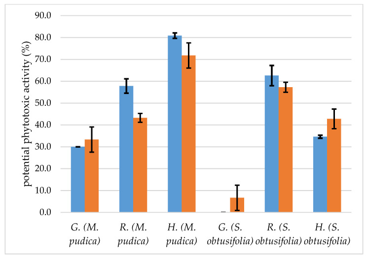
The results of the potential phytotoxic effect of C. goetheanus specimens B, and C are shown in Figure 2. We found that the essential oil samples showed different levels of intensity of seed germination inhibition, both for M. pudica and S. obtusifolia; for example, the specimen C essential oil had inhibition values of 33.33 ± 5.77% and 6.67 ± 5.77%, for M. pudica and S. obtusifolia, respectively. With regard to the essential oil sample of specimen B, the percentage of inhibition was equal to 30.00 ± 0.00% for M. pudica and showed no phytotoxic effect on the germination of S. obtusifolia. Potential phytotoxic effects were more intense for receptor species M. pudica. In other works in the literature, it is demonstrated that this invasive plant species is more susceptible to damage caused by substances present in EOs [57,58].
Figure 2.
Potential phytotoxic activity of C. goetheanus EOs from Calycolpus goetheanus.  specimens B, and
  specimens C. G = germination, R = radicle, and H = hypocotyl.
Regarding radicle elongation, the intensity of inhibition varied according to the receptor species, the specimen C essential oil inhibited the radicle elongation of M. pudica by 43.24 ± 2.03% and S. obtusifolia by 57.24 ± 2.28%, showing a greater effect on S. obtusifolia (Figure 2). Specimen B’s essential oil had a greater inhibitory effect for the radicle elongation of M. pudica with an inhibition of 57.83 ± 3.28% and 62.57 ± 4.63 for S. obtusifolia. In both cases analyzed, we can see that, for this variable studied, receptor plant S. obtusifolia was the most affected by the EOs. The results of this study, when compared to the literature, do not follow the same pattern of response; for example, in previous studies, the most affected receptor species was M. pudica [59,60]; however, these response patterns depend on factors other than the receptor species, such as the chemical profile of the essential oil [60,61,62,63].
The effects of EOs from specimens B and C on hypocotyl elongation followed a different pattern from the effects on radicle elongation, and at a different intensity of inhibition, with receptor species M. pudica being the most affected by both essential oil samples, the inhibition values were 80.87 ± 1.25 and 71.78 ± 5.75%, respectively (Figure 2), while S. obtusifolia showed lower susceptibility to the effects of EOs, with intensity levels of 34.63 ± 0.69% and 42.79 ± 4.50%, respectively (Figure 2). In the literature, a minimum inhibition of 50% is considered a satisfactory standard to evaluate the potentials of an essential oil [64], which was partially observed in this work (Figure 2).
According to Shao et al., [65], the 1,8-cineole compound obtained lower results for the inhibition of the root growth of Amaranthus retroflexus and Poa annua, when compared to the other two major constituents of the Seriphidium terrae-albae essential oil (α-thujone and β-thujone). Other authors also demonstrate the phytotoxic potential of 1,8-cineole on different species of receptor plants [66,67,68]; in addition, compounds such as δ-cadinene and (E)-Caryophyllene have also shown phytotoxic potential on several plant species; in addition, other compounds such as δ-cadinene and (E)-Caryophyllene have also shown phytotoxic potential on several plant species such as Mimosa pudica, Senna obtusifolia, Sinapis arvensis, Trifolium campestre, and Phalaris canariensis weeds [58,69], results similar to those of other authors [63,70]. Jaradat [71] points out that the Teucrium polium L. essential oil has α-cadinol as the component with the highest content (46.80%). According to the author, the natural chemicals of this species are known for their phytotoxic effects against different types of invasive species. According to Elshamy et al. [72], the Launaea spinosa EOs, which have γ-eudesmol as the third highest component (6.31%), showed phytotoxic activity against Portulaca oleracea.
2.5. In Silico Study
In our results, specimens B and C showed good post-emergence herbicidal activity against the species M. pudica L. and S. obtusifolia (L.) Irwin and Barneby were used as weed models. The HPPD protein has been reported as the molecular target of substances that have post-emergence emergence herbicidal activity [73,74,75,76]. Therefore, we used this protein as a target in order to investigate the molecular interactions and the affinity energy formed in the HPPD-compounds complexes (Figure 3). According to the literature, the majority are those compounds that have a concentration above 5% of the substance in the essential oil [77,78,79,80,81].
Figure 3.
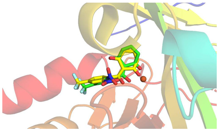
The structure obtained by redocking (yellow), overlapping the crystallographic structure (red) of HPPD complexed with NTBC.
To validate our docking protocol, we initially redocked the crystallographic ligand. For a docking protocol to be considered adequate, the RMSD value between the crystallographic ligand and the redocked ligand must be equal to or less than two angstroms [5,82,83,84].
To perform the docking methodology, we first performed the crystallographic ligand redocking to assess whether the software is able to reproduce the mode of interaction observed in the crystallographic structure of the protein. For this, the NTBC present in the PDB 6J63 was redocked and the results of the fitting poses were evaluated considering the RMSD value and the fitting score. The RMSD value between the redocked ligand and the crystallographic one was 1.85 Å (Figure 4). This result proves that the docking protocol used is suitable for the investigation of the molecular binding of the investigated complex.
Figure 4.
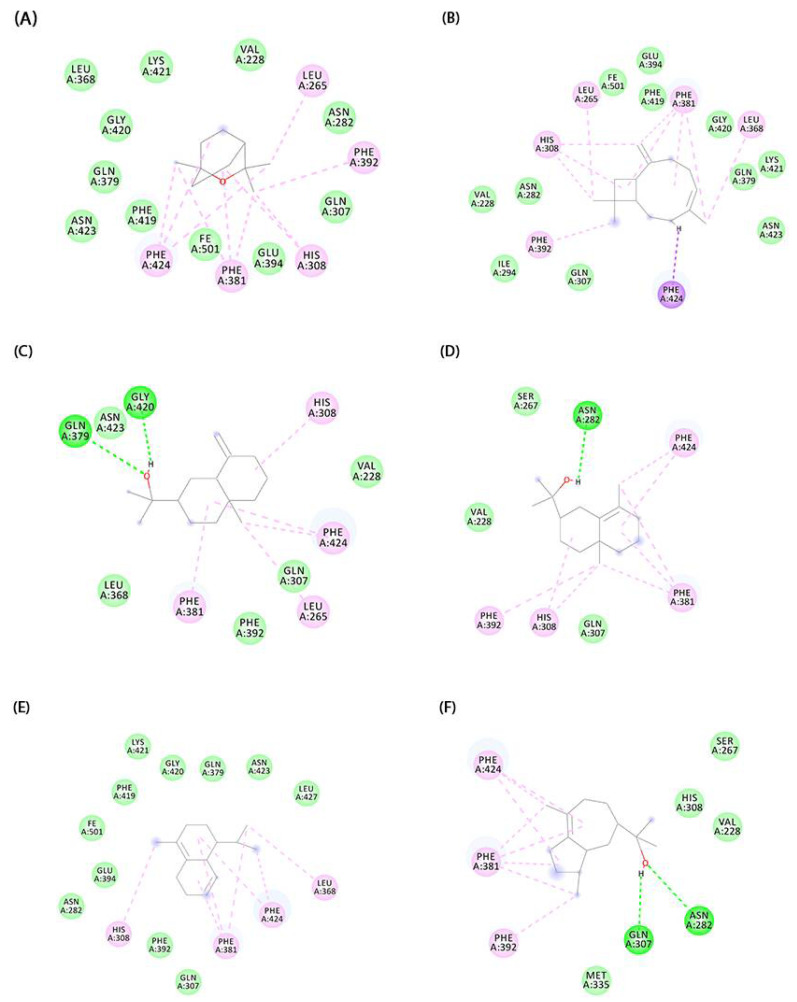
Docked conformation of molecules in the binding cavity of HPPD: In (A) we have the complex established with 1,8-Cineole, (B) (E)-Caryophyllene, (C) β-Eudesmol, (D) γ-Eudesmol, (E) δ-Cadinene, and (F) Bulnesol.
Then, molecular docking of the major compounds 2758 (1,8-cineole), 5281515 ((E)-Caryophyllene), 91457 (β-eudesmol), 6432005 (γ-eudesmol), 10657 (δ-cadinene), and 90785 (bulnesol) was performed. The affinity energy results are summarized in Table 2. In addition, the interactions established between the compounds and the HPPD active site are shown in Figure 4. From the simulated binding modes, it was possible to observe that the van der Waals and alkyl and pi-alkyl interactions were the main ones responsible for directing the receptor–ligand interaction. In some complexes, such as the one established between HPPD and α-cadinol, there was the formation of a hydrogen bond between Phe419 and the hydroxyl of the molecule. The difference in the mode of interaction and the distance between the ligands and Fe2+, present in the protein site, are capable of influencing the effectiveness of target inhibition [85,86]. Thus, the difference in EO inhibition capacity observed in phytotoxicity experiments may be related to the interaction of its major compounds with the protein site and its ability to chelate Fe2+.
Table 2.
Moldock scores obtained from the docking protocol using Molegro Virtual Docker 5.5.
| Molecule | MolDock Score | Rerank Score |
|---|---|---|
| 1,8-Cineole | −37.63 | −33.03 |
| (E)-Caryophyllene | −81.15 | −63.10 |
| β-Eudesmol | −73.23 | −55.07 |
| γ-Eudesmol | −72.77 | −63.24 |
| δ-Cadinene | −63.73 | −53.31 |
| Bulnesol | −85.15 | −68.20 |
3. Materials and Methods
3.1. Botanical Material
Aerial parts of three C. goetheanus specimens were collected in the coastal region of the State of Pará, in the city of Magalhães Barata, Brazil, the geographic coordinates of which are S 00°48′20.9′′ W 47°33′57.3′. C. goetheanus specimen (A) was collected on 4 October 2018 in a floodplain area on the left bank of the Curral River, and specimens (B) and (C) were collected on 20 September 2019, the first in a floodplain area on the right bank of the Curral River, while specimen (C) was collected in a secondary forest area (capoeira). The exsiccates were incorporated into the archive of Herbario Joao Murça Pires (MG) of Museu Paraense Emílio Goeldi, in the collection of Aromatic Plants of the Amazon, Belém, Pará and received records MG237476 (C. goetheanus A), MG237471 (C. goetheanus B), MG237475 (C. goetheanus C).
3.2. Preparation and Characterization of the Botanical Material
The samples of C. goetheanus leaves were dried in an oven with air circulation at 35 °C for 5 days, and then ground in a knife mill (Tecnal, model TE-631/3, Brazil). The moisture content was analyzed using an infrared moisture tester (ID50; GEHAKA, Duquesa de Goias, Real Parque, Sao Paulo, Brazil).
3.3. Extraction of EOs
The samples were subjected to hydrodistillation in modified Clevenger-type glass systems for 3 h, coupled to a refrigeration system to maintain the condensation water at around 12 °C. After the extraction, the oils were centrifuged for 5 min at 3000 rpm, de-hydrated with anhydrous sodium sulfate, and centrifuged again under the same conditions. Oil yield was calculated in mL/100 g. The oils were stored in amber glass ampoules, sealed with flame, and stored in a refrigerator at 5 °C [87].
3.4. Chemical Composition Analysis
The chemical compositions of the EOs of C. goetheanus (A, B, and C), were analyzed using a Shimadzu QP-2010 plus (Kyoto, Japan) a gas chromatography system equipped with an Rtx-5MS capillary column (30 m × 0.25 mm; 0.25 µm film thickness) (Restek Corporation, Bellefonte, PA, USA) coupled to a mass spectrometer (GC/MS) (Shimadzu, Kyoto, Japan). The program temperature was maintained at 60–240 °C at a rate of 3 °C/min, with an injector temperature of 250 °C, helium as the carrier gas (linear velocity of 32 cm/s, measured at 100 °C), and a splitless injection (1 μL of a 2:1000 hexane solution), using the same operating conditions as described in the literature [6,88,89,90]). The components were quantified using gas chromatography (GC) on a Shimadzu QP-2010 system (Kyoto, Japan), equipped with a flame ionization detector (FID) (Kyoto, Japan), under the same operating conditions as before, except for the carrier hydrogen gas. The retention index for all volatile constituents was calculated using a homologous series of n-alkanes (C8–C40) Sigma-Aldrich (St. Louis, MI, USA), according with Van den Dool and Kratz [91]. The components were identified by comparison (i) of the experimental mass spectra with those compiled in libraries (reference) and (ii) their retention indices to those found in the literature [32,33].
3.5. Trolox Equivalent Antioxidant Capacity (TEAC)
The ABTS•+ and DPPH• assays were methods used for the assessment of the antioxidant capacities of EOs. The antioxidant potential of the studied substances was determined according to their equivalence to the potent antioxidant, Trolox (6-hydroxy-2,5,7,8-tetramethylchromono-2-carboxylic acid; Sigma-Aldrich; 23881-3; São Paulo, Brazil), and a water-soluble synthetic vitamin E analogue.
3.5.1. The ABTS•+ Radical Scavenging Assay
The ABTS•+ Assay was determined according to the methodology adapted from Miller et al. [92], and modified by Re et al. [93]. ABTS•+ (2,2′-Azino-bis (3-ethylbenzothiazoline-6-sulfonic acid); Sigma-Aldrich; A1888; São Paulo, Brazil) was prepared using 7 mM ABTS•+ and 140 mM of potassium persulfate (K2O8S2; Sigma Aldrich; 216224; São Paulo, Brazil) incubated at room temperature without light for 16 h. Then, the solution was diluted with phosphate-buffered saline until it reached an absorbance of 0.700 (± 0.02) at 734 nm.
To measure the antioxidant capacity, 2.97 mL of the ABTS•+ solution was transferred to the cuvette, and the absorbance at 734 nm was determined using a Biospectro SP 22 spectrophotometer (São Paulo, Brazil). Then, 0.03 mL of the sample was added to the cuvette containing the ABTS•+ radical and, after 5 min, the second reading was performed. The synthetic antioxidant Trolox (6-hydroxy-2,5,7,8-tetramethylchromono-2-carboxylic acid; Sigma Aldrich; 23881-3; São Paulo, Brazil) was used as a standard solution for the calibration curve (y = 0.455x + 0.0002, where y represents the value of absorbance and x, the value of concentration, expressed as mM; R2 = 0.998). The results were expressed as mM. The values found for the samples were compared to the Trolox standard (1 mM).
3.5.2. DPPH• Radical Scavenging Assay
The test was carried out according to the method proposed by [94] To measure the antioxidant capacity, initially, the absorbance of DPPH• solution (2,2-diphenyl-1-picrylhydrazyl; Sigma-Aldrich; D9132; São Paulo, Brazil) 0.1 mM diluted in ethanol was determined. Subsequently, 0.6 mL of DPPH• solution, 0.35 mL of distilled water, and 0.05 mL of the sample were mixed and placed in a water bath at 37 °C for 30 min. Thereafter, the absorbances were determined in a spectrophotometer Bioespectro SP 22 (São Paulo, Brazil) at 517 nm. The synthetic antioxidant Trolox (6-hydroxy-2,5,7,8-tetramethylchromono-2-carboxylic acid; Sigma-Aldrich; 23881-3; São Paulo, Brazil) was used as a standard solution for the calibration curve (y = 0.2261x − 0.0094, where y represents the value of absorbance and x, the value of concentration, ex-pressed as mM; R2 = 0.9831). The results were expressed as mM. The values found for the samples were compared to the Trolox standard (1 mM).
3.6. Phytotoxic Potential Activity of the EOs
The phytotoxic potential bioassays, on the two species of receptor plants M. pudica and S. obtusifolia, were carried out with EOs of C. goetheanus, with only the EOs whose yield was ≥0.5 mL, that is, the samples A, and B, please, see Supplemental Material.
3.6.1. Seed Treatment
The phytotoxic activities were developed at the Agroindustry Laboratory of EM-BRAPA Amazonia Oriental, Belém, Pará, Brazil. The phytotoxic activity was evaluated in two bioindicator species that are weeds of common pasture areas in the Amazon region: M. pudica L. and S. obtusifolia (L.) Irwin and Barneby. Phytotoxic effects were analyzed on different parameters: percentage of seed germination and radicle and hypocotyl elongation. The seeds were collected in areas of cultivated pastures, in the degradation phase, in the municipality of Terra Alta-PA, underwent a cleaning process, and were treated in order to break dormancy, via immersion in concentrated sulfuric acid for 15 min [57,58].
3.6.2. Germination
The bioassays were performed as proposed by [57,58] with adaptations, in a BOD-type chamber, with controlled conditions of 25 °C and a photoperiod of 12 h, with monitoring for three days, daily counts, and elimination of germinated seeds. Seeds with a root length of 2 mm were considered germinated.
Each 9.00 cm diameter Petri dish was lined with a sheet of qualitative filter paper, where the test solutions were added only once, at the beginning of the bioassays, using 3 mL of the test solutions diluted in n-hexane. After the total evaporation of the solvent, 2.5 mL of distilled water was added; later, 10 seeds of the two receptor species (M. pudica, and S. obtusifolia) were added to each dish; the procedure was performed in triplicate.
3.6.3. Radicle and Hypocotyl Elongation
The radicle and hypocotyl elongation were performed in BOD-type chambers with a constant temperature of 25 °C and a photoperiod of 24 h. Each 9.0 cm diameter Petri dish received 3.0 mL of the test solution, lined with filter paper. The EOs were tested at the same concentrations as the germination bioassays. After evaporation of the solvent, a volume of 3 mL of distilled water was added, thus maintaining the original concentration.
The test solutions were added only once, at the beginning of the bioassays, and from then on only distilled water was added, whenever it was required to maintain the seedlings. In each of the plots, three pre-germinated seeds were placed for three days. At the end of the 7-day growth period, the length of the radicle and hypocotyl was measured. The control treatment consisted only of using distilled water. For all bioassays, the EOs were tested at concentrations of 1.0% (v/v), and the bioassays were performed in triplicate.
3.7. Prediction of Molecular Interactions
Molecular Docking
The protein 4-hydroxyphenylpyruvate dioxygenase (HPPD) has been reported as a molecular target for compounds with post-emergence herbicidal activity [73,74,75,76]. Because of this, we used this protein as a target for the major compounds in C. goetheanus essential oil. The three-dimensional structure of the HPPD protein was collected from the Protein Data Bank from PDB ID 6J63 [73]. The substances used in our studies were collected in PubChem from the CID’s compounds: 2758 (1,8-Cineole), 5281515 ((E)-Caryophyllene), 91457 (β-Eudesmol), 6432005 (γ-Eudesmol), 10657 (δ-Cadinene), and 90785 (Bulnesol). The molecular structure of these compounds was optimized with B3LYP/6-31G [95,96] using the Gaussian 09 [97]. To evaluate the molecular binding mode, the Molegro Virtual Docker 5.5 software [98,99,100] was used. The MolDock Score (GRID) scoring function was used with a Grid resolution of 0.30 Å. The protein binding site has a cavity with a volume of 388,096 and a surface of 1,076,482. The center is at X: 26.16, Y: −22.87, and Z: 4.28. The radius used to encompass the pocket binding was 12 Å. The MolDock SE algorithm was used for docking with the number of runs equal to 10, 1500 max interactions, and a max population size equal to 50. The maximum evaluation of 300 steps with a neighbor distance factor equal to 1 and energy threshold equal to 100 was used during the molecular docking simulation.
4. Conclusions
The chemical profile of EOs was characterized by the high content of hydrocarbon sesquiterpenes, especially (E)-caryophyllene (4.86 ± 13.61%), germacrene D (0.64 ± 6.34%), δ-cadinene (4.69 ± 8.01%), and oxygenated sesquiterpenes, mainly γ-eudesmol (1.56 ± 12.87%), α-cadinol (9.03%), and epi-α-muurolol, (5.69%). This significant sesquiterpene content may have influenced the strong elimination capacity of DPPH• free radicals observed in the EO of specimens A (0.79 ± 0.08 mM) and B (0.83 ± 0.02 mM) of C. goetheanus.
The recipient species Mimosa pudica presented greater sensitivity to the EOs of specimens B and C, with higher phytotoxic potential in hypocholic elongation with 80.87 ± 1.25% (specimen B) and 71.78 ± 5.75% (specimen C). This high inhibition potential may be peated to the presence of some terpenic compounds, such as δ-cadinene, 1,8-cineole, and (E)-Caryophyllene.
In the in silico study, specimens B and C showed good herbicide activity against the species M. pudica L. and S. obtusifolia, which may be associated with the difference in the inhibition capacity of the OE observed in the phytotoxicity experiments through the interaction of their major compounds with the protein site.
Acknowledgments
The authors C.d.J.P.F. and Â.A.B.d.M. thank CNPq for the scientific initiation scholarship. The author M.S.d.O.; thanks PCI-MCTIC/MPEG, as well as CNPq for the scholarship process number: (301194/2021-1).
Supplementary Materials
The following supporting information can be downloaded at: https://www.mdpi.com/article/10.3390/molecules27154678/s1, Experimental phytotoxicity tests of EOs, Figure S1.
Author Contributions
Conceptualization, C.d.J.P.F., Â.A.B.d.M. and O.O.F.; methodology, E.L.P.V., L.D.d.N., M.M.C., S.P., J.N.C., R.R.L., A.P.d.S.S.F. and M.S.d.O.; software, M.S.d.O.; formal analysis, E.H.d.A.A.; investigation, C.d.J.P.F.; writing—original draft preparation, C.d.J.P.F. and O.O.F.; writing—review and editing, M.S.d.O. and E.H.d.A.A.; visualization, S.P. and E.H.d.A.A.; supervision, E.H.d.A.A.; project administration, E.H.d.A.A.; funding acquisition, E.H.d.A.A. All authors have read and agreed to the published version of the manuscript.
Institutional Review Board Statement
Not applicable.
Informed Consent Statement
Not applicable.
Data Availability Statement
Not applicable.
Conflicts of Interest
The authors declare no conflict of interest.
Sample Availability
Samples of the compounds Calycolpus gotheanus are available from the authors.
Funding Statement
The APC was funded by Propesp/PAPQ—Programa de Apoio à Publicação Qualificada Edital 02/2022—PAPQ/PROPESP.
Footnotes
Publisher’s Note: MDPI stays neutral with regard to jurisdictional claims in published maps and institutional affiliations.
References
- 1.Asbahani A.E., Miladi K., Badri W., Sala M., Addi E.H.H.A., Casabianca H., El Mousadik A., Hartmann D., Jilale A., Renaud F.N.R., et al. Essential Oils: From Extraction to Encapsulation. Int. J. Pharm. 2015;483:220–243. doi: 10.1016/j.ijpharm.2014.12.069. [DOI] [PubMed] [Google Scholar]
- 2.Franco C.d.J.P., Ferreira O.O., Antônio Barbosa de Moraes Â., Varela E.L.P., do Nascimento L.D., Percário S., de Oliveira M.S., de Andrade E.H. Chemical Composition and Antioxidant Activity of Essential Oils from Eugenia patrisii Vahl, E. punicifolia (Kunth) DC.; and Myrcia Tomentosa (Aubl.) DC.; Leaf of Family Myrtaceae. Molecules. 2021;26:3292. doi: 10.3390/molecules26113292. [DOI] [PMC free article] [PubMed] [Google Scholar]
- 3.Hadidi M., Pouramin S., Adinepour F., Haghani S., Jafari S.M. Chitosan Nanoparticles Loaded with Clove Essential Oil: Characterization, Antioxidant and Antibacterial Activities. Carbohydr. Polym. 2020;236:116075. doi: 10.1016/j.carbpol.2020.116075. [DOI] [PubMed] [Google Scholar]
- 4.Bezerra F.W.F., de Oliveira M.S., Bezerra P.N., Cunha V.M.B., Silva M.P., da Costa W.A., Pinto R.H.H., Cordeiro R.M., da Cruz J.N., Chaves Neto A.M.J., et al. Extraction of Bioactive Compounds. In: Inamuddin, Asiri A.M., Suvardhan K., editors. Green Sustainable Process for Chemical and Environmental Engineering and Science. Elsevier; Amisterdan, The Netherlands: 2020. pp. 149–167. [Google Scholar]
- 5.De Oliveira M.S., Silva S.G., da Cruz J.N., Ortiz E., da Costa W.A., Bezerra F.W.F., Cunha V.M.B., Cordeiro R.M., de Jesus Chaves Neto A.M., de Aguiar Andrade E.H., et al. Supercritical CO2 Application in Essential Oil Extraction. In: Inamuddin R.M., Asiri A.M., editors. Industrial Applications of Green Solvents—Volume II. Materials Research Foundations; Millersville, PA, USA: 2019. pp. 1–28. [Google Scholar]
- 6.Santana de Oliveira M., da Cruz J.N., Almeida da Costa W., Silva S.G., Brito M.d.P., de Menezes S.A.F., de Jesus Chaves Neto A.M., de Aguiar Andrade E.H., de Carvalho Junior R.N. Chemical Composition, Antimicrobial Properties of Siparuna Guianensis Essential Oil and a Molecular Docking and Dynamics Molecular Study of Its Major Chemical Constituent. Molecules. 2020;25:3852. doi: 10.3390/molecules25173852. [DOI] [PMC free article] [PubMed] [Google Scholar]
- 7.Calo J.R., Crandall P.G., O’Bryan C.A., Ricke S.C. Essential Oils as Antimicrobials in Food Systems—A Review. Food Control. 2015;54:111–119. doi: 10.1016/j.foodcont.2014.12.040. [DOI] [Google Scholar]
- 8.Freires I., Denny C., Benso B., de Alencar S., Rosalen P. Antibacterial Activity of Essential Oils and Their Isolated Constituents against Cariogenic Bacteria: A Systematic Review. Molecules. 2015;20:7329–7358. doi: 10.3390/molecules20047329. [DOI] [PMC free article] [PubMed] [Google Scholar]
- 9.De Souza Neto J.D., Dos Santos E.K., Lucas E., Vetö N.M., Barrientos-Diaz O., Staggemeier V.G., Vasconcelos T., Turchetto-Zolet A.C. Advances and Perspectives on the Evolutionary History and Diversification of Neotropical myrteae (Myrtaceae) Bot. J. Linn. Soc. 2022;199:173–195. doi: 10.1093/botlinnean/boab095. [DOI] [Google Scholar]
- 10.Ferreira O.O., Cruz J.N., de Moraes Â.A.B., de Jesus Pereira Franco C., Lima R.R., dos Anjos T.O., Siqueira G.M., do Nascimento L.D., Cascaes M.M., de Oliveira M.S., et al. Essential Oil of the Plants Growing in the Brazilian Amazon: Chemical Composition, Antioxidants, and Biological Applications. Molecules. 2022;27:4373. doi: 10.3390/molecules27144373. [DOI] [PMC free article] [PubMed] [Google Scholar]
- 11.Proença C.E.B., Amorim B.S., Antonicelli M.C., Bünger M., Burton G.P., Caldas D.K.D., Costa I.R., Faria J.E.Q., Fernandes T., Gaem P.H., et al. Myrtaceae in Flora e Funga Do Brasil. [(accessed on 5 May 2022)]; Available online: https://floradobrasil.jbrj.gov.br/FB610263.
- 12.Fehlberg I., Ribeiro P.R., dos Santos I.B.F., dos Santos I.I.P., Guedes M.L.S., Ferraz C.G., Cruz F.G. Two New Sesquiterpenoids and One New P-Coumaroyl-Triterpenoid Derivative from Myrcia Guianensis. Phytochem. Lett. 2022;48:5–10. doi: 10.1016/j.phytol.2022.01.005. [DOI] [Google Scholar]
- 13.Castello A.C.D., Pereira A.S.S., Simões A.O., Koch I. Aspidosperma in Flora Do Brasil 2020. Jardim Botânico Do Rio de Janeiro. [(accessed on 19 July 2022)];2020 Available online: https://floradobrasil2020.jbrj.gov.br/FB40978.
- 14.Ferreira O.O., Neves da Cruz J., de Jesus Pereira Franco C., Silva S.G., da Costa W.A., de Oliveira M.S., de Aguiar Andrade E.H. First Report on Yield and Chemical Composition of Essential Oil Extracted from Myrcia Eximia DC (Myrtaceae) from the Brazilian Amazon. Molecules. 2020;25:783. doi: 10.3390/molecules25040783. [DOI] [PMC free article] [PubMed] [Google Scholar]
- 15.da Silva V.P., Alves C.C.F., Miranda M.L.D., Bretanha L.C., Balleste M.P., Micke G.A., Silveira E.V., Martins C.H.G., Ambrosio M.A.L.V., de Souza Silva T., et al. Chemical Composition and in Vitro Leishmanicidal, Antibacterial and Cytotoxic Activities of Essential Oils of the Myrtaceae Family Occurring in the Cerrado Biome. Ind. Crops Prod. 2018;123:638–645. doi: 10.1016/j.indcrop.2018.07.033. [DOI] [Google Scholar]
- 16.Habermann E., Pontes F.C., Pereira V.C., Imatomi M., Gualtieri S.C.J. Phytotoxic Potential of Young Leaves from Blepharocalyx salicifolius (Kunth) O. Berg (Myrtaceae) Brazilian J. Biol. 2016;76:531–538. doi: 10.1590/1519-6984.24114. [DOI] [PubMed] [Google Scholar]
- 17.Yasin M., Younis A., Javed T., Akram A., Ahsan M., Shabbir R., Ali M.M., Tahir A., El-Ballat E.M., Sheteiwy M.S., et al. River Tea Tree Oil: Composition, Antimicrobial and Antioxidant Activities, and Potential Applications in Agriculture. Plants. 2021;10:2105. doi: 10.3390/plants10102105. [DOI] [PMC free article] [PubMed] [Google Scholar]
- 18.Hazrati H., Saharkhiz M.J., Niakousari M., Moein M. Natural Herbicide Activity of Satureja Hortensis L. Essential Oil Nanoemulsion on the Seed Germination and Morphophysiological Features of Two Important Weed Species. Ecotoxicol. Environ. Saf. 2017;142:423–430. doi: 10.1016/j.ecoenv.2017.04.041. [DOI] [PubMed] [Google Scholar]
- 19.Zhou L., Li J., Kong Q., Luo S., Wang J., Feng S., Yuan M., Chen T., Yuan S., Ding C. Chemical Composition, Antioxidant, Antimicrobial, and Phytotoxic Potential of Eucalyptus Grandis × E. Urophylla Leaves Essential Oils. Molecules. 2021;26:1450. doi: 10.3390/molecules26051450. [DOI] [PMC free article] [PubMed] [Google Scholar]
- 20.Smeriglio A., Trombetta D., Cornara L., Valussi M., De Feo V., Caputo L. Characterization and Phytotoxicity Assessment of Essential Oils from Plant Byproducts. Molecules. 2019;24:2941. doi: 10.3390/molecules24162941. [DOI] [PMC free article] [PubMed] [Google Scholar]
- 21.Werrie P.-Y., Durenne B., Delaplace P., Fauconnier M.-L. Phytotoxicity of Essential Oils: Opportunities and Constraints for the Development of Biopesticides. A Review. Foods. 2020;9:1291. doi: 10.3390/foods9091291. [DOI] [PMC free article] [PubMed] [Google Scholar]
- 22.Dantas Costa A.M., Palomaris D., De Souza M., Cavalcante V., Lúcia De Araújo V., Ramos A.T., Maruo V.M. Plantas Tóxicas De Interesse Pecuário Em Região De Ecótono Amazônia E Cerrado. Parte Ii: Araguaína, Norte Do Tocantins. Acta Vet. Bras. 2011:317–324. [Google Scholar]
- 23.Barbosa J.D., da Silveira J.A.S., Albernaz T.T., da Silva e Silva N., dos Santos Belo Reis A., Oliveira C.M.C., Riet-Correa G., Duarte M.D. Lesões de Pele Causadas Pelos Espinhos de Mimosa Pudica (Leg. Mimosoideae) Nos Membros de Bovinos e Ovinos No Estado Do Pará. Pesqui. Vet. Bras. 2009;29:435–438. doi: 10.1590/S0100-736X2009000500014. [DOI] [Google Scholar]
- 24.Carneiro N.S., Alves C.C.F., Alves J.M., Egea M.B., Martins C.H.G., Silva T.S., Bretanha L.C., Balleste M.P., Micke G.A., Silveira E.V., et al. Chemical Composition, Antioxidant and Antibacterial Activities of Essential Oils from Leaves and Flowers of Eugenia Klotzschiana Berg (Myrtaceae) An. Acad. Bras. Cienc. 2017;89:1907–1915. doi: 10.1590/0001-3765201720160652. [DOI] [PubMed] [Google Scholar]
- 25.Pascoal A.C.R.F., Lourenço C.C., Sodek L., Tamashiro J.Y., Franchi G.C., Nowill A.E., Stefanello M.É.A., Salvador M.J. Essential Oil from the Leaves of Campomanesia guaviroba (DC.) Kiaersk. (Myrtaceae): Chemical Composition, Antioxidant and Cytotoxic Activity. J. Essent. Oil Res. 2011;23:34–37. doi: 10.1080/10412905.2011.9700479. [DOI] [Google Scholar]
- 26.Landrum L.R. Two New Species of Calycolpus (Myrtaceae) from Brazil. Brittonia. 2008;60:252–256. doi: 10.1007/s12228-008-9033-0. [DOI] [Google Scholar]
- 27.Gaem Paulo H. Calycolpus in Flora e Funga Do Brasil. [(accessed on 5 May 2022)]; Available online: https://floradobrasil.jbrj.gov.br/FB10263.
- 28.GAEM P.H. Calycolpus in Flora Do Brasil 2020. Jardim Botânico Do Rio de Janeiro. [(accessed on 19 July 2022)];2020 Available online: https://floradobrasil2020.jbrj.gov.br/FB10263.
- 29.Rosário A.S.D., Secco R.D.S., Amaral D.D.D., Santos J.U.M.D., Bastos M.D.N.D.C. Flórula Fanerogâmica Das Restingas Do Estado Do Pará. Ilhas de Algodoal e Maiandeua-2. Myrtaceae A.L. de Jussieu. Série Ciências Nat. 2005;1:31–42. [Google Scholar]
- 30.Pereira R.A., Zoghbi M.D.G.B., Bastos M.D.N.D.C. Essential Oils of Twelve Species of Myrtaceae Growing Wild in the Sandbank of the Resex Maracanã, State of Pará, Brazil. J. Essent. Oil Bear. Plants. 2010;13:440–450. doi: 10.1080/0972060X.2010.10643847. [DOI] [Google Scholar]
- 31.Dos Santos E.L., Lima A.M., Moura V.F.D.S., Setzer W.N., da Silva J.K.R., Maia J.G.S., Carneiro J.D.S., Figueiredo P.L.B. Seasonal and Circadian Rhythm of a 1,8-Cineole Chemotype Essential Oil of Calycolpus Goetheanus From Marajó Island, Brazilian Amazon. Nat. Prod. Commun. 2020;15:1–9. doi: 10.1177/1934578X20933055. [DOI] [Google Scholar]
- 32.Mondello L. Mass Spectra of Flavors and Fragrances of Natural and Synthetic Compounds. Volume 10. Wiley Blackwell; Hoboken, NJ, USA: 2015. pp. 4–5. [Google Scholar]
- 33.Adams R.P. Identification of Essential Oil Components by Gas Chromatograpy/Mass Spectrometry. 4th ed. Allured Publishing Corporation; Carol Stream, IL, USA: 2007. pp. 804–806. [Google Scholar]
- 34.Cai Z.-M., Peng J.-Q., Chen Y., Tao L., Zhang Y.-Y., Fu L.-Y., Long Q.-D., Shen X.-C. 1,8-Cineole: A Review of Source, Biological Activities, and Application. J. Asian Nat. Prod. Res. 2021;23:938–954. doi: 10.1080/10286020.2020.1839432. [DOI] [PubMed] [Google Scholar]
- 35.Rossi Y.E., Palacios S.M. Insecticidal Toxicity of Eucalyptus Cinerea Essential Oil and 1,8-Cineole against Musca Domestica and Possible Uses According to the Metabolic Response of Flies. Ind. Crops Prod. 2015;63:133–137. doi: 10.1016/j.indcrop.2014.10.019. [DOI] [Google Scholar]
- 36.Murata S., Shiragami R., Kosugi C., Tezuka T., Yamazaki M., Hirano A., Yoshimura Y., Suzuki M., Shuto K., Ohkohchi N., et al. Antitumor Effect of 1, 8-Cineole against Colon Cancer. Oncol. Rep. 2013;30:2647–2652. doi: 10.3892/or.2013.2763. [DOI] [PubMed] [Google Scholar]
- 37.Fidyt K., Fiedorowicz A., Strządała L., Szumny A. β -Caryophyllene and β -Caryophyllene Oxide-Natural Compounds of Anticancer and Analgesic Properties. Cancer Med. 2016;5:3007–3017. doi: 10.1002/cam4.816. [DOI] [PMC free article] [PubMed] [Google Scholar]
- 38.Moreira R.R.D., Santos A.G.D., Carvalho F.A., Perego C.H., Crevelin E.J., Crotti A.E.M., Cogo J., Cardoso M.L.C., Nakamura C.V. Antileishmanial Activity of Melampodium Divaricatum and Casearia Sylvestris Essential Oils on Leishmania Amazonensis. Rev. Inst. Med. Trop. Sao Paulo. 2019;61:1–7. doi: 10.1590/s1678-9946201961033. [DOI] [PMC free article] [PubMed] [Google Scholar]
- 39.De Oliveira C.C., de Oliveira C.V., Grigoletto J., Ribeiro L.R., Funck V.R., Grauncke A.C.B., de Souza T.L., Souto N.S., Furian A.F., Menezes I.R.A., et al. Anticonvulsant Activity of β-Caryophyllene against Pentylenetetrazol-Induced Seizures. Epilepsy Behav. 2016;56:26–31. doi: 10.1016/j.yebeh.2015.12.040. [DOI] [PubMed] [Google Scholar]
- 40.Brito L.F., Oliveira H.B.M., Neves Selis N., Souza C.L.S., Júnior M.N.S., Souza E.P., Silva L.S.C.D., Souza Nascimento F., Amorim A.T., Campos G.B., et al. Anti-inflammatory Activity of β -caryophyllene Combined with Docosahexaenoic Acid in a Model of Sepsis Induced by Staphylococcus Aureus in Mice. J. Sci. Food Agric. 2019;99:5870–5880. doi: 10.1002/jsfa.9861. [DOI] [PubMed] [Google Scholar]
- 41.Zheljazkov V.D., Kacaniova M., Dincheva I., Radoukova T., Semerdjieva I.B., Astatkie T., Schlegel V. Essential Oil Composition, Antioxidant and Antimicrobial Activity of the Galbuli of Six Juniper Species. Ind. Crops Prod. 2018;124:449–458. doi: 10.1016/j.indcrop.2018.08.013. [DOI] [Google Scholar]
- 42.Casiglia S., Bruno M., Bramucci M., Quassinti L., Lupidi G., Fiorini D., Maggi F. Kundmannia sicula (L.) DC: A Rich Source of Germacrene D. J. Essent. Oil Res. 2017;29:437–442. doi: 10.1080/10412905.2017.1338625. [DOI] [Google Scholar]
- 43.Guo X., Shang X., Li B., Zhou X.Z., Wen H., Zhang J. Acaricidal Activities of the Essential Oil from Rhododendron nivale Hook. f. and Its Main Compund, δ-Cadinene against Psoroptes Cuniculi. Vet. Parasitol. 2017;236:51–54. doi: 10.1016/j.vetpar.2017.01.028. [DOI] [PubMed] [Google Scholar]
- 44.Pérez-López A., Cirio A.T., Rivas-Galindo V.M., Aranda R.S., De Torres N.W. Activity against Streptococcus Pneumoniae of the Essential Oil and δ-Cadinene Isolated from Schinus Molle Fruit. J. Essent. Oil Res. 2011;23:25–28. doi: 10.1080/10412905.2011.9700477. [DOI] [Google Scholar]
- 45.Hui L.-M., Zhao G.-D., Zhao J.-J. δ-Cadinene Inhibits the Growth of Ovarian Cancer Cells via Caspase-Dependent Apoptosis and Cell Cycle Arrest. Int. J. Clin. Exp. Pathol. 2015;8:6046–6056. [PMC free article] [PubMed] [Google Scholar]
- 46.Plengsuriyakarn T., Karbwang J., Na-Bangchang K. Anticancer Activity Using Positron Emission Tomography-Computed Tomography and Pharmacokinetics of β -Eudesmol in Human Cholangiocarcinoma Xenografted Nude Mouse Model. Clin. Exp. Pharmacol. Physiol. 2015;42:293–304. doi: 10.1111/1440-1681.12354. [DOI] [PubMed] [Google Scholar]
- 47.Kotawong K., Chaijaroenkul W., Muhamad P., Na-Bangchang K. Cytotoxic Activities and Effects of Atractylodin and β-Eudesmol on the Cell Cycle Arrest and Apoptosis on Cholangiocarcinoma Cell Line. J. Pharmacol. Sci. 2018;136:51–56. doi: 10.1016/j.jphs.2017.09.033. [DOI] [PubMed] [Google Scholar]
- 48.Wootton-Beard P.C., Moran A., Ryan L. Stability of the Total Antioxidant Capacity and Total Polyphenol Content of 23 Commercially Available Vegetable Juices before and after in Vitro Digestion Measured by FRAP, DPPH, ABTS and Folin-Ciocalteu Methods. Food Res. Int. 2011;44:217–224. doi: 10.1016/j.foodres.2010.10.033. [DOI] [Google Scholar]
- 49.Santos D.N., De Souza L.L., De Oliveira C.A.F., Silva E.R., Oliveira A.L. De Arginase Inhibition, Antibacterial and Antioxidant Activities of Pitanga Seed (Eugenia uniflora L.) Extracts from Sustainable Technologies of High Pressure Extraction. Food Biosci. 2015;12:93–99. doi: 10.1016/j.fbio.2015.09.001. [DOI] [Google Scholar]
- 50.Ferreira O.O., Franco C.D.J.P., Varela E.L.P., Silva S.G., Cascaes M.M., Percário S., de Oliveira M.S., Andrade E.H.D.A. Chemical Composition and Antioxidant Activity of Essential Oils from Leaves of Two Specimens of Eugenia Florida DC. Molecules. 2021;26:5848. doi: 10.3390/molecules26195848. [DOI] [PMC free article] [PubMed] [Google Scholar]
- 51.Singh H.P., Kaur S., Negi K., Kumari S., Saini V., Batish D.R., Kohli R.K. Assessment of in Vitro Antioxidant Activity of Essential Oil of Eucalyptus Citriodora (Lemon-Scented Eucalypt; Myrtaceae) and Its Major Constituents. LWT–Food Sci. Technol. 2012;48:237–241. doi: 10.1016/j.lwt.2012.03.019. [DOI] [Google Scholar]
- 52.Diniz Do Nascimento L., Antônio Barbosa De Moraes A., Santana Da Costa K., Marcos J., Galúcio P., Taube P.S., Leal Costa M., Neves Cruz J., Helena De Aguiar Andrade E., Guerreiro De Faria L.J. Bioactive Natural Compounds and Antioxidant Activity of Essential Oils from Spice Plants: New Findings and Potential Applications. Biomolecules. 2020;10:988. doi: 10.3390/biom10070988. [DOI] [PMC free article] [PubMed] [Google Scholar]
- 53.Lima R.K., Cardoso M.D.G., Andrade M.A., Guimarães P.L., Batista L.R., Nelson D.L. Bactericidal and Antioxidant Activity of Essential Oils from Myristica fragrans Houtt and Salvia microphylla H.B.K. JAOCS, J. Am. Oil Chem. Soc. 2012;89:523–528. doi: 10.1007/s11746-011-1938-1. [DOI] [Google Scholar]
- 54.Hussain A.I., Anwar F., Iqbal T., Bhatti I.A. Antioxidant Attributes of Four Lamiaceae Essential Oils. Pakistan J. Bot. 2011;43:1315–1321. [Google Scholar]
- 55.Kundu A., Saha S., Walia S., Ahluwalia V., Kaur C. Antioxidant Potential of Essential Oil and Cadinene Sesquiterpenes of Eupatorium Adenophorum. Toxicol. Environ. Chem. 2013;95:127–137. doi: 10.1080/02772248.2012.759577. [DOI] [Google Scholar]
- 56.Assaeed A., Elshamy A., El Gendy A.E.N., Dar B., Al-Rowaily S., Abd-ElGawad A. Sesquiterpenes-Rich Essential Oil from above Ground Parts of Pulicaria Somalensis Exhibited Antioxidant Activity and Allelopathic Effect Onweeds. Agronomy. 2020;10:399. doi: 10.3390/agronomy10030399. [DOI] [Google Scholar]
- 57.de Oliveira M.S., da Costa W.A., Pereira D.S., Botelho J.R.S., de Alencar Menezes T.O., de Aguiar Andrade E.H., da Silva S.H.M., da Silva Sousa Filho A.P., de Carvalho R.N. Chemical Composition and Phytotoxic Activity of Clove (Syzygium aromaticum) Essential Oil Obtained with Supercritical CO2. J. Supercrit. Fluids. 2016;118:185–193. doi: 10.1016/j.supflu.2016.08.010. [DOI] [Google Scholar]
- 58.Gurgel E.S.C., de Oliveira M.S., Souza M.C., da Silva S.G., de Mendonça M.S., Souza Filho A.P.d.S. Chemical Compositions and Herbicidal (Phytotoxic) Activity of Essential Oils of Three Copaifera Species (Leguminosae-Caesalpinoideae) from Amazon-Brazil. Ind. Crops Prod. 2019;142:111850. doi: 10.1016/j.indcrop.2019.111850. [DOI] [Google Scholar]
- 59.Rodrigues I.M.C., Souza Filho A.P.S., Ferreira F.A., Demuner A.J. Prospecção Química de Compostos Produzidos Por Senna Alata Com Atividade Alelopática. Planta Daninha. 2010;28:1–12. doi: 10.1590/S0100-83582010000100001. [DOI] [Google Scholar]
- 60.Souza Filho A.P.S., Mourão Jr. M. Padrão de Resposta de Mimosa Pudica e Senna Obtusifolia à Atividade Potencialmente Alelopática de Espécies de Poaceae. Planta Daninha. 2010;28:927–938. doi: 10.1590/S0100-83582010000500001. [DOI] [Google Scholar]
- 61.Ootani M.A., dos Reis M.R., Cangussu A.S.R., Capone A., Fidelis R.R., Oliveira W., Barros H.B., Portella A.C.F., de Souza Aguiar R., dos Santos W.F. Phytotoxic Effects of Essential Oils in Controlling Weed Species Digitaria Horizontalis and Cenchrus Echinatus. Biocatal. Agric. Biotechnol. 2017;12:59–65. doi: 10.1016/j.bcab.2017.08.016. [DOI] [Google Scholar]
- 62.Gatto L.J., Fabri N.T., Souza A.M.D., Fonseca N.S.T.D., Furusho A.D.S., Miguel O.G., Dias J.D.F.G., Zanin S.M.W., Miguel M.D. Chemical Composition, Phytotoxic Potential, Biological Activities and Antioxidant Properties of Myrcia hatschbachii D. Legrand Essential Oil. Braz. J. Pharm. Sci. 2020;56:1–9. doi: 10.1590/s2175-97902019000318402. [DOI] [Google Scholar]
- 63.Cândido A.C.S., Scalon S.P.Q., Silva C.B., Simionatto E., Morel A.F., Stüker C.Z., Matos M.F.C., Peres M.T.L.P. Chemical Composition and Phytotoxicity of Essential Oils of Croton doctoris S. Moore (Euphorbiaceae) Brazilian J. Biol. 2022;82:1–11. doi: 10.1590/1519-6984.231957. [DOI] [PubMed] [Google Scholar]
- 64.Verdeguer M., Sánchez-Moreiras A.M., Araniti F. Phytotoxic Effects and Mechanism of Action of Essential Oils and Terpenoids. Plants. 2020;9:1571. doi: 10.3390/plants9111571. [DOI] [PMC free article] [PubMed] [Google Scholar]
- 65.Shao H., Hu Y., Han C., Wei C., Zhou S., Zhang C., Zhang C. Chemical Composition and Phytotoxic Activity of Seriphidium terrae-albae (Krasch) Poljakov (Compositae) Essential Oil. Chem. Biodivers. 2018;15:e1800348. doi: 10.1002/cbdv.201800348. [DOI] [PubMed] [Google Scholar]
- 66.Alipour M., Saharkhiz M.J. Phytotoxic Activity and Variation in Essential Oil Content and Composition of Rosemary (Rosmarinus officinalis L.) during Different Phenological Growth Stages. Biocatal. Agric. Biotechnol. 2016;7:271–278. doi: 10.1016/j.bcab.2016.07.003. [DOI] [Google Scholar]
- 67.Sharma M., Arora K., Sachdev R.K. Bio-Herbicidal Potential of Essential Oil of Callistemon—A Review. Indian J. Ecol. 2021;48:252–260. [Google Scholar]
- 68.Caputo L., Trotta M., Romaniello A., De Feo V. Chemical Composition and Phytotoxic Activity of Rosmarinus Officinalis Essential Oil. Nat. Prod. Commun. 2018;13:1367–1370. doi: 10.1177/1934578X1801301033. [DOI] [Google Scholar]
- 69.Amri I., Hanana M., Jamoussi B., Hamrouni L. Essential Oils of Pinus Nigra J.F. Arnold Subsp. Laricio Maire: Chemical Composition and Study of Their Herbicidal Potential. Arab. J. Chem. 2017;10:S3877–S3882. doi: 10.1016/j.arabjc.2014.05.026. [DOI] [Google Scholar]
- 70.Araniti F., Sánchez-Moreiras A.M., Graña E., Reigosa M.J., Abenavoli M.R. Terpenoid Trans-Caryophyllene Inhibits Weed Germination and Induces Plant Water Status Alteration and Oxidative Damage in Adult Arabidopsis. Plant Biol. 2017;19:79–89. doi: 10.1111/plb.12471. [DOI] [PubMed] [Google Scholar]
- 71.Jaradat N.A. Review of the Taxonomy, Ethnobotany, Phytochemistry, Phytotherapy and Phytotoxicity of Germander Plant (Teucrium polium L.) Asian J. Pharm. Clin. Res. 2015;8:13–19. [Google Scholar]
- 72.Elshamy A.I., Abd-ElGawad A.M., El-Amier Y.A., El Gendy A.E.G., Al-Rowaily S.L. Interspecific Variation, Antioxidant and Allelopathic Activity of the Essential Oil from Three Launaea Species Growing Naturally in Heterogeneous Habitats in Egypt. Flavour Fragr. J. 2019;34:316–328. doi: 10.1002/ffj.3512. [DOI] [Google Scholar]
- 73.Lin H.Y., Yang J.F., Wang D.W., Hao G.F., Dong J.Q., Wang Y.X., Yang W.C., Wu J.W., Zhan C.G., Yang G.F. Molecular Insights into the Mechanism of 4-Hydroxyphenylpyruvate Dioxygenase Inhibition: Enzyme Kinetics, X-Ray Crystallography and Computational Simulations. FEBS J. 2019;286:975–990. doi: 10.1111/febs.14747. [DOI] [PubMed] [Google Scholar]
- 74.Wang H., Wang L., Zhang X., Bai S., Jin T., Liu W., Wang J. Unravelling Phytotoxicity and Mode of Action of Tripyrasulfone, a Novel Herbicide. J. Agric. Food Chem. 2021;69:7168–7177. doi: 10.1021/acs.jafc.1c01294. [DOI] [PubMed] [Google Scholar]
- 75.Wang H., Liu B., Lei P., Zhu J., Chen L., He Q., He J. Improving the Herbicide Resistance of 4-Hydroxyphenylpyruvate Dioxygenase SpHPPD by Directed Evolution. Enzyme Microb. Technol. 2022;154:109964. doi: 10.1016/j.enzmictec.2021.109964. [DOI] [PubMed] [Google Scholar]
- 76.Neuckermans J., Lequeue S., Mertens A., Branson S., Schwaneberg U., De Kock J. High-Throughput Quantification of Ochronotic Pigment Formation in Escherichia Coli to Evaluate the Potency of Human 4-Hydroxyphenylpyruvate Dioxygenase Inhibitors in Multi-Well Format. MethodsX. 2021;8:101181. doi: 10.1016/j.mex.2020.101181. [DOI] [PMC free article] [PubMed] [Google Scholar]
- 77.Cascaes M.M., Carneiro O.D.S., Nascimento L.D.D., de Moraes Â.A.B., de Oliveira M.S., Cruz J.N., Guilhon G.M.S.P., Andrade E.H.D.A. Essential Oils from Annonaceae Species from Brazil: A Systematic Review of Their Phytochemistry, and Biological Activities. Int. J. Mol. Sci. 2021;22:12140. doi: 10.3390/ijms222212140. [DOI] [PMC free article] [PubMed] [Google Scholar]
- 78.Ramos da Silva L.R., Ferreira O.O., Cruz J.N., de Jesus Pereira Franco C., Oliveira dos Anjos T., Cascaes M.M., Almeida da Costa W., Helena de Aguiar Andrade E., Santana de Oliveira M. Lamiaceae Essential Oils, Phytochemical Profile, Antioxidant, and Biological Activities. Evid. Based Complement. Altern. Med. 2021;2021:6748052. doi: 10.1155/2021/6748052. [DOI] [PMC free article] [PubMed] [Google Scholar]
- 79.Mesquita K.D.S.M., Feitosa B.D.S., Cruz J.N., Ferreira O.O., Franco C.D.J.P., Cascaes M.M., Oliveira M.S.D., Andrade E.H.D.A. Chemical Composition and Preliminary Toxicity Evaluation of the Essential Oil from Peperomia circinnata Link Var. circinnata. (Piperaceae) in Artemia Salina Leach. Molecules. 2021;26:7359. doi: 10.3390/molecules26237359. [DOI] [PMC free article] [PubMed] [Google Scholar]
- 80.de Oliveira M.S., Cruz J.N., Ferreira O.O., Pereira D.S., Pereira N.S., Oliveira M.E.C., Venturieri G.C., Guilhon G.M.S.P., Souza Filho A.P.D.S., Andrade E.H.D.A. Chemical Composition of Volatile Compounds in Apis Mellifera Propolis from the Northeast Region of Pará State, Brazil. Molecules. 2021;26:3462. doi: 10.3390/molecules26113462. [DOI] [PMC free article] [PubMed] [Google Scholar]
- 81.Cruz J.N., de Oliveira M.S., Andrade E.H.D.A., Rodrigues Lima R. Molecular Modeling Approaches Can Reveal the Molecular Interactions Established between a Biofilm and the Bioactive Compounds of the Essential Oil of Piper Divaricatum. Molecules. 2022;27:4199. doi: 10.3390/molecules27134199. [DOI] [PMC free article] [PubMed] [Google Scholar]
- 82.da Rocha Galucio N.C., de Araújo Moysés D., Pina J.R.S., Marinho P.S.B., Júnior P.C.G., Cruz J.N., Vale V.V., Khayat A.S., do Rosario Marinho A.M. Antiproliferative, Genotoxic Activities and Quantification of Extracts and Cucurbitacin B Obtained from Luffa operculata (L.) Cogn. Arab. J. Chem. 2022;15:103589. doi: 10.1016/j.arabjc.2021.103589. [DOI] [Google Scholar]
- 83.Neto R.D.A., Santos C.B., Henriques S.V., Machado L.D.O., Cruz J.N., da Silva C.H.D.P., Federico L.B., Oliveira E.H.D., de Souza M.P., da Silva P.N., et al. Novel Chalcones Derivatives with Potential Antineoplastic Activity Investigated by Docking and Molecular Dynamics Simulations. J. Biomol. Struct. Dyn. 2022;40:2204–2216. doi: 10.1080/07391102.2020.1839562. [DOI] [PubMed] [Google Scholar]
- 84.Castro A.L.G., Cruz J.N., Sodré D.F., Correa-Barbosa J., Azonsivo R., de Oliveira M.S., de Sousa Siqueira J.E., da Rocha Galucio N.C., de Oliveira Bahia M., Burbano R.M.R., et al. Evaluation of the Genotoxicity and Mutagenicity of Isoeleutherin and Eleutherin Isolated from Eleutherine Plicata Herb. Using Bioassays and in Silico Approaches. Arab. J. Chem. 2021;14:103084. doi: 10.1016/j.arabjc.2021.103084. [DOI] [Google Scholar]
- 85.Zhang B., Zhang L., He L., Yang X., Shi Y., Liao S., Yang S., Cheng J., Ren T. Interactions of Fipronil within Fish and Insects: Experimental and Molecular Modeling Studies. J. Agric. Food Chem. 2018;66:5756–5761. doi: 10.1021/acs.jafc.8b00573. [DOI] [PubMed] [Google Scholar]
- 86.Gao Y., He L., Zhu J., Cheng J., Li B., Liu F., Mu W. The Relationship between Features Enabling SDHI Fungicide Binding to the Sc-Sdh Complex and Its Inhibitory Activity against Sclerotinia Sclerotiorum. Pest Manag. Sci. 2020;76:2799–2808. doi: 10.1002/ps.5827. [DOI] [PubMed] [Google Scholar]
- 87.Ribeiro F.P., Santana de Oliveira M., de Oliveira Feitosa A., Santana Barbosa Marinho P., Moacir do Rosario Marinho A., de Aguiar Andrade E.H., Favacho Ribeiro A. Chemical Composition and Antibacterial Activity of the Lippia Origanoides Kunth Essential Oil from the Carajás National Forest, Brazil. Evid. Based Complement. Altern. Med. 2021;2021:1–8. doi: 10.1155/2021/9930336. [DOI] [PMC free article] [PubMed] [Google Scholar]
- 88.Cascaes M.M., Silva S.G., Cruz J.N., Santana de Oliveira M., Oliveira J., de Moraes A.A.B., da Costa F.A.M., da Costa K.S., Diniz do Nascimento L., Helena de Aguiar Andrade E. First Report on the Annona Exsucca DC. Essential Oil and in Silico Identification of Potential Biological Targets of Its Major Compounds. Nat. Prod. Res. 2021;35:1–4. doi: 10.1080/14786419.2021.1893724. [DOI] [PubMed] [Google Scholar]
- 89.Souza da Silva Júnior O., de Jesus Pereira Franco C., Barbosa de Moraes A.A., Cruz J.N., Santana da Costa K., Diniz do Nascimento L., Helena de Aguiar Andrade E. In Silico Analyses of Toxicity of the Major Constituents of Essential Oils from Two Ipomoea L. Species. Toxicon. 2021;195:111–118. doi: 10.1016/j.toxicon.2021.02.015. [DOI] [PubMed] [Google Scholar]
- 90.Nascimento L.D.D., Silva S.G., Cascaes M.M., Costa K.S.D., Figueiredo P.L.B., Costa C.M.L., Andrade E.H.D.A., de Faria L.J.G. Drying Effects on Chemical Composition and Antioxidant Activity of Lippia Thymoides Essential Oil, a Natural Source of Thymol. Molecules. 2021;26:2621. doi: 10.3390/molecules26092621. [DOI] [PMC free article] [PubMed] [Google Scholar]
- 91.van Den Dool H., Dec Kratz P. A Generalization of the Retention Index System Including Linear Temperature Programmed Gas—Liquid Partition Chromatography. J. Chromatogr. A. 1963;11:463–471. doi: 10.1016/S0021-9673(01)80947-X. [DOI] [PubMed] [Google Scholar]
- 92.Miller M., Rao J.K.M., Wlodawer A., Gribskov M.R. A Left-Handed Crossover Involved in Amidohydrolase Catalysis. Crystal Structure of Erwinia Chrysanthemi l-Asparaginase with Bound l-Aspartate. FEBS Lett. 1993;328:275–279. doi: 10.1016/0014-5793(93)80943-O. [DOI] [PubMed] [Google Scholar]
- 93.Re R., Pellegrini N., Proteggente A., Pannala A., Yang M., Rice-Evans C. Antioxidant Activity Applying an Improved ABTS Radical Cation Decolorization Assay. Free Radic. Biol. Med. 1999;26:1231–1237. doi: 10.1016/S0891-5849(98)00315-3. [DOI] [PubMed] [Google Scholar]
- 94.Blois M.S. Antioxidant Determinations by the Use of a Stable Free Radical. Nature. 1958;181:1199–1200. doi: 10.1038/1811199a0. [DOI] [Google Scholar]
- 95.Becke A.D. Density-functional Thermochemistry. III. The Role of Exact Exchange. J. Chem. Phys. 1993;98:5648–5652. doi: 10.1063/1.464913. [DOI] [Google Scholar]
- 96.Lee C., Yang W., Parr R.G. Development of the Colle-Salvetti Correlation-Energy Formula into a Functional of the Electron Density. Phys. Rev. B. 1988;37:785–789. doi: 10.1103/PhysRevB.37.785. [DOI] [PubMed] [Google Scholar]
- 97.Frisch M.J., Trucks G.W., Schlegel H.B., Scuseria G.E., Robb M.A., Cheeseman J.R., Scalmani G., Barone V., Petersson G.A., Nakatsuji H., et al. Gaussian 16 Revision 16.A.03. Gaussian Inc.; Wallingford, CT, USA: 2016. [Google Scholar]
- 98.Thomsen R., Christensen M.H. MolDock: A New Technique for High-Accuracy Molecular Docking. J. Med. Chem. 2006;49:3315–3321. doi: 10.1021/jm051197e. [DOI] [PubMed] [Google Scholar]
- 99.Santana de Oliveira M., Pereira da Silva V.M., Cantão Freitas L., Gomes Silva S., Nevez Cruz J., Aguiar Andrade E.H. Extraction Yield, Chemical Composition, Preliminary Toxicity of Bignonia nocturna (Bignoniaceae) Essential Oil and in Silico Evaluation of the Interaction. Chem. Biodivers. 2021;18:e2000982. doi: 10.1002/cbdv.202000982. [DOI] [PubMed] [Google Scholar]
- 100.Costa E.B., Silva R.C., Espejo-Román J.M., Neto M.F.d.A., Cruz J.N., Leite F.H.A., Silva C.H.T.P., Pinheiro J.C., Macêdo W.J.C., Santos C.B.R. Chemometric Methods in Antimalarial Drug Design from 1,2,4,5-Tetraoxanes Analogues. SAR QSAR Environ. Res. 2020;31:677–695. doi: 10.1080/1062936X.2020.1803961. [DOI] [PubMed] [Google Scholar]
Associated Data
This section collects any data citations, data availability statements, or supplementary materials included in this article.
Supplementary Materials
Data Availability Statement
Not applicable.


