Introduction
Leg pain, sacroiliac pain and backache are common complaints of pregnant women. Hip pain due to transient osteoporosis (TOH) is seen in third trimester, while incidence of orthopaedic injuries is 1–6%. Femoral neck fractures contribute less than 1% [1]. Femoral neck fractures are due to high velocity injuries when it happens in a normal bone. It is very rare for young pregnant patients to have such fracture without a trauma unless the bone quality is affected.
Here we report the orthopaedic and obstetric management of a primigravida at 29 weeks 5 days who had a fracture neck of femur without trauma.
Case Report
Twenty-six-year-old primigravida at 29-week and 5-day gestation presented with pain in the left hip for 3 weeks and inability to walk for 3 days. Three days prior to admission, she experienced exacerbation of pain and difficulty in lifting left leg. She had subclinical hypothyroidism diagnosed in first trimester and was on levothyroxine sodium 25 mcg supplement. She had gestational diabetes and was on insulin and oral hypoglycaemic agents. She did not give history of any steroid use, smoking or alcohol abuse. She had normal pulse rate, respiratory rate and blood pressure. Her uterus was 30 weeks with normal foetal heart sounds and without any signs of abruption. Her body weight was 75 kg and BMI 32.
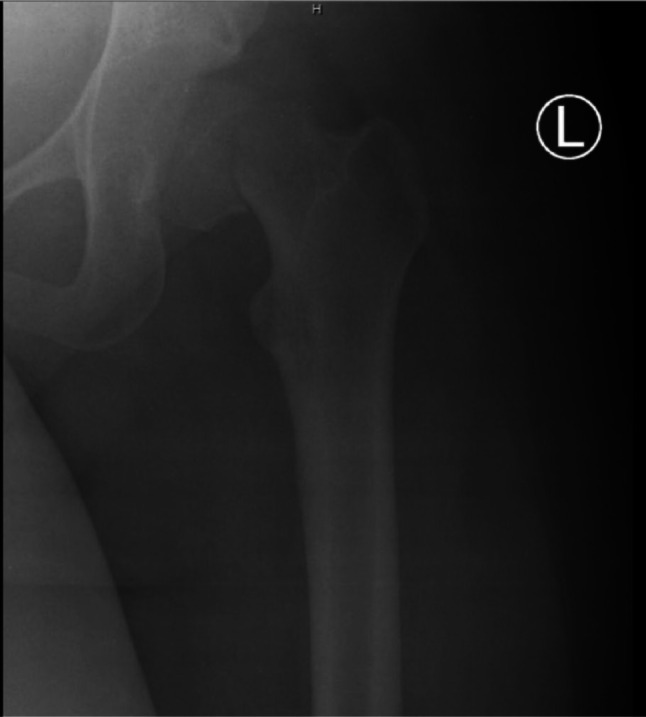
On examination her left lower limb was shortened and externally rotated. Plain radiographs showed a displaced subcapital fracture of the neck of the left femur (Fig. 1) with an inferiorly subluxed femoral head. Though we did not do a DEXA scan to assess bone mineral density, the bone density of fractured hip was less than a normal young lady of the same age as per the plain radiograph using Singh index.
Fig. 1.

Displaced subcapital fracture left femoral neck
Obstetric ultrasound done showed an appropriate for gestation grown foetus, adequate liquor and a posterior fundal placenta. Complete haemogram, coagulation profile, thyroid function tests and sugar monitoring were normal. Rheumatoid factor, antinuclear antibody and C-reactive protein were normal which ruled out inflammatory arthritis-associated osteoporosis. Serum calcium level was 9.2 mmol and vitamin D level was 23 ng/ml. Renal parameters, serum phosphorus, alkaline phosphatase and parathyroid hormone levels were also normal. After explaining the possible diagnosis of pathological fracture and risk of preterm labour she was taken up for surgery within 24 h.
She underwent closed reduction and internal fixation with two 32-mm partially threaded noncannulated stainless steel screws under spinal anaesthesia. Post-operatively she was mobilised from day one except for weight bearing on operated limb and was given vitamin D correction. Gradual partial weight bearing of operated limb was encouraged at 6 weeks post-surgery.
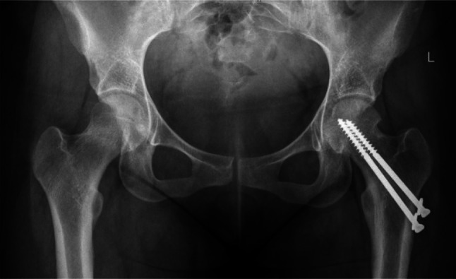
She presented with labour pains at 38-week gestation. Caesarean section was done under regional anaesthesia. A term male baby appropriate for gestational age, 2.54 kg with normal Apgar scores, was delivered. Plain radiographs showed a well-healed fracture at 3 months. She started full weight bearing on operated limb at 3 months from index surgery. There was no evidence of avascular necrosis or arthritis at 26 months of follow-up (Fig. 2). Informed consent was obtained from the patient prior to reporting.
Fig. 2.

United fracture femoral neck at 26 months
Discussion
Neck of femur fracture is a common osteoporotic fracture in elderly population occurring after a trivial fall. In our patient we ruled out causes of osteoporosis like hyperparathyroidism, Cushing’s syndrome, inflammatory arthritis, renal osteodystrophy by doing appropriate blood tests. Patient did not have any habits of alcohol abuse, carbonated juice addiction or smoking which predispose to osteoporosis. Moreover there was no history of trauma prior to the fracture. Low vitamin D levels and subclinical hypothyroidism are risk factors for osteoporosis [2, 3]. Our patient had these two risk factors to associate with TOH.
The Osteoporosis Self-Assessment Tool for Asians (OSTA) has been developed to identify patients with high osteoporosis risk in elderly group. The OSTA index is calculated with the formula: [body weight (kg) − age (years)] × 0.2. The OSTA values are classified as follows: < − 4, high risk; − 4 to − 1, intermediate risk; > − 1, low risk.
In our case, the OSTA index is 0.98 and hence our patient is in the low-risk category since she was young. Singh index was calculated on plain radiographs by identifying the absence of trabecular pattern of the proximal femur. Normal bone is given an index 6 and severe osteoporosis is index 1. In our case, the index is 2 which is osteoporotic. As it is unlikely for young patients to undergo bone mineral density testing prior to pregnancy, it is difficult to confirm the diagnosis of an underlying osteoporosis in these patients. As no other risk factors were identified and considering the Singh index of 2, we conclude transient osteoporosis of hip (TOH) as the underlying pathology in our patient by exclusion.
TOH is a self-limiting condition that presents spontaneously in the third trimester with sudden-onset pain in the hip which resolves in 6–14 months. In detected cases of TOH, avoiding vigorous activity, use of nonweight bearing measures and physiotherapy decrease the risk of fracture. 12.1% of TOH patients were identified to have hip fractures [4]. TOH and avascular necrosis (AVN) of femoral head have similar clinical presentation and radiographic findings in the early stage. MRI can be used to distinguish both during pregnancy. Nonunion and avascular necrosis are common complications in patients with fracture of the neck of femur with underlying TOH. In our patient the fracture united in 3 months and did not show any signs of AVN at 26 months of follow-up. There was no foetal or maternal morbidity in our case unlike the high reported incidence of maternal and foetal adverse outcomes.
Conclusion
This case illustrates the rare occurrence of a fracture without any history of trauma in a pregnant patient with underlying unidentified TOH. There is a greater need for awareness among health professionals to start vitamin D supplementation in pregnant women from ten weeks of pregnancy. Further studies are required to evaluate the need for routine vitamin D screening in pregnant patients with subclinical hypothyroidism to identify patients with underlying TOH and establish association.
Acknowledgment
We acknowledge the contribution of the orthopaedic and anaesthesia team in management of this case.
Dr. Tinu Philip
is currently working as Senior Specialist in the Royal Oman Police Hospital, Sultanate of Oman. She is a graduate of Kottayam Medical College, Kerala, and completed her post-graduation from the Institute of Obstetrics and Gynaecology, Madras Medical College, Chennai, in 2007. She held the post of Vice Dean Undergraduate Studies for three years in Believers Church Medical College Hospital and was involved in various administrative activities along with clinical work. Her interests include medical education, clinical research, high-risk obstetrics, improving antenatal care and gynaecology services in low-resource settings.
Funding
This research received no specific grant from any funding agency in the public, commercial or not for profit sectors.
Declarations
Conflict of interest
We declare that we have no conflicts of interest relevant to this article.
Informed Consent
Informed consent was obtained from the participant of the study before publication.
Footnotes
Tinu Philip is an Associate Professor, Department of Obstetrics and Gynaecology, Believers Church Medical College Hospital, Tiruvalla, Kerala, India; James C. George is an Associate Professor, Department of Orthopaedics, Believers Church Medical College Hospital, Tiruvalla; Kunjamma Roy is a Professor, Department of Obstetrics and Gynaecology, Believers Church Medical College Hospital, Tiruvalla, Kerala, India; Rekha G Muricken is an Associate Professor, Department of Obstetrics and Gynaecology, Believers Church Medical College Hospital, Tiruvalla, Kerala, India; Kuruvilla P Chacko is an Professor, Department of Obstetrics and Gynaecology, Believers Church Medical College Hospital, Tiruvalla, Kerala, India.
Publisher's Note
Springer Nature remains neutral with regard to jurisdictional claims in published maps and institutional affiliations.
References
- 1.Harold JA, Isaacson E, Palatnik A. Femoral fracture in pregnancy: a case series and review of clinical management. Int J Women's Health. 2019;11:267–271. doi: 10.2147/IJWH.S198345. [DOI] [PMC free article] [PubMed] [Google Scholar]
- 2.Paoletta M, Moretti A, Liguori S, et al. Transient osteoporosis of the hip and subclinical hypothyroidism: an unusual dangerous duet? Case report and pathogenetic hypothesis. BMC Musculoskelet Disord. 2020;21:543. doi: 10.1186/s12891-020-03574-x. [DOI] [PMC free article] [PubMed] [Google Scholar]
- 3.Zhou X, Li Z, Li B, Guo S, Yao M. Expression and Clinical Significance of Serum 25-OH-D in pregnant women with SCH (Subclinical Hypothyroidism) and GDM (Gestational Diabetes Mellitus) Pak J Med Sci. 2018;34(5):1278–1282. doi: 10.12669/pjms.345.15719. [DOI] [PMC free article] [PubMed] [Google Scholar]
- 4.Hadji P, Boekhoff J, Hahn M, et al. Pregnancy-associated transient osteoporosis of the hip: results of a case-control study. Arch Osteoporos. 2017;12(1):11. doi: 10.1007/s11657-017-0310-y. [DOI] [PubMed] [Google Scholar]


